

Two cases of breast carcinoma with osteoclastic giant cells: are the osteoclastic giant cells pro-tumoural differentiation of macrophages?
- PMID: 20731838
- PMCID: PMC2936386
- DOI: 10.1186/1746-1596-5-55
Two cases of breast carcinoma with osteoclastic giant cells: are the osteoclastic giant cells pro-tumoural differentiation of macrophages?
Abstract
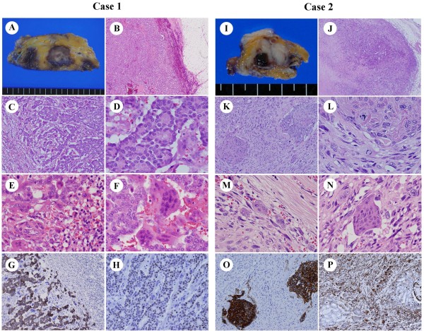
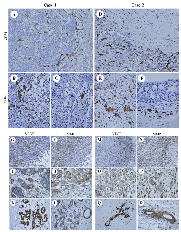
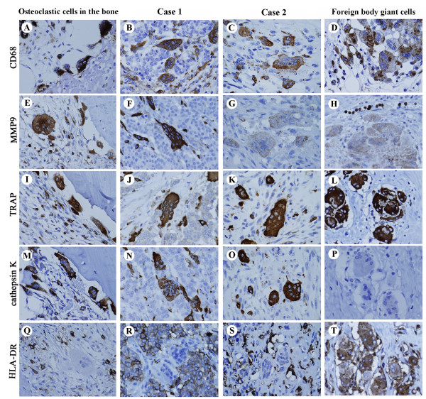
Breast carcinoma with osteoclastic giant cells (OGCs) is characterized by multinucleated OGCs, and usually displays inflammatory hypervascular stroma. OGCs may derive from tumor-associated macrophages, but their nature remains controversial. We report two cases, in which OGCs appear in common microenvironment despite different tumoural histology. A 44-year-old woman (Case 1) had OGCs accompanying invasive ductal carcinoma, and an 83-year-old woman (Case 2) with carcinosarcoma. Immunohistochemically, in both cases, tumoural and non-tumoural cells strongly expressed VEGF and MMP12, which promote macrophage migration and angiogenesis. The Chalkley count on CD-31-stained sections revealed elevated angiogenesis in both cases. The OGCs expressed bone-osteoclast markers (MMP9, TRAP, cathepsin K) and a histiocyte marker (CD68), but not an MHC class II antigen, HLA-DR. The results indicate a pathogenesis: regardless of tumoural histology, OGCs derive from macrophages, likely in response to hypervascular microenvironments with secretion of common cytokines. The OGCs have acquired bone-osteoclast-like characteristics, but lost antigen presentation abilities as an anti-cancer defense. Appearance of OGCs may not be anti-tumoural immunological reactions, but rather pro-tumoural differentiation of macrophage responding to hypervascular microenvironments induced by breast cancer.
Figures
Similar articles
-
Breast carcinoma with osteoclastic giant cells: case report and review of the literature.Int J Clin Exp Pathol. 2014 Mar 15;7(4):1788-91. eCollection 2014. Int J Clin Exp Pathol. 2014. PMID: 24817980 Free PMC article. Review.
-
Neuroendocrine differentiation in breast carcinoma with osteoclast-like giant cells. Report of a case and review of the literature.Int J Surg. 2014;12 Suppl 2:S8-S11. doi: 10.1016/j.ijsu.2014.08.392. Epub 2014 Sep 6. Int J Surg. 2014. PMID: 25204617 Review.
-
Breast carcinoma with osteoclast-like giant cells: A cytological-pathological correlation with a literature review.Ann Diagn Pathol. 2018 Apr;33:1-5. doi: 10.1016/j.anndiagpath.2017.11.003. Epub 2017 Nov 9. Ann Diagn Pathol. 2018. PMID: 29566940 Review.
-
Pleomorphic lobular carcinoma of the breast with osteoclast-like giant cells: a case report and review of the literature.Diagn Pathol. 2018 Aug 28;13(1):62. doi: 10.1186/s13000-018-0744-6. Diagn Pathol. 2018. PMID: 30153845 Free PMC article. Review.
-
Mammary carcinoma with osteoclast-like giant cells: a case report.Int J Clin Exp Pathol. 2014 Dec 1;7(12):9038-43. eCollection 2014. Int J Clin Exp Pathol. 2014. PMID: 25674284 Free PMC article.
Cited by
-
Invasive breast carcinoma with osteoclast-like stromal giant cells: A case report.World J Clin Cases. 2023 Mar 6;11(7):1521-1527. doi: 10.12998/wjcc.v11.i7.1521. World J Clin Cases. 2023. PMID: 36926394 Free PMC article.
-
Idiopathic granulomatous mastitis associated with risperidone-induced hyperprolactinemia.Diagn Pathol. 2012 Jan 5;7:2. doi: 10.1186/1746-1596-7-2. Diagn Pathol. 2012. PMID: 22221904 Free PMC article.
-
Multifocal invasive ductal breast cancer with osteoclast-like giant cells: a case report.J Med Case Rep. 2011 Feb 27;5:85. doi: 10.1186/1752-1947-5-85. J Med Case Rep. 2011. PMID: 21352587 Free PMC article.
-
"Giants in a Microcosm": Multinucleated Giant Cells Populating an Invasive Micropapillary Carcinoma of the Breast.Int J Surg Pathol. 2015 Dec;23(8):654-5. doi: 10.1177/1066896915605616. Epub 2015 Sep 13. Int J Surg Pathol. 2015. PMID: 26370736 Free PMC article. No abstract available.
-
MMP-12 and Periodontitis: Unraveling the Molecular Pathways of Periodontal Tissue Destruction.J Inflamm Res. 2024 Oct 28;17:7793-7806. doi: 10.2147/JIR.S480466. eCollection 2024. J Inflamm Res. 2024. PMID: 39494211 Free PMC article. Review.
References
-
- Fattaneh A, Tavassolo B, Devilee P. World Health Organization Classification of Tumours. Pathology and Genetics of Tumor of the breast and female genital organs. Lyon, IARC Press; 2003.
-
- Agnanits NT, Rosen PP. Rosen's breast pathology. Philadelphia, Lippincot Williams & Wilkins; 2001.
Publication types
MeSH terms
Substances
LinkOut - more resources
Full Text Sources
Medical
Research Materials
Miscellaneous
