

Phenotyping breast cancer cell lines EM-G3, HCC1937, MCF7 and MDA-MB-231 using 2-D electrophoresis and affinity chromatography for glutathione-binding proteins
- PMID: 20731849
- PMCID: PMC2933630
- DOI: 10.1186/1471-2407-10-449
Phenotyping breast cancer cell lines EM-G3, HCC1937, MCF7 and MDA-MB-231 using 2-D electrophoresis and affinity chromatography for glutathione-binding proteins
Abstract
Background: Transformed phenotypes are common to cell lines derived from various cancers. Proteome profiling is a valuable tool that may reveal uncharacteristic cell phenotypes in transformed cells. Changes in expression of glutathione S-transferases (GSTs) and other proteins interacting with glutathione (GSH) in model cell lines could be of particular interest.
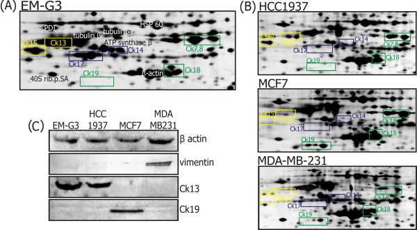
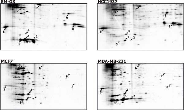
Methods: We compared the phenotypes of breast cell lines EM-G3, HCC1937, MCF7 and MDA-MB-231 using 2-D electrophoresis (2-DE). We further separated GSH-binding proteins from the cell lines using affinity chromatography with GSH-Sepharose 4B, performed 2-DE analysis and identified the main protein spots.
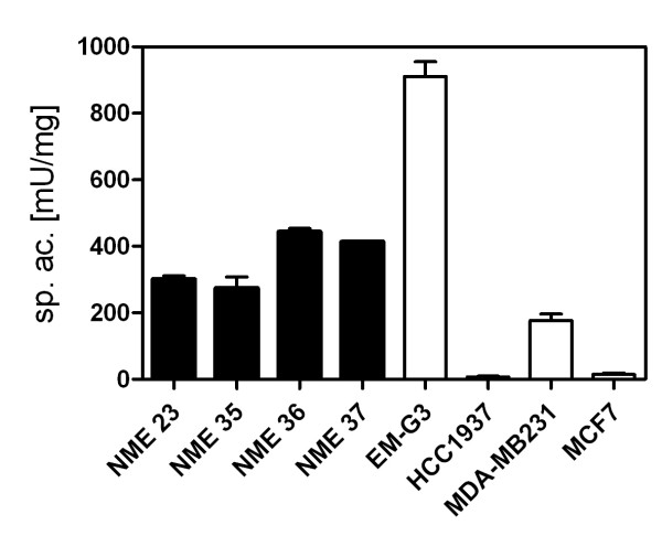
Results: Correlation coefficients among 2-DE gels from the cell lines were lower than 0.65, pointing to dissimilarity among the cell lines. Differences in primary constituents of the cytoskeleton were shown by the 2-D protein maps and western blots. The spot patterns in gels of GSH-binding fractions from primary carcinoma-derived cell lines HCC1937 and EM-G3 were similar to each other, and they differed from the spot patterns of cell lines MCF7 and MDA-MB-231 that were derived from pleural effusions of metastatic mammary carcinoma patients. Major differences in the expression of GST P1-1 and carbonyl reductase [NADPH] 1 were observed among the cell lines, indicating differential abilities of the cell lines to metabolize xenobiotics.
Conclusions: Our results confirmed the applicability of targeted affinity chromatography to proteome profiling and allowed us to characterize the phenotypes of four breast cancer cell lines.
Figures
Similar articles
-
Detecting acute neurotoxicity during platinum chemotherapy by neurophysiological assessment of motor nerve hyperexcitability.BMC Cancer. 2010 Aug 23;10:451. doi: 10.1186/1471-2407-10-451. BMC Cancer. 2010. PMID: 20731872 Free PMC article.
-
The combination of capecitabine and oxaliplatin in metastatic colorectal cancer.Clin Colorectal Cancer. 2003 Feb;2(4):205-9. doi: 10.1016/s1533-0028(11)70328-0. Clin Colorectal Cancer. 2003. PMID: 12620138 Review. No abstract available.
-
Should capecitabine replace infusional fluorouracil and leucovorin when combined with oxaliplatin in metastatic colorectal cancer?J Clin Oncol. 2007 Sep 20;25(27):4165-7. doi: 10.1200/JCO.2007.11.6582. Epub 2007 Aug 20. J Clin Oncol. 2007. PMID: 17709796 No abstract available.
-
Oxaliplatin, irinotecan and capecitabine as first-line therapy in metastatic colorectal cancer (mCRC): a dose-finding study and pharmacogenomic analysis.Br J Cancer. 2010 Mar 16;102(6):987-94. doi: 10.1038/sj.bjc.6605595. Epub 2010 Mar 9. Br J Cancer. 2010. PMID: 20216541 Free PMC article. Clinical Trial.
-
Capecitabine/Oxaliplatin combinations in advanced colorectal cancer: summary of recent randomized studies.Clin Colorectal Cancer. 2005 Nov;5(4):242-4. doi: 10.1016/s1533-0028(11)70189-x. Clin Colorectal Cancer. 2005. PMID: 16356300 Review. No abstract available.
Cited by
-
Quantitative CK19 biomarker detection in breast cancer cell lines.J Med Life. 2022 Feb;15(2):188-195. doi: 10.25122/jml-2021-1101. J Med Life. 2022. PMID: 35419102 Free PMC article.
-
Fibroblasts prepared from different types of malignant tumors stimulate expression of luminal marker keratin 8 in the EM-G3 breast cancer cell line.Histochem Cell Biol. 2012 May;137(5):679-85. doi: 10.1007/s00418-012-0918-3. Epub 2012 Jan 24. Histochem Cell Biol. 2012. PMID: 22270320
-
LanCL proteins are not Involved in Lanthionine Synthesis in Mammals.Sci Rep. 2017 Jan 20;7:40980. doi: 10.1038/srep40980. Sci Rep. 2017. PMID: 28106097 Free PMC article.
-
Aptamer-Based Recognition of Breast Tumor Cells: A New Era for Breast Cancer Diagnosis.Int J Mol Sci. 2024 Jan 10;25(2):840. doi: 10.3390/ijms25020840. Int J Mol Sci. 2024. PMID: 38255914 Free PMC article.
-
Classification of cancer cell lines using matrix-assisted laser desorption/ionization time‑of‑flight mass spectrometry and statistical analysis.Int J Mol Med. 2017 Oct;40(4):1096-1104. doi: 10.3892/ijmm.2017.3083. Epub 2017 Jul 27. Int J Mol Med. 2017. PMID: 28765873 Free PMC article.
References
-
- Lacroix M, Leclercq G. Relevance of breast cancer cell lines as models for breast tumours: an update. Breast Cancer Res Treat. 2004;83:249–289. doi: 10.1023/B:BREA.0000014042.54925.cc. - DOI - PubMed
-
- Slany A, Haudek VJ, Zwickl H, Gundacker NC, Grusch M, Weiss TS, Seir K, Rodgarkia-Dara C, Hellerbrand C, Gerner C. Cell characterization by proteome profiling applied to primary hepatocytes and hepatocyte cell lines Hep-G2 and Hep-3B. J Proteome Res. 2010;9:6–21. doi: 10.1021/pr900057t. - DOI - PubMed
Publication types
MeSH terms
Substances
LinkOut - more resources
Full Text Sources
Medical
Research Materials
Miscellaneous
