. 2010 Nov 4:1359:1-13.
doi: 10.1016/j.brainres.2010.08.044. Epub 2010 Aug 21.

Epidermal growth factor targeting of bacteriophage to the choroid plexus for gene delivery to the central nervous system via cerebrospinal fluid

Affiliations

Epidermal growth factor targeting of bacteriophage to the choroid plexus for gene delivery to the central nervous system via cerebrospinal fluid

Ana Maria Gonzalez et al. Brain Res. .

Abstract

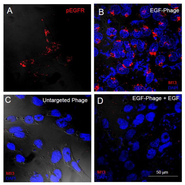
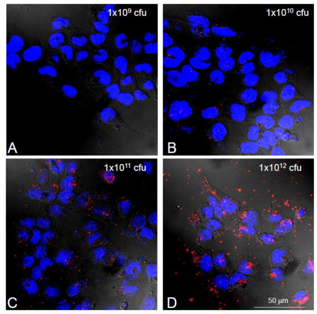
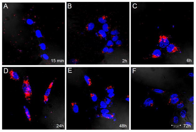
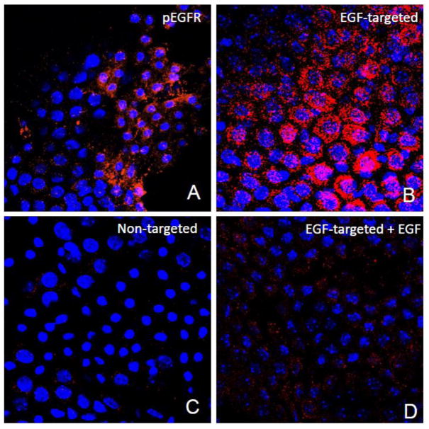
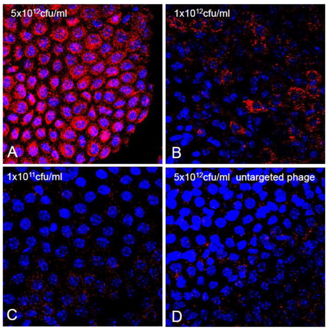
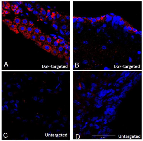
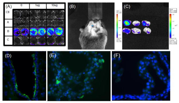
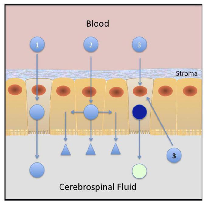
Because the choroid plexus normally controls the production and composition of cerebrospinal fluid and, as such, its many functions of the central nervous system, we investigated whether ligand-mediated targeting could deliver genes to its secretory epithelium. We show here that when bacteriophages are targeted with epidermal growth factor, they acquire the ability to enter choroid epithelial cells grown in vitro as cell cultures, ex vivo as tissue explants or in vivo by intracerebroventricular injection. The binding and internalization of these particles activate EGF receptors on targeted cells, and the dose- and time-dependent internalization of particles is inhibited by the presence of excess ligand. When the phage genome is further reengineered to contain like green fluorescent protein or firefly luciferase under control of the cytomegalovirus promoter, gene expression is detectable in the choroid plexus and ependymal epithelium by immunohistochemistry or by noninvasive imaging, respectively. Taken together, these data support the hypothesis that reengineered ligand-mediated gene delivery should be considered a viable strategy to increase the specificity of gene delivery to the central nervous system and bypass the blood-brain barrier so as to exploit the biological effectiveness of the choroid plexus as a portal of entry into the brain.


Conflict of interest statement

The authors declare no conflicts of interest.

Figures

Figure 1
Figure 1. Internalization of EGF-targeted phage is specific in CP cells in vitro
TR-CSFB cells were incubated with EGF-targeted (Panels A, B & D) or non targeted phage (Panel C) (1×10¹²cfu/ml) for 2h at 37C. The cells were acid washed to remove any particles on the cell surface, fixed and then immuno-stained with antibodies against phosphorylated EGF-R (Panel A) or M13 phage (Panels B-D) as described in the text. Binding of primary antibodies was detected using 594 Alexa-donkey anti goat (Panel A) or 488 Alexa-goat anti rabbit antibodies (Panels B-D). Positive immunostaining for the phosphorylated form of the EGFR receptor is observed in TR-CSFB cells after incubation with specific antibodies (Panel A). Internalization of EGF-phage is observed in the cytoplasm of these cells (Panel B). In contrast, no internalization is observed when the cells are incubated with untargeted phage (Panel C). Internalization of EGF targeted phage is blocked in the presence of an excess of EGF (Panel D). Cells were counterstained with DAPI to visualize the cell nuclei. pEGFR: Red; M13 phage: Red; Cell nuclei: Blue.
Figure 2
Figure 2. Internalization of EGF-Phage is dose dependant in CP cells in vitro
TR-CSFB cells were incubated with increasing concentrations of EGF-targeted phage for 2h, at 37C. Cells were immuno-stained with antibodies against M13 phage and detected using 488 Alexa goat labeled secondary antibodies. EGF-phage is internalized by TR-CSFB cells at high titers of phage (>1×10¹¹ cfu/ml). Cells were counterstained with DAPI to visualize the cell nuclei. M13 phage: Red; Cell nuclei: Blue.
Figure 3
Figure 3. Internalization of EGF-Phage is time dependant in CP cells in vitro
TR-CSFB cells were incubated with EGF-targeted phage for 2h at 37C, media replaced and cells returned to the 37C incubator. Cells were acid washed, immuno-stained with M13 antibodies and visualized using 488 Alexa labeled secondary antibodies. Cells were counterstained with DAPI to visualize the cell nuclei. By 2h EGF-targeted phage is already internalized and by 6h significant amounts of EGF-phage have translocated to the peri-nuclear area. By 48h, there is a reduction in intracellular M13 staining although phage particles are still detectable by 72h. M13 phage: Red; Cell nuclei: Blue.
Figure 4
Figure 4. Internalization of EGF-targeted phage is specific in CP explants
Choroid plexus were dissected from the lateral and 4th ventricle and incubated with EGF-targeted (Panels A, B &D) or non targeted phage (Panel C) at a concentration of 1×10¹²cfu/ml for 2h at 37C. To remove any particles on the cell surface, the explants were washed with PBS containing Tween 20, fixed and then immunostained with antibodies against phosphorylated EGFR (Panel A) or M13 phage (Panels B-D). Binding of primary antibodies was detected using 594 Alexa-donkey anti goat (Panel A) or 488 Alexa-goat anti rabbit antibodies (Panels B-D). Positive immunostaining for the phosphorylated form of the EGFR receptor was observed in the choroid epithelial cells after incubation with specific antibodies (Panel A). Internalization of EGF-phage is observed in the cytoplasm of the epithelial cells (Panel B). In contrast, no immunoreactivity is observed when the explants are incubated with untargeted phage (Panel C). Internalization of EGF-targeted phage is blocked in the presence of an excess of EGF (Panel D). Tissue explants were counterstained with DAPI to visualize the cell nuclei. pEGFR: Red; M13 phage: Red; Cell nuclei: Blue.
Figure 5
Figure 5. Internalization of EGF-targeted phage is dose dependant in CP explants
Choroid plexus explants were incubated with increasing concentrations of EGF-targeted phage for 2h, at 37C and processed as described in the text. Cells were immunostained with antibodies against M13 phage and the binding detected using 488 Alexa goat labeled secondary antibodies. At low titers (1×10¹¹ cfu/ml), the internalization EGF-targeted phage is almost undetectable but significant increase of immunoreactive M13 internalization is observed at 1×10¹² cfu/ml. At a concentration of 5×10¹² cfu/ml, almost all of choroid epithelial cells in the explants are immunopositive. Cells were counterstained with DAPI to visualize the cell nuclei. M13 phage: Red; Cell nuclei: Blue.
Figure 6
Figure 6. EGF-targeted phage is internalised 24h after ICV injection
EGF-targeted phage (Panels A & B) or untargeted phage (Panels C & D) were injected into the lateral ventricle and 24h later rats were killed and brain sections immunostained with anti-M13 antibodies. M13 immunoreactivity is observed in epithelial cells of the choroid plexus (Panel A) and the ependyma (Panel B). No immunoreactivity is observed in these tissues when untargeted phage was injected ICV. (Red: M13; Blue: DAPI).
Figure 7
Figure 7. Gene expression after ICV injection
Panel (A) In vitro transduction: The EGF target PC3 cells were incubated with 1× 10¹¹ untargeted or EGF-targeted phage that were both re-engineered as described in the text to contain the firefly luciferase (f-luc) gene under the control of the CMV promoter. Duplicate wells of cells were treated with 0, 1ug or 10 ug of camptothecin and either incubated with no phage (Ctl), EGF-targeted phage containing GFP gene (A), EGF-targeted phage containing f-luc gene (B) or untargeted phage containing the f-luc gene (C). After incubating cells for 48 hrs, a 5 minute incubation with luciferin enabled a Lumina CCD Imaging system to visualize gene expression in cells treated with EGF-targeted phage (Row B) and, as expected the response was dose dependant on cells treated with camptothecin. No signal was observed in the absence of targeting (Row C) or when EGF-targeted particles do not contain the luciferase gene Row A. Panel B-F: Effects of ICV injection of EGF-targeted phage in vivo. Mice were injected icv with 1×10¹² untargeted or EGF-targeted phage-luc and in Panels B and D, transgene expression of luciferase monitored on 48 hours later by Lumina CCD Imaging either non invasively (Panel B) or after brain dissection (Panel C). To identify the cellular localization of this gene expression similar experiments were performed with EGF-targeted phage-gfp (Panel D and E) or untargeted phage-gfp (Panel F) which demonstrated cell expression of GFP in ependymal cells and in isolated cells in the choroid plexus. Whereas a 4-fold lower background signal could be quantified after luciferase activity imaging, immunostaining localized this signal to macrophages and monocytes at the site of injection and not in ependyma or choroid plexus (Panel F).
Figure 8
Figure 8. Drug Delivery to the Central Nervous System Via Choroid Plexus
There are three modes of drug delivery to the CNS using the choroid plexus as a portal of entry. The first (1) is direct whereby molecules are targeted and directly translocated across the CP epithelial barrier to find their targets from within the CNS. A second (2) is illustrated by entering and trateing the CP epithelium so that its capacity the produce and composition of CSF is modified. These then find their targets in the CNS parenchyma. Finally the third strategy, in the example here using gene delivery, results in longer term changes of the choroid plexus, allows the epithelial cell to produce novel therapeutic factors itself and as such can confer the epithelium with an ability to change the production and composition of CSF. This latter process and the feasibility with luminal targeting (3) is demonstrated in the results presented here.

Similar articles

Cited by

References

    1. Aida Y, Pabst MJ. Removal of endotoxin from protein solutions by phase separation using Triton X-114. J Immunol Methods. 1990;132:191–5. - PubMed
    1. Bajocchi G, Feldman SH, Crystal RG, Mastrangeli A. Direct in vivo gene transfer to ependymal cells in the central nervous system using recombinant adenovirus vectors. Nat Genet. 1993;3:229–34. - PubMed
    1. Barry MA, Dower WJ, Johnston SA. Toward cell-targeting gene therapy vectors: selection of cell-binding peptides from random peptide-presenting phage libraries. Nat Med. 1996;2:299–305. - PubMed
    1. Bickel U, Yoshikawa T, Pardridge WM. Delivery of peptides and proteins through the blood-brain barrier. Adv Drug Deliv Rev. 2001;46:247–79. - PubMed
    1. Brown PD, Davies SL, Speake T, Millar ID. Molecular mechanisms of cerebrospinal fluid production. Neuroscience. 2004;129:957–70. - PMC - PubMed

Publication types

Substances