Review
. 2010 Nov;128(2):375-84.
doi: 10.1016/j.pharmthera.2010.08.001. Epub 2010 Aug 21.

The development of MDA-7/IL-24 as a cancer therapeutic

Affiliations
Review

The development of MDA-7/IL-24 as a cancer therapeutic

Paul Dent et al. Pharmacol Ther. 2010 Nov.

Abstract

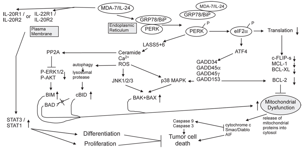
The cytokine melanoma differentiation associated gene 7 (mda-7) was identified by subtractive hybridization as a protein whose expression increased during the induction of terminal differentiation, and that was either not expressed or was present at low levels in tumor cells compared to non-transformed cells. Based on conserved structure, chromosomal location and cytokine-like properties, MDA-7, was classified as a member of the interleukin (IL)-10 gene family and designated as MDA-7/IL-24. Multiple studies have demonstrated that expression of MDA-7/IL-24 in a wide variety of tumor cell types, but not in corresponding equivalent non-transformed cells, causes their growth arrest and rapid cell death. In addition, MDA-7/IL-24 has been noted to radiosensitize tumor cells which in part is due to the generation of reactive oxygen species (ROS) and ceramide that cause endoplasmic reticulum stress and suppress protein translation. Phase I clinical trial data has shown that a recombinant adenovirus expressing MDA-7/IL-24 (Ad.mda-7 (INGN-241)) was safe and had measurable tumoricidal effects in over 40% of patients, strongly arguing that MDA-7/IL-24 could have significant therapeutic value. This review describes what is presently known about the impact of MDA-7/IL-24 on tumor cell biology and its potential therapeutic applications.


Figures

Fig. 1
Fig. 1
Molecular pathways by which MDA-7/IL-24 regulates cell viability and cell growth. MDA/IL-24 has two major targets in cells: the IL-20/IL-22 receptor complexes and the HSP70 family chaperone GRP78/BiP. MDA-7/IL-24 binding to its cognate receptors activates STAT family transcription factors and activation of these factors can promote differentiation and proliferation in a cell type dependent manner. STAT transcription factors play no role in MDA-7/IL-24 toxicity. MDA-7/IL-24 binds to GRP78/BiP; it is possible that entry of bacterial synthesized GST-MDA-7 into tumor cells is mediated by binding to cell surface GRP78/BiP. The majority of GRP78/BiP is present in the endoplasmic reticulum and is bound to PKR-like endoplasmic reticulum kinase (PERK); the chaperone inhibits PERK kinase activity. MDA-7/IL-24 disrupts the association of GRP78/BiP with PERK permitting PERK to phosphorylate eIF2α; phospho-eIF2α suppresses the translation of the majority of cellular proteins resulting in the rapid loss of protective proteins that have short half lives such as MCL-1 and BCL-XL, and via ATF4 promotes the transcription of a specific subset of genes e.g. GADD34 that promote apoptosis. PERK signaling promotes increased LASS6 (ceramide synthase 6) levels that promote increased Ca2+ mobilization leading to elevated ROS levels. Increased ceramide/Ca2+/ROS activate JNK and p38 signaling that promotes activation of the toxic BH3 domain proteins BAX and BAK.
Fig. 2
Fig. 2
GST-MDA-7 causes LC3-GFP vesicularization in transformed cells in a PERK-dependent manner. A, (i), GBM6 and U251 cells were plated in four-well chamber glass slides in triplicate and 12 h after plating transfected with a plasmid to express LC3-GFP and in parallel co-transfected with either a vector control plasmid (CMV) or with a plasmid to express dnPERK. Twelve hours after transfection, cells were treated with GST or GST-MDA-7 (100 nmol/L). Twenty-four hours after GST-MDA-7 exposure, the GBM6 and U251 cells were examined under visual light (visible) or under fluorescent light (LC3-GFP). Representative images from the triplicate plating (n = 2). (ii), U251 cells were plated in four-well chamber glass slides in triplicate and 12 h after plating transfected with a plasmid to express LC3-GFP and in parallel co-transfected with either a vector control plasmid to express a nonspecific scrambled siRNA (siSCR) or plasmids to knockdown expression of Beclin-1 (siBeclin-1) or ATG5 (siATG5). Parallel studies also transfected cells with plasmids to express scrambled siRNA and untagged GFP. Twelve hours after transfection, cells were treated with GST or GST-MDA-7 (100 nmol/L). Twenty-four hours after GST-MDA-7 exposure, the U251 cells were examined under fluorescent light (LC3-GFP and GFP). Representative images from the triplicate plating (n = 3). Immunoblotting, cells transfected with siRNA constructs to modulate the expression of ATG5 and Beclin-1 were immunoblotted to determine the expression of Beclin-1 and ATG5 48 h after transfection. Cells treated with GST-MDA-7 and GST were immunoblotted 48 h after treatment to determine the expression of Beclin-1, ATG5, the cleavage status of LC3 and GAPDH (n = 2). B, Transformed MEFs (WT; deleted for PERK, PERK−/−) 24 h after plating were treated with GST or GST-MDA-7 (100 nmol/L). Twenty-four hours after GST-MDA-7 treatment, cells were isolated and subjected to SDS-PAGE to determine the expression of Beclin-1, ATG5, the cleavage status of LC3 and GAPDH (n = 2). C, GBM6 and U251 cells were plated in four-well chamber glass slides in triplicate and 12 h after plating transfected with either a vector control plasmid to express a nonspecific scrambled siRNA or plasmids to knockdown expression of Beclin-1 or of ATG5. Twelve hours after transfection, cells were treated with GST or GST-MDA-7 (30 nmol/L). Forty-eight hours after GST-MDA-7 exposure, the viability of the GBM6 and U251 cells was determined by: terminal deoxynucleotidyl transferase-mediated dUTP nick end labeling assay or; by Annexin V-PI flow cytometry on isolated cells (±SE; n = 3). Data shown are for GBM6 (terminal deoxynucleotidyl transferase-mediated dUTP nick end labeling) and U251 cells was determined by Annexin V/PI flow cytometry (±SE; n = 2). Data reproduced with permission from Yacoub et al. Caspase-, cathepsin and PERK-dependent regulation of MDA-7/IL-24-induced cell killing in primary human glioma cells. Mol Cancer Ther 7 (2008), 297–313.
Fig. 3
Fig. 3
Role of Beclin-1 and ATG5 in Ad.mda-7-induced apoptosis. DU-145 cells were transfected with the indicated siRNAs, and cell viability was determined by MTT assay (A), Caspase-Glo 3/7 assay for caspase-3 expression (B), and colony formation assays (C) 48 hours after infection with Ad.mda-7 (10, 50, or 100 pfu/cell) or Ad.vec (100 pfu/cell). C, colony formation assays in monolayer culture. Colonies were fixed, stained, and counted (>50 cells) 2 weeks after plating. D, Expression profile of Beclin-1 and ATG5 at the mRNA level 48 h after Ad.mda-7 infection (50 or 100 pfu/cell) of DU-145 cells. ★, P < 0.05; ★★, P < 0.001, compared with control. Data reproduced with permission from Bhutia et al. Mechanism of autophagy to apoptosis switch triggered in prostate cancer cells by antitumor cytokine melanoma differentiation associated gene 7/interleukin-24. Cancer Res. 70 (2010), 3667–3676.

Similar articles

Cited by

References

    1. Bhutia SK, Dash R, Das SK, Azab B, Su ZZ, Lee SG, et al. Mechanism of autophagy to apoptosis switch triggered in prostate cancer cells by antitumor cytokine melanoma differentiation-associated gene 7/interleukin-24. Cancer Res. 2010;70:3667–3676. - PMC - PubMed
    1. Bursch W. The autophagosomal ± lysosomal compartment in programmed cell death. Cell Death and Differ. 2001;8:569–581. - PubMed
    1. Caudell EG, Mumm JB, Poindexter N, Ekmekcioglu S, Mhashilkar AM, Yang XH, et al. The protein product of the tumor suppressor gene, melanoma differentiation-associated gene 7, exhibits immunostimulatory activity and is designated IL-24. J Immunol. 2002;168:6041–6046. - PubMed
    1. Chada S, Mhashilkar AM, Ramesh R, Mumm JB, Sutton RB, Bocangel D, et al. Bystander activity of Ad-mda7: human MDA-7 protein kills melanoma cells via an IL-20 receptor-dependent but STAT3-independent mechanism. Mol Ther. 2004a;10:1085–1095. - PubMed
    1. Chada S, Sutton RB, Ekmekcioglu S, Ellerhorst J, Mumm JB, Leitner WW, et al. MDA-7/IL-24 is a unique cytokine-tumor suppressor in the IL-10 family. Int Immunopharmacol. 2004b;4:649–667. - PubMed

Publication types

MeSH terms