. 2010 Nov 10;170(4):1261-9.
doi: 10.1016/j.neuroscience.2010.08.019. Epub 2010 Aug 21.

Roles of estrogen receptors alpha and beta in sexually dimorphic neuroprotection against glutamate toxicity

Affiliations

Roles of estrogen receptors alpha and beta in sexually dimorphic neuroprotection against glutamate toxicity

D N Bryant et al. Neuroscience. .

Abstract

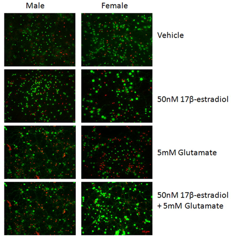
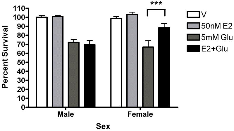
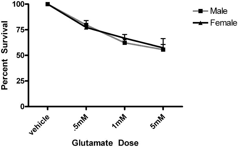
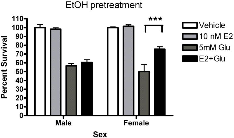
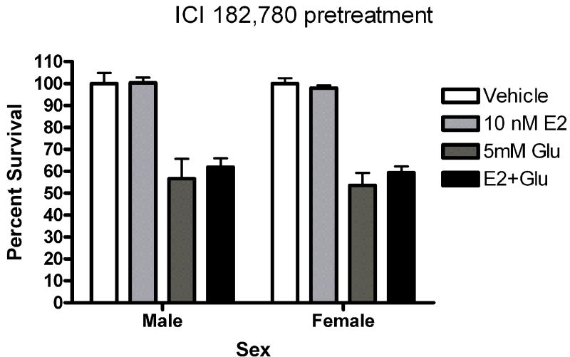
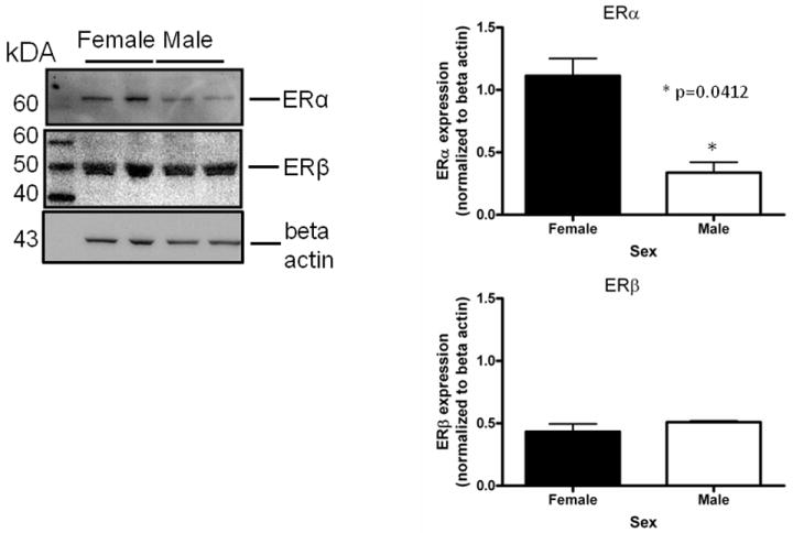
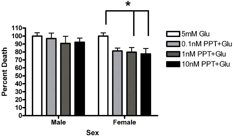
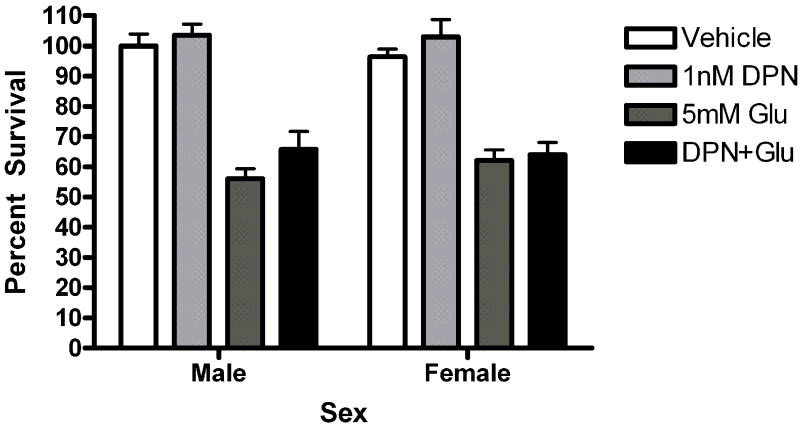
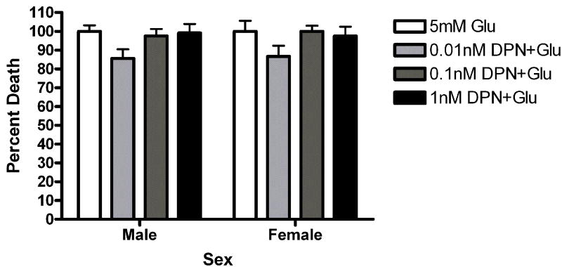
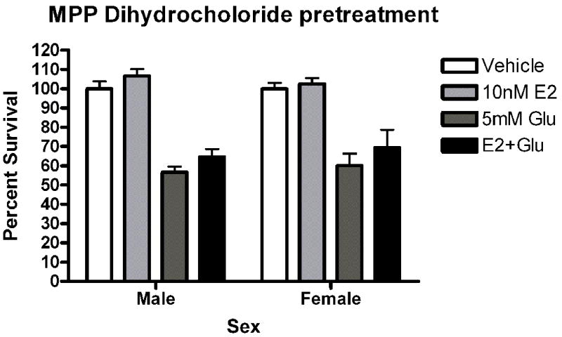
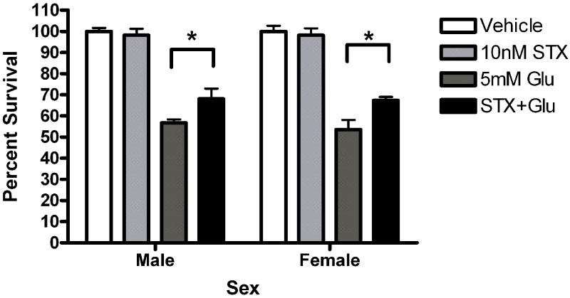
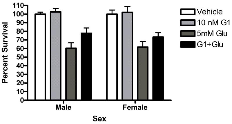
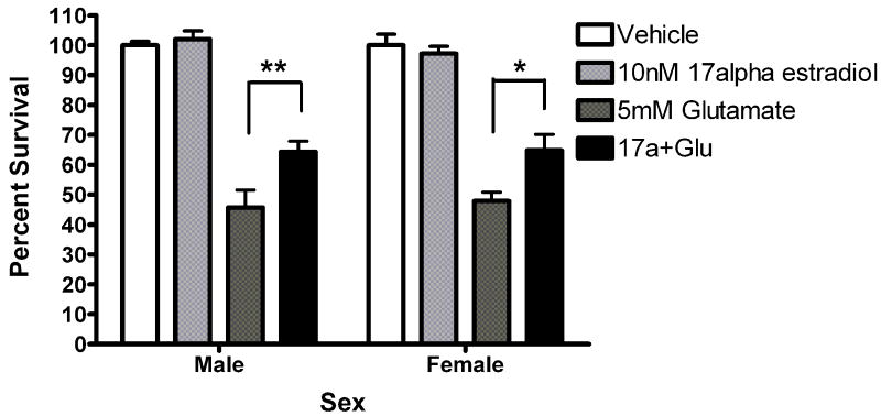
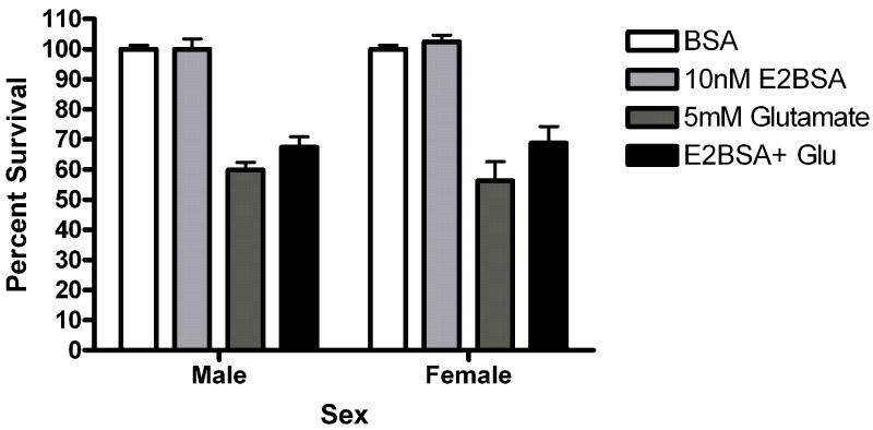
Although most agree that 17β-estradiol is neuroprotective via a variety of mechanisms, less is known about the role that biological sex plays in receptor-mediated estradiol neuroprotection. To address this issue we isolated primary cortical neurons from rat pups sorted by sex and assessed the ability of estradiol to protect the neurons from death induced by glutamate. Five-minute pretreatment with 10-50 nM 17β-estradiol protected female but not male neurons from glutamate toxicity 24 h later. Both estrogen receptor alpha (ERα) and estrogen receptor beta (ERβ) are expressed in these cultures. Experiments using an ERα selective agonist or antagonist indicate that this receptor is important for neuroprotection in female cortical neurons. The ERβ selective agonist conveys a small degree of neuroprotection to both male and female cortical neurons. Interestingly, we found that 17α estradiol and the novel membrane estrogen receptor (mER) agonist STX, but not bovine serum albumin conjugated estradiol or the GPR30 agonist G1 were neuroprotective in both male and female neurons. Taken together these data highlight a role for ERα in sexually dimorphic neuroprotection.


Figures

Figure 1
Figure 1
50nM estradiol protects female but not male neurons from glutamate toxicity. At 10DIV, male and female primary cortical neurons were exposed to 0.005% ethanol or 50nM estradiol for 5 minutes prior to a five minute exposure to 5mM glutamate. After 1 wash in complete Neurobasal media, cells received fresh media and were returned to the incubator for 24 hours. A. Representative images from the Live/Dead assay (Invitrogen). Living neurons are stained green by Calcein AM. Dead cells are stained red by the ethidium bromide homodimer. B. 5 minute pre-exposure to 50nM estradiol protects female neurons (***Bonferroni t (glutamate vs. estradiol+ glutamate) =3.911, p<0.001) but not male neurons from glutamate toxicity (n=5).
Figure 1
Figure 1
50nM estradiol protects female but not male neurons from glutamate toxicity. At 10DIV, male and female primary cortical neurons were exposed to 0.005% ethanol or 50nM estradiol for 5 minutes prior to a five minute exposure to 5mM glutamate. After 1 wash in complete Neurobasal media, cells received fresh media and were returned to the incubator for 24 hours. A. Representative images from the Live/Dead assay (Invitrogen). Living neurons are stained green by Calcein AM. Dead cells are stained red by the ethidium bromide homodimer. B. 5 minute pre-exposure to 50nM estradiol protects female neurons (***Bonferroni t (glutamate vs. estradiol+ glutamate) =3.911, p<0.001) but not male neurons from glutamate toxicity (n=5).
Figure 2
Figure 2
Glutamate toxicity is not sexually dimorphic in our experimental protocol. Male and female neurons were exposed to varying doses (.5mM, 1mM and 5mM) for 5 minutes. Neurons were handled as described in figure 1 and the methods section. There was no sex difference in percent survival (n=3).
Figure 3
Figure 3
ICI 182, 780 blocks estrogen neuroprotection in female neurons. Male and female neurons were exposed to (A) 0.01% ethanol or (B) 1uM ICI for 15 minutes prior to a 5 minute exposure to 0.001% ethanol or 10nM estradiol. Neurons we subsequently exposed to 5mM glutamate for 5 minutes. (A) Estradiol (10 nM) protected female neurons but not male neurons from glutamate toxicity in ethanol pretreated neurons (***Bonferroni post hoc t (glutamate vs. estradiol+ glutamate) =4.977, p<0.001, n=4). (B) ICI pretreatment blocked estradiol protection in female neurons (n=4).
Figure 3
Figure 3
ICI 182, 780 blocks estrogen neuroprotection in female neurons. Male and female neurons were exposed to (A) 0.01% ethanol or (B) 1uM ICI for 15 minutes prior to a 5 minute exposure to 0.001% ethanol or 10nM estradiol. Neurons we subsequently exposed to 5mM glutamate for 5 minutes. (A) Estradiol (10 nM) protected female neurons but not male neurons from glutamate toxicity in ethanol pretreated neurons (***Bonferroni post hoc t (glutamate vs. estradiol+ glutamate) =4.977, p<0.001, n=4). (B) ICI pretreatment blocked estradiol protection in female neurons (n=4).
Figure 4
Figure 4
Female neurons contain more immunoreactive ERα than male neurons at 10DIV. Whole cell lysates from female and male neurons were immunoblotted using antibodies against ERα (6F11, Novacastra) ERβ (PA1-310B, Thermo) and beta actin (Sigma). Densitometric analysis revealed that there was a statistically significant difference in the ratio of ERα to beta actin (*t=4.773, p=0.0412, n=2) between female and male neurons.
Figure 5
Figure 5
The ERα selective ligand PPT is neuroprotective in female neurons only. Male and female neurons treated with the ERα selective ligand PPT (A, B) or the ERβ selective ligand DPN (C, D) for 5 minutes prior to a 5 minute glutamate exposure. (A) PPT was neuroprotective in female neurons Bonferroni t (glutamate vs. ppt+ glutamate)=2.610, p<0.05, n=3) but not male neurons. (B) PPT was neuroprotective in female neurons at 10nM (t=2.42, p<0.05) and 1nM (t=6.96, p<0.05) but not 0.01nM concentrations. (C) At a 1 nM concentration, DPN did not protect male or female neurons (n=4). (D) Neurons were exposed to three concentrations of (1nM, 0.1nM and 0.01nM) of dPN. A two way (Sex X Treatment) ANOVA indicated that there was an overall effect of treatment in neurons exposed to DPN (F=4.02, p=0.01). Post hoc tests were not statistically significant, however.
Figure 5
Figure 5
The ERα selective ligand PPT is neuroprotective in female neurons only. Male and female neurons treated with the ERα selective ligand PPT (A, B) or the ERβ selective ligand DPN (C, D) for 5 minutes prior to a 5 minute glutamate exposure. (A) PPT was neuroprotective in female neurons Bonferroni t (glutamate vs. ppt+ glutamate)=2.610, p<0.05, n=3) but not male neurons. (B) PPT was neuroprotective in female neurons at 10nM (t=2.42, p<0.05) and 1nM (t=6.96, p<0.05) but not 0.01nM concentrations. (C) At a 1 nM concentration, DPN did not protect male or female neurons (n=4). (D) Neurons were exposed to three concentrations of (1nM, 0.1nM and 0.01nM) of dPN. A two way (Sex X Treatment) ANOVA indicated that there was an overall effect of treatment in neurons exposed to DPN (F=4.02, p=0.01). Post hoc tests were not statistically significant, however.
Figure 6
Figure 6
The selective ERα antagonist MPP blocks estradiol neuroprotection. (A) Male and female neurons were pretreated with 1uM MPP dihydrochloride for 15 minutes prior to exposure to estradiol and glutamate. Estradiol was not protective in female neurons under these conditions (n=4).
Figure 7
Figure 7
(A) STX and 17α-estradiol protect male and female neurons. Neurons were exposed to 10 nM STX for 5 minutes prior to a 5 minute exposure to 5mM glutamate. STX protected both male (*Bonferroni post hoc t (glutamate vs. STX+ glutamate) =2.557,p<0.05, n=3) and female (*Bonferroni post hoc t (glutamate vs. STX+ glutamate) =3.076,p<0.05, n=3) neurons from glutamate toxicity.(7b) Neurons were exposed to 10nM of the non-steroidal GPR30 agonist G1for 5 minutes prior to exposure to glutamate. G1 was not neuroprotective (n=3) (7c) Neurons were exposed to 10 nM of 17α estradiol for 5 minutes prior to a 5 minute exposure to 5mM glutamate. 17α- estradiol is neuroprotective in male (post hoc tglutamate vs 17α- estradiol+glutamate=3.507,p<0.01) and female (post hoc tglutamate vs 17α-estradiol+glutamate=3.18, p<0.05) neurons (n=3). (7d) Neurons pre-exposed to 10nM bovine serum albumin conjugated estradiol for 5 minutes were not protected from glutamate toxicity (n=3).
Figure 7
Figure 7
(A) STX and 17α-estradiol protect male and female neurons. Neurons were exposed to 10 nM STX for 5 minutes prior to a 5 minute exposure to 5mM glutamate. STX protected both male (*Bonferroni post hoc t (glutamate vs. STX+ glutamate) =2.557,p<0.05, n=3) and female (*Bonferroni post hoc t (glutamate vs. STX+ glutamate) =3.076,p<0.05, n=3) neurons from glutamate toxicity.(7b) Neurons were exposed to 10nM of the non-steroidal GPR30 agonist G1for 5 minutes prior to exposure to glutamate. G1 was not neuroprotective (n=3) (7c) Neurons were exposed to 10 nM of 17α estradiol for 5 minutes prior to a 5 minute exposure to 5mM glutamate. 17α- estradiol is neuroprotective in male (post hoc tglutamate vs 17α- estradiol+glutamate=3.507,p<0.01) and female (post hoc tglutamate vs 17α-estradiol+glutamate=3.18, p<0.05) neurons (n=3). (7d) Neurons pre-exposed to 10nM bovine serum albumin conjugated estradiol for 5 minutes were not protected from glutamate toxicity (n=3).

References

    1. Alkayed NJ, Harukuni I, Kimes AS, London ED, Traystman RJ, Hurn PD. Gender-linked brain injury in experimental stroke. Stroke. 1998;29:159–165. - PubMed
    1. Barchiesi F, Jackson EK, Imthurn B, Fingerle J, Gillespie DG, Dubey RK. Differential regulation of estrogen receptor subtypes alpha and beta in human aortic smooth muscle cells by oligonucleotides and estradiol. J Clin Endocrinol Metab. 2004;89:2373–2381. - PubMed
    1. Behl C, Widmann M, Trapp T, Holsboer F. 17-beta estradiol protects neurons from oxidative stress-induced cell death in vitro. Biochem Biophys Res Commun. 1995;216:473–482. - PubMed
    1. Behl C, Skutella T, Lezoualc’h F, Post A, Widmann M, Newton CJ, Holsboer F. Neuroprotection against oxidative stress by estrogens: structure-activity relationship. Mol Parmacol. 1997;51:535–541. - PubMed
    1. Bishop J, Simpkins JW. Estradiol treatment increases viability of glioma and neuroblastoma cells in vitro. Mol Cell Neurosci. 1994;5:303–308. - PubMed

Publication types

MeSH terms