

Feline spinal cord diseases
- PMID: 20732602
- PMCID: PMC7114573
- DOI: 10.1016/j.cvsm.2010.05.005
Feline spinal cord diseases
Abstract
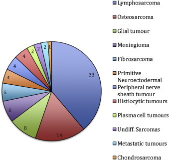
The objective of this article is to review the recent literature that reports on the most common diseases affecting the spinal cord of cats, and to draw some general conclusions that will be useful to formulate diagnosis and prognosis for feline spinal patients. The most common types of feline spinal cord diseases documented were inflammatory/infectious diseases, and feline infectious peritonitis was the most common disease, representing approximately 50% of all feline myelitis. Neoplasms were documented in approximately 25% of cases; lymphosarcoma was the most common tumor affecting the spinal cord of cats, with reported prevalence between 28% and 40%. Cats diagnosed with spinal lymphosarcoma were significantly younger (median age 4 years) than cats with other spinal cord tumors (median age 10 years). Cats with clinical signs of intervertebral disc disease had a median age of 8 years, and 67% had Hansen type I disc protrusions. The most commonly affected intervertebral disc was at the L4 to L5 intervertebral disc space. Fibrocartilaginous embolism-affected older cats (median age 10 years), seemed to predominate in the cervicothoracic intumescence, and clinical signs were markedly lateralized, especially when the cervical region was affected.
Copyright 2010 Elsevier Inc. All rights reserved.
Figures


Similar articles
-
The paralyzed cat. Neuroanatomic diagnosis and specific spinal cord diseases.J Feline Med Surg. 2009 May;11(5):361-72. doi: 10.1016/j.jfms.2009.03.004. J Feline Med Surg. 2009. PMID: 19389636 Free PMC article. Review.
-
Prevalence of diseases of the spinal cord of cats.J Vet Intern Med. 2004 Nov-Dec;18(6):851-8. doi: 10.1892/0891-6640(2004)18<851:podots>2.0.co;2. J Vet Intern Med. 2004. PMID: 15638269
-
Feline spinal lymphosarcoma: a retrospective evaluation of 23 cats.J Vet Intern Med. 1994 Mar-Apr;8(2):99-104. doi: 10.1111/j.1939-1676.1994.tb03205.x. J Vet Intern Med. 1994. PMID: 8046683
-
Clinical reasoning in feline spinal disease: which combination of clinical information is useful?J Feline Med Surg. 2020 Jun;22(6):521-530. doi: 10.1177/1098612X19858447. Epub 2019 Jun 28. J Feline Med Surg. 2020. PMID: 31251096 Free PMC article.
-
Fibrocartilaginous embolic myelopathy in small animals.Vet Clin North Am Small Anim Pract. 2010 Sep;40(5):859-69. doi: 10.1016/j.cvsm.2010.05.003. Vet Clin North Am Small Anim Pract. 2010. PMID: 20732595 Review.
Cited by
-
Vertebral Osteosarcoma in Two Cats-Diagnosis, Treatment, and Outcome.Animals (Basel). 2023 Nov 10;13(22):3478. doi: 10.3390/ani13223478. Animals (Basel). 2023. PMID: 38003096 Free PMC article.
-
Probable lumbar acute non-compressive nucleus pulposus extrusion in a cat with acute onset paraparesis.J Feline Med Surg. 2012 Oct;14(10):764-7. doi: 10.1177/1098612X12450110. Epub 2012 Jun 1. J Feline Med Surg. 2012. PMID: 22661021 Free PMC article.
-
Neurogenic Bladder in Dogs, Cats and Humans: A Comparative Review of Neurological Diseases.Animals (Basel). 2022 Nov 22;12(23):3233. doi: 10.3390/ani12233233. Animals (Basel). 2022. PMID: 36496754 Free PMC article. Review.
-
Presumptive acute non-compressive nucleus pulposus extrusion in 11 cats: clinical features, diagnostic imaging findings, treatment and outcome.J Feline Med Surg. 2017 Jan;19(1):21-26. doi: 10.1177/1098612X15605150. Epub 2016 Jul 10. J Feline Med Surg. 2017. PMID: 26377703 Free PMC article.
-
Prevalence, clinical presentation and MRI of intervertebral disc herniations in cats.J Feline Med Surg. 2022 Dec;24(12):e443-e452. doi: 10.1177/1098612X221121893. Epub 2022 Sep 29. J Feline Med Surg. 2022. PMID: 36172921 Free PMC article.
References
-
- Marioni-Henry K., Vite C., Newton A., et al. Prevalence of diseases of the spinal cord of cats. J Vet Intern Med. 2004;18:851–858. - PubMed
-
- Marioni-Henry K., Van Winkle T.J., Smith S.H., et al. Tumors affecting the spinal cord of cats: 85 cases (1980-2005) J Am Vet Med Assoc. 2008;232:237–243. - PubMed
Publication types
MeSH terms
LinkOut - more resources
Full Text Sources
Medical
Miscellaneous
