. 2010 Oct 29;285(44):33614-22.
doi: 10.1074/jbc.M110.140004. Epub 2010 Aug 23.

Cyclic AMP phosphodiesterase 4D (PDE4D) Tethers EPAC1 in a vascular endothelial cadherin (VE-Cad)-based signaling complex and controls cAMP-mediated vascular permeability

Affiliations

Cyclic AMP phosphodiesterase 4D (PDE4D) Tethers EPAC1 in a vascular endothelial cadherin (VE-Cad)-based signaling complex and controls cAMP-mediated vascular permeability

Sarah N Rampersad et al. J Biol Chem. .

Abstract

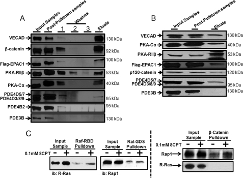
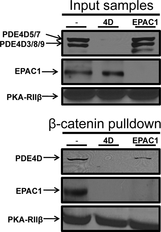
Vascular endothelial cell (VEC) permeability is largely dependent on the integrity of vascular endothelial cadherin (VE-cadherin or VE-Cad)-based intercellular adhesions. Activators of protein kinase A (PKA) or of exchange protein activated by cAMP (EPAC) reduce VEC permeability largely by stabilizing VE-Cad-based intercellular adhesions. Currently, little is known concerning the nature and composition of the signaling complexes that allow PKA or EPAC to regulate VE-Cad-based structures and through these actions control permeability. Using pharmacological, biochemical, and cell biological approaches we identified and determined the composition and functionality of a signaling complex that coordinates cAMP-mediated control of VE-Cad-based adhesions and VEC permeability. Thus, we report that PKA, EPAC1, and cyclic nucleotide phosphodiesterase 4D (PDE4D) enzymes integrate into VE-Cad-based signaling complexes in human arterial endothelial cells. Importantly, we show that protein-protein interactions between EPAC1 and PDE4D serve to foster their integration into VE-Cad-based complexes and allow robust local regulation of EPAC1-based stabilization of VE-Cad-based adhesions. Of potential translational importance, we mapped the EPAC1 peptide motif involved in binding PDE4D and show that a cell-permeable variant of this peptide antagonizes EPAC1-PDE4D binding and directly alters VEC permeability. Collectively, our data indicate that PDE4D regulates both the activity and subcellular localization of EPAC1 and identify a novel mechanism for regulated EPAC1 signaling in these cells.


Figures

FIGURE 1.
FIGURE 1.
cAMP-elevating agents or selective activators of PKA or EPAC decrease human arterial VEC permeability. The impact of isoproterenol (Iso; 1 μm), Fsk (1 μm), 6-Bz-cAMP (3 μm), 8-CPT-cAMP (10 μm), Ro (10 μm), cilostamide (Cil; 1 μm), or combinations of some of these agents on HAEC permeability was measured using transit of FITC-dextran over a 1-h time span (“Experimental Procedures”). Values are means ± S.E. from seven experiments and expressed as the percentage (%) of decreases in permeability compared with control values (dimethyl sulfoxide, 0.5% v/v (Control)). *, significant differences between treatments and control; **, significant differences between treatments and Fsk (1 μm) alone (p < 0.05); NS, not significant.
FIGURE 2.
FIGURE 2.
Activators of PKA or EPAC and selective knockdown of these cAMP effectors differentially impact the integrity of VE-Cad-based HAEC structures and cell permeability. A, confluent HAEC monolayer cultures were incubated with dimethyl sulfoxide (0.5% v/v (Vehicle)) or with test agents (as indicated) for 10 min. Following these incubations, HAEC were separated into three groups for analysis. Group 1, Row A: cells were immediately fixed (paraformaldehyde, 4% w/v). Group 2, Row B: cells were treated with EGTA (5 mm, 20 min) and subsequently fixed as is Group 1. Group 3, Row C: after removal of EGTA, cells were either supplemented with a Ca2+ (2 mm) solution containing no other additions or supplemented with test agents for 2 h and then fixed as in Group 1. VE-Cad-based structures were visualized by staining with anti-VE-Cad (BV6 clone). Fluorescent images were acquired and processed as described under “Experimental Procedures.” The impact of treatments on the integrity of VE-Cad-based structures was determined by comparing the number of contiguous BV6-stained plasma membrane segments with lengths greater than 100 pixels in eight individual random sections from at least six separate experiments. A representative image of results obtained in each treatment paradigm is shown, and the statistical bases of claims are presented under “Experimental Procedures.” B, HAEC were transfected with PKA-Cα-, EPAC1-, or PDE4D-selective siRNAs or a control siRNA and were subsequently allowed to propagate for 48 h. The levels of the relevant target proteins in HAEC transfected with the selective siRNAs were determined by immunoblot analysis. C, The impact of selective PKA, EPAC1, or PDE4D knockdown on basal HAEC permeability and permeability subsequent to incubation with the PKA-, EPAC-, or PDE4D-selective agents described in A was determined as described in the legend for Fig. 1. D, following this growth phase, HAEC were incubated with either saline or VEGF-supplemented saline for 20 min, after which they were fixed as described in A for Group 1.
FIGURE 3.
FIGURE 3.
cAMP-signaling proteins are integral components of VE-Cad-based signaling complexes in human arterial VECs. Following lysis of HAEC in a detergent-supplemented Tris-based buffer (see “Experimental Procedures”), VE-Cad-based cellular complexes were isolated by adsorption of proteins to an immobilized Fc-VE-Cad chimeric protein (A) or an immobilized GST-β-catenin chimeric protein (B) as described under “Experimental Procedures.” Proteins eluted from Fc-VE-Cad or GST-β-catenin were identified by immunoblot (ib) analysis with selective antisera. C, activation of R-Ras or Rap1 was measured by Raf- or Ral-GDS pulldown assays, respectively, and levels of these proteins present in GST-β-catenin pulldown assays were determined as above.
FIGURE 4.
FIGURE 4.
Effects of RNAi-based knockdown of PKA-Cα, EPAC1, or PDE4D on the stability of human arterial VEC VE-Cad- and actin-based structures. HAEC were transfected with EPAC1- or PDE4D-selective siRNAs or a control siRNA as described in the legend for Fig. 2. Following lysis, the transfected HAEC VE-Cad-based cellular complexes were isolated by adsorption to an immobilized GST-β-catenin chimeric protein. Proteins eluted from GST-β-catenin were identified by immunoblot analysis and quantified by densitometry (see “Experimental Procedures”).
FIGURE 5.
FIGURE 5.
Impact on HAEC permeability and VE-Cad-based structures of interfering with PDE4D-EPAC1 interactions using an EPAC1-based, PDE4D-displacing peptide. A, incubation of a series of 26 individual peptides encoding the sequence 362VLVLERASQGAGPSRPPTPGRNRYT386 in which individual amino acids sequentially substituted with alanine were probed with GST-PDE4D3 fusion proteins (10 μg/ml) to allow identification of the residues important for the interaction between PDE4D and EPAC1 was carried out as described in Ref. and under “Experimental Procedures.” B and C, HAEC incubated for 2 h with a steroylated EPAC1-based peptide encoding amino acids Val362 to Thr386, designed to disrupt the interaction between EPAC1 and PDE4D (defined herein as a disrupting peptide (DP)), or with a scrambled version of this peptide (defined herein as Control peptide) were either lysed and subjected to β-catenin pulldown analysis as described under “Experimental Procedures” and subsequently investigated by immunoblot analysis (B) or fixed and analyzed by immunofluorescence (C).

Similar articles

Cited by

References

    1. Dejana E., Tournier-Lasserve E., Weinstein B. M. (2009) Dev. Cell 16, 209–221 - PubMed
    1. Vestweber D. (2008) Arterioscler. Thromb. Vasc. Biol. 28, 223–232 - PubMed
    1. Galan Moya E. M., Le Guelte A., Gavard J. (2009) Cell. Signal. 21, 1727–1737 - PubMed
    1. Gavard J. (2009) FEBS Lett. 583, 1–6 - PubMed
    1. Tan W., Palmby T. R., Gavard J., Amornphimoltham P., Zheng Y., Gutkind J. S. (2008) FASEB J. 22, 1829–1838 - PubMed

Publication types

MeSH terms