. 2010 Oct 29;285(44):33662-70.
doi: 10.1074/jbc.M110.156075. Epub 2010 Aug 23.

Testicular expression of Adora3i2 in Adora3 knockout mice reveals a role of mouse A3Ri2 and human A3Ri3 adenosine receptors in sperm

Affiliations

Testicular expression of Adora3i2 in Adora3 knockout mice reveals a role of mouse A3Ri2 and human A3Ri3 adenosine receptors in sperm

Lindsey A Burnett et al. J Biol Chem. .

Abstract

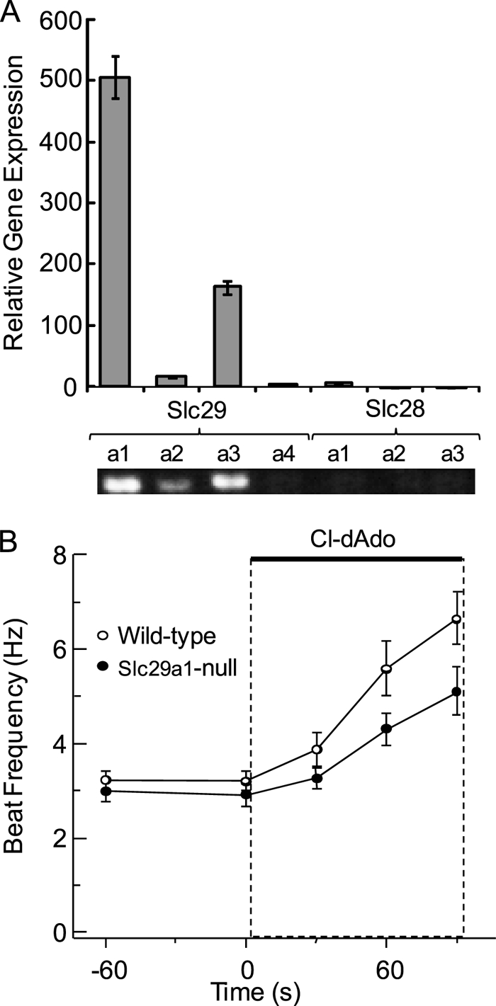
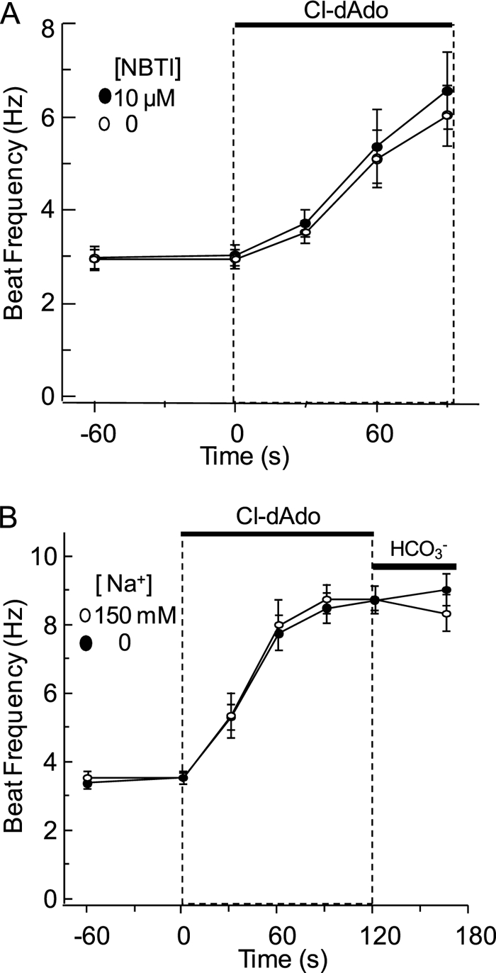
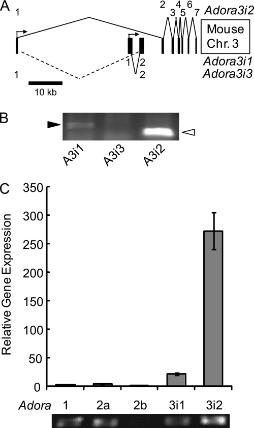
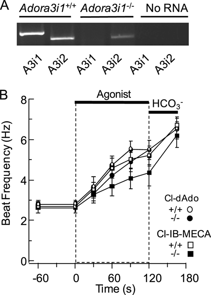
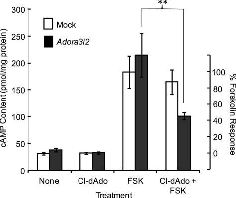
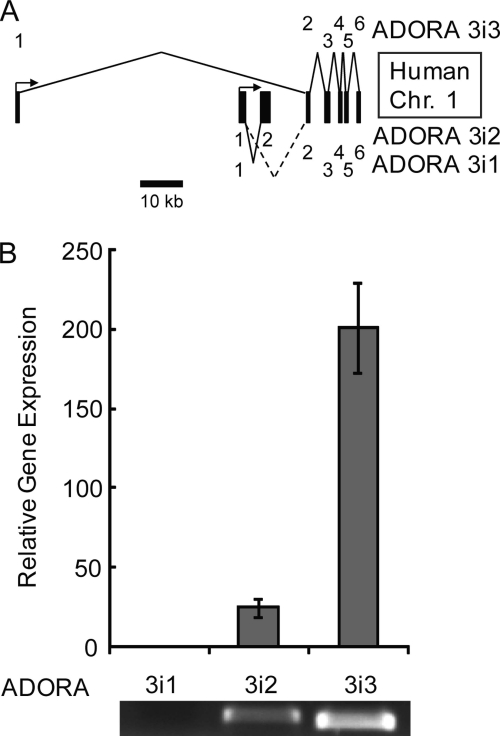
Adenosine is a candidate modulator of sperm motility in the female reproductive tract that increases sperm flagellar beat frequency in vitro. Past work suggested that this acceleration may involve equilibrative (ENT) and concentrative (CNT) nucleoside transporters. Here we show that Slc29a1 (ENT-1) is the predominant nucleoside transporter expressed in the mouse testis. Unexpectedly, the beat of Slc29a1-null sperm still accelerates in response to 2-chloro-2'-deoxyadenosine (Cl-dAdo). Moreover, in wild-type sperm neither blockade of CNTs by removal of external Na(+), nor inhibition of ENTs with nitrobenzylthioionosine, prevents acceleration of the sperm beat by Cl-dAdo. In contrast, pertussis toxin produces strong blockade, indicating involvement of a Gα(i/o)-coupled adenosine receptor. Although agonists selective for adenosine receptors A1R, A2aR, and A2bR are ineffective, A3R-selective agonists Cl-IB-MECA and IB-MECA do accelerate the beat. Consistent with this pharmacological profile, the predominant Adora transcripts in the testis are products of the nested Adora3i1 and Adora3i2 genes. Surprisingly, Cl-IB-MECA and Cl-dAdo still accelerate the beat of Adora3i1-null sperm indicating that the remaining Adora3i2 transcript produces an A3R that functions in sperm. When cloned Adora3i2 is heterologously expressed in tsA-201 cells, Cl-dAdo decreases forskolin-evoked accumulation of cAMP, indicating that Adora3i2 specifies a functional A3Ri2 adenosine receptor that couples through Gα(i). Database mining reveals that mouse Adora3i2 is expressed primarily in testis, almost exclusively in spermatids. Expression of the orthologous ADORA3i3 transcript also is most prominent in human testis; presumably producing an A3Ri3 receptor that is functional in sperm and that may be a target for development of male-directed contraceptives.


Figures

FIGURE 1.
FIGURE 1.
Testicular expression of Slc28a and Slc29a, acceleration of Slc29a1-null sperm by Cl-dAdo. A, relative expression of Slc28a and Slc29a genes in the testis was determined by quantitative RT-PCR of testicular RNA with gene-specific primers. PCR products were verified by sequencing, and predicted product length was verified by agarose gel electrophoresis (lower panel). B, individual wild-type and Slc29a1−/− sperm were perfused for 45 s with Medium HS, then (at t = 0) for 90 s with HS containing 25 μm Cl-dAdo. Shown are mean beat frequencies for cells sampled at −60, 0, 30, 60, and 90 s (n = 21–60 cells in three independent experiments). Mean acceleration determined by regression analysis of the initial 60 s of response.
FIGURE 2.
FIGURE 2.
Cl-dAdo action resists ENT inhibitor NBTI and blockade of CNT by Na+ removal. A, individual wild-type sperm were perfused for 60 s with Medium HS containing 0 or 10 μm NBTI, then (at t = 0) for 90 s with the same medium fortified with 25 μm Cl-dAdo. Shown are mean beat frequencies for cells sampled −60, 0, 30, 60, and 90 s (n = 12–27 cells in three independent experiments). B, individual wild-type sperm were perfused for 60 s with Medium HS (150 mm Na+), or with nominally Na+-free HS (0 mm Na+). At t = 0, the perfusing media were fortified with 25 μm Cl-dAdo. At t = 120 s these media were replaced with medium fortified with 15 mm NaHCO3 or 15 mm tetraethylammonium bicarbonate. Mean beat frequencies for cells sampled at t = −60, 0, 30, 60, 90, 120, and 165 s (n = 21–45 cells in three independent experiments) and acceleration were determined as above.
FIGURE 3.
FIGURE 3.
Acceleration of the flagellar beat by A3R agonist Cl-IB-MECA. Individual wild-type sperm were perfused for 120 s with Medium HS containing 0–30 μm Cl-IB-MECA as indicated, then for 45 s with HS fortified with 15 mm NaHCO3. Shown are mean beat frequencies for cells sampled at 0, 30, 60, 90, 120, and 165 s (n = 24–36 cells in three independent experiments). Mean acceleration determined by regression analysis of the initial 60 s of response was 0.08 ± 0.05, 2.3 ± 0.9, 2.8 ± 1.1, and 4.8 ± 1.0 Hz min−1. Perfusion with 30 μm Cl-IB-MECA for 90 s increased beat frequency significantly (p = 0.021).
FIGURE 4.
FIGURE 4.
Adora gene expression in the adult testis. A, organization of the mouse Adora3 gene, present on chromosome 3: 105673825–105726959. Approximate scale indicated by scale bar. Numbers at the right indicate exon useages. B, PCR for full-length Adora3 transcripts in the testis; Adora3i1 (black arrowhead) and Adora3i2 (white arrowhead) were amplified. C, relative expression of Adora transcripts in the testis reported by quantitative RT-PCR of testicular cDNA with isoform-specific primers for Adora1, Adora2a, Adora2b, Adora3i2, and primer for the conserved coding region of Adora3i1 and Adora3i3. PCR products were confirmed by sequencing and predicted product length was verified by agarose gel electrophoresis (lower panel).
FIGURE 5.
FIGURE 5.
A3R agonists accelerate the beat of wild-type and Adora3i1−/− sperm. A, Adora3+/+ and Adora3−/− testis were probed for the presence of full-length transcripts of Adora3i1 and Adora3i2 (A3i1 and A3i2). B, individual sperm were perfused for 60 s with Medium HS, then (at t = 0) for 120 s with HS containing 25 μm Cl-dAdo or 30 μm Cl-IB-MECA, then for 60 s with Medium HS fortified with 15 mm NaHCO3. Shown are mean beat frequencies for sperm of wild-type (n = 18–24 cells in three experiments) and Adora3i1−/− mice (n = 12–30 cells in three experiments), sampled at t = −60, 0, 30, 60, 90, 120, and 180 s. Mean acceleration determined by regression analysis of the initial 60 s of response to Cl-dAdo and Cl-IB-MECA. Wild-type and Adora3i1−/− sperm showed no significant difference in beat frequency after 90 s of perfusion with Cl-dAdo (p = 0.23), but the beat was slightly slower (p < 0.01) for Adora3i1−/− than for wild-type sperm after 90 s of perfusion with Cl-IB-MECA.
FIGURE 6.
FIGURE 6.
Pertussis toxin blocks acceleration by Cl-dAdo. Wild-type sperm were treated for 30 min in Medium HS alone (none), or with Ptx (4 μg/ml), heat-inactivated (iPtx; 4 μg/ml), or Ptx B-oligomer (B-oligo; 4 μg/ml). Populations of loosely-tethered sperm from each treatment group were bathed for 2–5 min with Medium HS alone, or with HS fortified with Cl-dAdo (25 μm), Cl-IB-MECA (30 μm), or NaHCO3 (15 mm). Shown are mean beat frequencies (n = 40–60 cells in 5 experiments, except n = 18–30 cells in three experiments for the B-oligo treatment group). Mean frequencies for untreated controls challenged with Cl-dAdo (6.77 ± 0.53 Hz) or Cl-IB-MECA (5.86 ± 0.56 Hz) were significantly higher than for Ptx-treated cells (3.70 ± 0.15 and 3.17 ± 0.17 Hz). Asterisk indicates p < 0.001.
FIGURE 7.
FIGURE 7.
Adenosine reduces forskolin-evoked cAMP rise in cultured cells expressing Adora3i2. tsA-201 cells expressing cloned mouse Adora3i2 tagged with RFP (Adora3i2) or expressing cytosolic GFP alone (Mock) were treated with medium that was supplemented at (t = 0) with either: no additions (None), 30 μm Cl-dAdo, 3 μm forskolin (FSK), or with 3 μm forskolin after addition of 30 μm Cl-dAdo at t = −5 min (Cl-dAdo + FSK). At t = 15 min, assays were terminated and cAMP content of the neutralized extracts was determined by radioimmunoassay. Protein content was determined on assay of parallel cultures. The right axis shows responses normalized to the mean basal content and the mean content of cells treated with forskolin alone. The cAMP content of Adora3i2-expressing cells was reduced significantly (∼60%) for cells receiving Cl-dAdo prior to addition of forskolin compared with cells challenged with forskolin alone. Double asterisk indicates p < 0.002.
FIGURE 8.
FIGURE 8.
ADORA3 gene expression in the human testis. A, organization of the human ADORA3 gene, present on chromosome 1:112025971-112106597. Approximate scale indicated by scale bar. Numbers at the right indicate exon useages. B, relative expression of ADORA3 transcripts in the testis reported by quantitative RT-PCR of testicular RNA with gene-specific primers. PCR products were confirmed by sequencing, and predicted product length was verified by agarose gel electrophoresis (lower panel).

Similar articles

Cited by

References

    1. Carlson A. E., Quill T. A., Westenbroek R. E., Schuh S. M., Hille B., Babcock D. F. (2005) J. Biol. Chem. 280, 32238–32244 - PubMed
    1. Carlson A. E., Westenbroek R. E., Quill T., Ren D., Clapham D. E., Hille B., Garbers D. L., Babcock D. F. (2003) Proc. Natl. Acad. Sci. U.S.A. 100, 14864–14868 - PMC - PubMed
    1. Hess K. C., Jones B. H., Marquez B., Chen Y., Ord T. S., Kamenetsky M., Miyamoto C., Zippin J. H., Kopf G. S., Suarez S. S., Levin L. R., Williams C. J., Buck J., Moss S. B. (2005) Dev. Cell 9, 249–259 - PMC - PubMed
    1. Jin J., Jin N., Zheng H., Ro S., Tafolla D., Sanders K. M., Yan W. (2007) Biol. Reprod. 77, 37–44 - PubMed
    1. Quill T. A., Ren D., Clapham D. E., Garbers D. L. (2001) Proc. Natl. Acad. Sci. U.S.A. 98, 12527–12531 - PMC - PubMed

Publication types