

doi: 10.1093/jmicro/dfq064.
Epub 2010 Aug 23.
A novel method for large-scale immuno-SEM using protein G coupled polystyrene beads
Affiliations
- PMID: 20732882
- PMCID: PMC3156677
- DOI: 10.1093/jmicro/dfq064
Item in Clipboard
A novel method for large-scale immuno-SEM using protein G coupled polystyrene beads
J Electron Microsc (Tokyo).
2010.
Abstract
Polystyrene beads with a mean diameter of 0.76 μm were coupled with protein G and then anti-type II collagen IgG or anti-chondroitin-4-sulphate IgG were tagged to protein G. Antibody-tagged beads were applied to articular cartilage and labelled beads were counted in each sample. Antibody-tagged beads labelled significantly higher than IgG isotype control. We propose immuno-SEM using protein G coupled beads as a valuable method for micrometre range observation for specific protein distribution on surfaces of tissues or organs. This will provide information about structure as well as antigenicity on the surface at the same time.
Figures
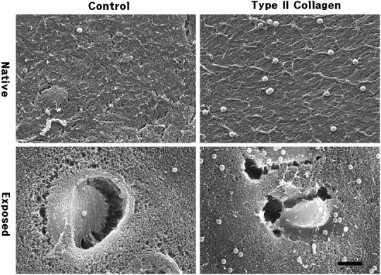
Immunolabelled beads for type II collagen on native synovial surface and cut exposed surface of articular cartilage in rabbit femur condyle. Anti-type II collagen IgG-tagged beads labelled significantly higher than IgG untagged control. Asterisk indicates lacuna; scale bar = 5 μm.
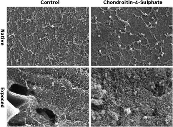
Immunolabelled beads for chondroitin-4-sulphate on native synovial surface and cut exposed surface of articular cartilage in rabbit femur condyle. Anti-chondroitin-4-sulphate IgG-coated beads labelled significantly higher than IgG untagged control. Asterisk indicates lacuna; scale bar = 5 μm.
Similar articles
-
Ultrasensitive and rapid enzyme immunoassay (thin aqueous layer immune complex transfer enzyme immunoassay) for antibody IgG to HIV-1 p17 antigen.J Clin Lab Anal. 1998;12(3):179-89. doi: 10.1002/(SICI)1098-2825(1998)12:3<179::AID-JCLA9>3.0.CO;2-4. J Clin Lab Anal. 1998. PMID: 9591706 Free PMC article.
-
The glycosaminoglycans of human articular cartilage: molecular weight distribution of chondroitin sulphate in different layers in the adult individual.Clin Orthop Relat Res. 1978 Jul-Aug;(134):364-70. Clin Orthop Relat Res. 1978. PMID: 729263
-
Preparation of Au coated polystyrene beads and their application in an immunoassay.J Immunol Methods. 2006 Dec 20;317(1-2):163-70. doi: 10.1016/j.jim.2006.10.004. Epub 2006 Nov 10. J Immunol Methods. 2006. PMID: 17107687
-
Cross-linked type I and type II collagenous matrices for the repair of full-thickness articular cartilage defects--a study in rabbits.Biomaterials. 2003 Aug;24(19):3255-63. doi: 10.1016/s0142-9612(03)00143-1. Biomaterials. 2003. PMID: 12763453
-
Changes in chondroitin sulphate structure induced by joint disease.Acta Orthop Scand Suppl. 1995 Oct;266:107-10. Acta Orthop Scand Suppl. 1995. PMID: 8553837 Review. No abstract available.
Cited by
-
Correlating Fluorescence and High-Resolution Scanning Electron Microscopy (HRSEM) for the study of GABAA receptor clustering induced by inhibitory synaptic plasticity.Sci Rep. 2017 Oct 23;7(1):13768. doi: 10.1038/s41598-017-14210-5. Sci Rep. 2017. PMID: 29061992 Free PMC article.
References
-
- Dennis J E, Cohen N, Goldberg V M, Caplan A I. Targeted delivery of progenitor cells for cartilage repair. J. Orthop. Res. 2004;22:735–741. - PubMed
-
- Biggs M J, Richards R G, Wilkinson C D, Dalby M J. Focal adhesion interactions with topographical structures: a novel method for immuno-SEM labelling of focal adhesions in S-phase cells. J. Microsc. 2008;231:28–37. - PubMed
-
- Iwano M, Che F S, Takayama S, Fukui K, Isogai A. Three-dimensional architecture of ribosomal DNA within barley nucleoli revealed with electron microscopy. Scanning. 2003;25:257–263. - PubMed
-
- Liang Y, Sasaki K. Expression of adhesion molecules relevant to leukocyte migration on the microvilli of liver peritoneal mesothelial cells. Anat. Rec. 2000;258:39–46. - PubMed
Publication types
MeSH terms
Substances
Grants and funding
LinkOut - more resources
Full Text Sources
