. 2010 Dec;24(12):5013-23.
doi: 10.1096/fj.10-165381. Epub 2010 Aug 23.

Oligomerization of Cavbeta subunits is an essential correlate of Ca2+ channel activity

Affiliations

Oligomerization of Cavbeta subunits is an essential correlate of Ca2+ channel activity

Qi Zong Lao et al. FASEB J. 2010 Dec.

Abstract

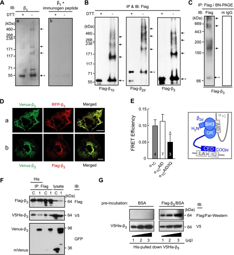
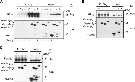
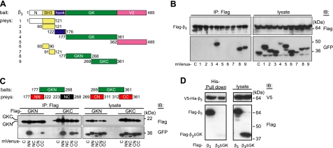
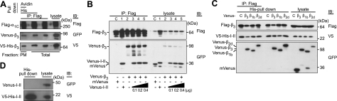
Voltage-gated calcium channels conduct Ca(2+) ions in response to membrane depolarization. The resulting transient increase in cytoplasmic free calcium concentration is a critical trigger for the initiation of such vital responses as muscle contraction and transcription. L-type Ca(v)1.2 calcium channels are complexes of the pore-forming α(1C) subunit associated with cytosolic Ca(v)β subunits. All major Ca(v)βs share a highly homologous membrane associated guanylate kinase-like (MAGUK) domain that binds to α(1C) at the α-interaction domain (AID), a short motif in the linker between transmembrane repeats I and II. In this study we show that Ca(v)β subunits form multimolecular homo- and heterooligomeric complexes in human vascular smooth muscle cells expressing native calcium channels and in Cos7 cells expressing recombinant Ca(v)1.2 channel subunits. Ca(v)βs oligomerize at the α(1C) subunits residing in the plasma membrane and bind to the AID. However, Ca(v)β oligomerization occurs independently on the association with α(1C). Molecular structures responsible for Ca(v)β oligomerization reside in 3 regions of the guanylate kinase subdomain of MAGUK. An augmentation of Ca(v)β homooligomerization significantly increases the calcium current density, while heterooligomerization may also change the voltage-dependence and inactivation kinetics of the channel. Thus, oligomerization of Ca(v)β subunits represents a novel and essential aspect of calcium channel regulation.


Figures

Figure 1.
Figure 1.
Oligomerization of Cavβ subunits. A) Immunoblotting (IB) of β3 in human aortic smooth muscle cells with an anti-β3 antibody after denaturation for 5 min at 95°C with or without prior treatment with DTT (a). To demonstrate the specificity of the antibody, in control (b), an anti-β3 antibody was preincubated with its immunogen peptide at room temperature for 1 h prior to the same immunoblotting procedure. B) Oligomerization of recombinant Cavβs expressed in Cos7 cells. Flag-labeled β1b, β2d, or β3 (0.5 μg each) was expressed in Cos7 cells and immunoprecipitated (IP) and immunoblotted with an anti-Flag antibody with or without prior DTT treatment. C) An aliquot of the Flag-β3 immunoprecipitate was analyzed without denaturation by blue native PAGE, followed by immunoblotting with an anti-Flag antibody (left panel) or with a rabbit anti-mouse IgG antibody (m IgG; right panel). Arrows mark oligomers; asterisk indicates monomer. D) Colocalization of Venus-β3 and RFP-β3 (a) or Venus-β3 and Flag-β3 (b) coexpressed in Cos7 cells. Images are confocal laser-scanning micrographs of representative cells obtained with YFP (Venus-β3) and RFP cubes (RFP-β3 and Flag-β3/Red-X). Merged images illustrate colocalization of differentially labeled β3 subunits. Scale bars = 20 μm. E) FRET between Venus- and Cerulean-tagged β2d subunits coexpressed in Cos1 cells with α2δ-1 and α1C (shaded bar) or its Ala-mutants α1CAID (α1C with disrupted AID region; open bar) or α1CAID/IQ (the same plus deleted LA/IQ region; solid bar). Inset: sketch of known interactions of α1C (gray) and β2d (blue) (24). FRET efficiency was measured as described previously (1) in live cells in the plasma membrane region of interest at −10 mV, 22°C. Number of tested cells is shown in the bars. *P < 0.05. F) Cavβ oligomers contain ≥3 β subunits. Flag-β3 and V5-His-β3 (0.5 μg each) were coexpressed in the absence (lane C, control) or presence (lane 1) of Venus-β3 (0.8 μg); the anti-Flag-IP fraction was subsequently pulled down on a His-binding matrix and immunoblotted with an anti-GFP antibody. G) Far-Western blotting. V5-His-β3 was pulled down by His; aliquots were separated by SDS-PAGE and probed by Far-Western blotting (see Materials and Methods) using 3% BSA (top right, control) or purified Flag-β3 in 3% BSA (top right). V5-His-β3 was verified by immunoblotting with an anti-V5 antibody after the Far-Western procedure (bottom panel). All plasmid DNA amounts in this study are reported per transfection per 106 cells/100-mm dish; 3 μg of Flag-β3 plasmid was used for purification of Flag-β3.
Figure 2.
Figure 2.
Cavβs form both homo- and heterooligomers. A) Venus-labeled β3 (lanes C1, 1) β1b (lanes C2, 2), β2d (lanes C3, 3) (0.8 μg each), or mVenus (control, lane C4; 0.4 μg) were expressed in the absence (lanes C1–C3) or presence (lanes 1–3, C4) of Flag-β3 (0.5 μg) in Cos7 cells, immunoprecipitated by an anti-Flag antibody, and immunoblotted with anti-Flag and anti-GFP antibody. B, C) Venus-labeled Cavβs were also coimmunoprecipitated with Flag-β2d (B) or Flag-β1b (C). (Note that the presence of low-molecular-mass bands recognized by an anti-GFP antibody may represent degraded Cavβs).
Figure 3.
Figure 3.
Multiple determinants of Cavβ oligomerization. A) Domain structure of β3 (485 aa) with regard to the fragments tested for binding to β3. B) Co-IP of Flag-β3 with indicated mVenus-labeled fragments of β3 (lanes 1–9) and mVenus (lane C, control). C) Co-IP of Flag-labeled GKN and GKC with mVenus-labeled NN, NC, CN, and CC fragments or mVenus (lane C). D) Deletion of GK from the full-length β3 subunit abolished its binding with other β3 molecules. V5-His-β3 and Flag-β3 (left lanes) or Flag-β3ΔGK (right lanes) were coexpressed in Cos7 cells (see lysates, right panel). Unlike Flag-β3, Flag-β3ΔGK was not bound to V5-His-β3, as revealed by His pulldown (left panel). We used 0.5 μg of each plasmid for cotransfection.
Figure 4.
Figure 4.
Cavβ oligomers bind to α1C at the α1C I-II linker. A) Oligomers of β3 exist in the plasma-membrane-bound α1C2δ/β3 channel complexes. Flag-α1C, α2δ, V5-His-β3, and Venus-β3 were coexpressed in Cos7 cells, and the plasma-membrane proteins were biotinylated. Anti-Flag IP of cell lysate isolated β3 bound to α1C. His pulldown confirmed the interaction of V5-His and Venus-β3 associated with α1C, while avidin pulldown confirmed it in the plasma-membrane fraction of α1C. B) Oligomerization of β3 is not affected by AID. Co-IP of homooligomers of Flag-β3 and Venus-β3 in the absence (lanes C, 2) or presence of increasing amounts of Venus-I-II linker containing AID (25) (0.1–0.4 μg/transfection, lanes 3–5). C) Evidence that I-II linker binds to Cavβ homo- and heterooligomers. Co-IP of Flag-β3 and V5-His-I-II linker with Venus-labeled β3, β1b, or β2d. Constructs were coexpressed in Cos7 cells. His pulldown was performed to isolate β subunits bound to AID, and the subsequent anti-Flag co-IP of the isolated β subunits was performed to check whether they were oligomers or monomers. D) Lack of oligomerization of I-II linker. His pulldown of V5-His-I-II linker and Venus-I-II linker coexpressed in Cos7 cells revealed the absence of Venus-I-II linker in the precipitate. Plasmids used for cotransfection were as following: Flag-α1C (1.2 μg), α2δ (1.0 μg), V5-His-β3 (0.5 μg), Venus-CaVβs (0.8 μg), V5-His-I-II linker (0.3 μg), and Venus-I-II linker (0.4 μg or as indicated).
Figure 5.
Figure 5.
Functional significance of Cavβ oligomerization. A-C) GKC increases oligomerization of β3. A) mVenus (lane 1) or indicated Venus-labeled fragments (lanes 2–9) were coexpressed with a mixture of Flag-β3 and Venus-β3. The β3 complexes were immunoprecipitated by an anti-Flag antibody and immunoblotted by an anti-GFP antibody. B) Flag-β3 (0.5 μg) was coexpressed with mVenus (lane 1) or increasing amounts of Venus-GKC (lanes 2, 3). Top panel: anti-Flag IP and immunoblotting without DTT treatment. Arrows mark oligomers; asterisk indicates monomer. C) Mixture of Flag-β3 (0.3 μg) and Venus-β3 (0.5 μg) plasmids was supplemented with mVenus (lane C, control) or increasing amounts of Venus-GKC vector (0.05–1.2 μg/transfection, lanes 1–6). Flag-β3 was immunoprecipitated from Cos7 cells by an anti-Flag antibody and immunoblotted with Flag or GFP antibodies as indicated. Red arrow indicates an apparent maximum augmentation of β3 oligomerization. D, E) Coexpression of GKC stimulates β31C association in the plasma membrane. D) Flag-α1C, α2δ, and Venus-β3 (1.2, 1.0, and 0.8 μg, respectively) were coexpressed in Cos7 cells in the presence of mVenus (control) or increasing amounts of Venus-GKC (0.01–0.4 μg/transfection). Plasma membrane fraction of the Flag coimmunoprecipitates was isolated by avidin pulldown, as described in Fig. 4A, and the flow-through fractions were analyzed for cytosolic complexes. Resulting immunoblot is representative of 5 independent experiments, which exhibited similar levels of band intensity. E) Band intensities of β3 were normalized to the respective band intensities of α1C to estimate the β31C ratio in the channel complex. Histogram is average of 5 measurements with Image J to quantify the band intensities (mean±sd); Students' t test was used for statistical analysis in comparison to control. *P < 0.05; paired 2-tailed distribution. F, G) Augmentation of β3 homooligomerization enhances the calcium current density. Cos7 cells were transfected with α1C (1.2 μg), α2δ (1 μg), and either 0.8 (black circles) or 1.6 μg (open circles) of β3, GKC (0.4 μg, gray), or a mixture of 0.8 μg β3 and 0.4 μg GKC (red). Shown are respective current-voltage relationships (F) and representative traces of the Ca2+ currents recorded in response to a stepwise shift of membrane potential from the holding potential of −90 to +20 mV and scaled to the peak current (G). H, I) Effect of heterooligomerization on current-voltage relationship (H) and kinetics of inactivation of the Ca2+ current (I). Cos7 cells were transfected as above except using 0.4 μg each β3 and β2d (blue). For comparison, respective β2d channel data are shown in magenta (25). J) Lack of calcium channel activity (at +20 mV) in Cos7 cell expressing α1C (1.2 μg) and α2δ (1 μg).

Similar articles

Cited by

References

    1. Kobrinsky E., Abrahimi P., Duong S. Q., Thomas S., Harry J. B., Patel C., Lao Q. Z., Soldatov N. M. (2009) Effect of Cavβ subunits on structural organization of Cav1.2 calcium channels. PLoS ONE 4, e5587. - PMC - PubMed
    1. Lipscombe D., Madison D. V., Poenie M., Reuter H., Tsien R. Y., Tsien R. W. (1988) Spatial distribution of calcium channels and cytosolic calcium transients in growth cones and cell bodies of sympathetic neurons. Proc. Natl. Acad. Sci. U. S. A. 85, 2398–2402 - PMC - PubMed
    1. Navedo M. F., Amberg G. C., Votaw V. S., Santana L. F. (2005) Constitutively active L-type Ca2+ channels. Proc. Natl. Acad. Sci. U. S. A. 102, 11112–11117 - PMC - PubMed
    1. Westenbroek R. E., Ahlijanian M. K., Catterall W. A. (1990) Clustering of L-type Ca2+ channels at the base of major dendrites in hippocampal pyramidal neurons. Nature 347, 281–284 - PubMed
    1. Catterall W. A. (2000) Structure and regulation of voltage-gated Ca2+ channels. Annu. Rev. Cell Dev. Biol. 16, 521–555 - PubMed

Publication types

LinkOut - more resources