. 2010 Oct;154(2):678-90.
doi: 10.1104/pp.110.161968. Epub 2010 Aug 23.

ATP-binding cassette transporter G26 is required for male fertility and pollen exine formation in Arabidopsis

Affiliations

ATP-binding cassette transporter G26 is required for male fertility and pollen exine formation in Arabidopsis

Teagen D Quilichini et al. Plant Physiol. 2010 Oct.

Abstract

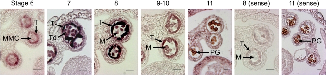
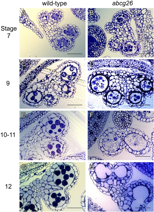
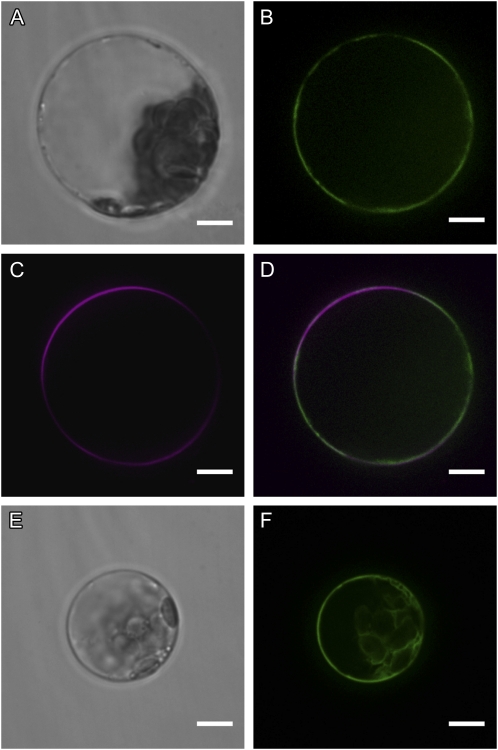
The highly resistant biopolymer, sporopollenin, gives the outer wall (exine) of spores and pollen grains their unparalleled strength, shielding these structures from terrestrial stresses. Despite a limited understanding of the composition of sporopollenin, it appears that the synthesis of sporopollenin occurs in the tapetum and requires the transport of one or more sporopollenin constituents to the surface of developing microspores. Here, we describe ABCG26, a member of the ATP-binding cassette (ABC) transporter superfamily, which is required for pollen exine formation in Arabidopsis (Arabidopsis thaliana). abcg26 mutants are severely reduced in fertility, with most siliques failing to produce seeds by self-fertilization and mature anthers failing to release pollen. Transmission electron microscopy analyses revealed an absence of an exine wall on abcg26-1 mutant microspores. Phenotypic abnormalities in pollen wall formation were first apparent in early uninucleate microspores as a lack of exine formation and sporopollenin deposition. Additionally, the highest levels of ABCG26 mRNA were in the tapetum, during early pollen wall formation, sporopollenin biosynthesis, and sporopollenin deposition. Accumulations resembling the trilamellar lipidic coils in the abcg11 and abcg12 mutants defective in cuticular wax export were observed in the anther locules of abcg26 mutants. A yellow fluorescent protein-ABCG26 protein was localized to the endoplasmic reticulum and plasma membrane. Our results show that ABCG26 plays a critical role in exine formation and pollen development and are consistent with a model by which ABCG26 transports sporopollenin precursors across the tapetum plasma membrane into the locule for polymerization on developing microspore walls.


Figures

Figure 1.
Figure 1.
Identification of Arabidopsis ABCG26 T-DNA insertion alleles. A, Diagrammatic representation of ABCG26 with T-DNA insertions in exon 5 (abcg26-1, SALK_062317) and the 5′ untranslated region (abcg26-2, SAIL_318_B09; abcg26-3, SAIL_885_F06). Black boxes represent exons, and the positions of T-DNA insertions are indicated by triangles. Arrowheads labeled P8 to P11 represent primer positions used in B. B, RT-PCR detection of ABCG26 transcripts in wild-type (Col-0) and abcg26-1 flowers. ACTIN2 expression was monitored as a control. C, Mature wild-type (left) and abcg26-1 mutant (right) plant morphology. Terminal siliques are shown (insets). D and E, Mature wild-type and abcg26-1 mutant flowers, respectively. Bars = 200 μm.
Figure 2.
Figure 2.
Flower preferred expression of ABCG26 splice variants. A, Diagrammatic representation of ABCG26 splice variants ABCG26.1 (At3g13220.1) and ABCG26.2 (At3g13220.2). Boxes and lines represent exons and introns, respectively. B, Expression of ABCG26 splice variants in various organs, assayed by quantitative RT-PCR. Expressed levels in the measured plant organs are shown as fold change relative to ABCG26.1 flower expression (set at 100) and were normalized using ACTIN2 as a reference gene. FLWR, Flower; 7D, 7-d-old seedling; YS, young stem; YL, young leaf; MS, mature stem; ML, mature leaf; MR, mature root. Error bars represent sd of three technical replicates; biological replication gave similar results.
Figure 3.
Figure 3.
Genetic complementation of the abcg26-1 mutation. A, Characterization of fertility in the wild type (Col-0), abg26 mutants, and abcg26-1 mutants containing the full-length ABCG26.1 or ABCG26.2 transgene under the control of the cauliflower mosaic virus 35S promoter. Fertility was measured according to seed production per silique along the primary stem. Small siliques contained 1.3 ± 0.4 seeds (n = 20), medium siliques contained 13.5 ± 0.8 seeds, and large siliques contained 48.3 ± 2.3 seeds. B, Left to right, representative terminal siliques from wild-type plants, abg26 mutant plants, abcg26-1 mutant plants expressing the 35S:ABCG26.1 transgene, and abcg26-1 mutant plants expressing the 35S:ABCG26.2 transgene.
Figure 4.
Figure 4.
ABCG26 tissue-specific expression pattern during microspore development. In situ hybridization was used to localize ABCG26 mRNA in wild-type (Col-0) developing flower bud sections using an ABCG26 gene-specific antisense probe and a control ABCG26 sense probe. Dark staining demarks sites of probe hybridization. From left to right, stage 6 locules contained microspore mother cells; stages 7 and 8 had locules with tetrads and free microspores; free microspores were seen in stages 9 and 10; and in stage 11 the tapetum was degenerating. Anther developmental stages are numbered according to Sanders et al. (1999). M, Microspore; MMC, microspore mother cell; PG, pollen grain; T, tapetum; Td, tetrad. Bars = 20 μm.
Figure 5.
Figure 5.
Microspore development in wild-type (Col-0) and abcg26-1 mutant anthers. Anther developmental stages are according to Sanders et al. (1999). Images in the left panels (A, C, E, and G) show wild-type anthers and those in the right panels (B, D, F, and H) show abcg26-1 anthers. A and B, Microspore development appeared identical in stage 7 wild-type and abcg26-1 anthers, when a callose wall encases tetrads of microspores. C and D, Following the release of free microspores from tetrads at stage 8, abcg26-1 microspores showed signs of degradation not observed in wild-type microspores. E and F, In stages 10 and 11, characterized by tapetum degeneration, severe abnormalities in abcg26-1 microspores were visible in comparison with wild-type microspores. G and H, At stage 12, pollen grains were not observed in abcg26-1 mutant anthers, whereas tricellular pollen grains were present in wild-type anthers. Bars = 50 μm.
Figure 6.
Figure 6.
Pollen wall structure in wild-type (Col-0) and abcg26-1 mutant plants. A and B, Scanning electron micrographs of pollen from wild-type (A) and abcg26-1 mutant (B) plants. C to F, Transmission electron micrographs of chemically fixed sections taken from wild-type (C and E) and abcg26-1 mutant (D and F) anthers between stages 9 and 10 of anther development (late unicellular stage). C and D, Low-magnification images showing tapetum cells, locules, and free microspores. E and F, High-magnification images showing pollen wall ultrastructure and locular inclusions (unlabeled arrows), which were only observed in the abcg26-1 mutant. Ba, Bacula; DEx, defective exine; Ex, exine; Lo, locule; M, microspore; Ne I, nexine I; T, tapetum; Te, tectum. Bars = 5 μm (A–D) and 100 nm (E and F).
Figure 7.
Figure 7.
Transmission electron micrographs of microspore primexine and exine formation in wild-type (Col-0) and abcg26-1 anthers. Pollen wall development between stages 5 and 8 of anther development is shown in sections taken from wild-type (A, C, E, and G) and abcg26-1 (B, D, F, and H) flowers. A and B, Microspore mother cells in stage 5. C and D, Tetrads in stage 7. E and F, Early unicellular microspores. G and H, Middle unicellular stage microspores. Ba, Bacula; CW, callose wall; Lo, locule; M, microspore; MMC, microspore mother cell; Ne I, nexine I, PM, plasma membrane; Te, tectum. Arrowheads indicate probacula. Bars = 500 nm (A–H) and 2 μm (I and J).
Figure 8.
Figure 8.
Transmission electron micrographs of locular inclusions in abcg26-1 mutant anthers at the uninucleate stage of development. A and D, Chemically fixed abcg26-1 mutant buds. B, C, E, and F, High-pressure frozen fixed abcg26-1 stamens. All samples were embedded in Spurr’s resin. Lo, Locule; M, microspore; T, tapetum. Arrows in A to C indicate locule inclusions. Bars = 500 nm (A–C) and 100 nm (D–F).
Figure 9.
Figure 9.
Subcellular localization of an EYFP:ABCG26 fusion protein in Arabidopsis mesophyll protoplasts. A to D, Transmitted light (A) and confocal microscopy of protoplast transiently expressing EYFP-ABCG26 (B), stained with FM4-64 (C), and an overlay of image B and C (D). E and F, Transmitted light (E) and confocal microscopy of a protoplast transiently expressing EYFP-ABCG26 at high levels (F). Bars = 5 μm.

Similar articles

Cited by

References

    1. Aarts MG, Hodge R, Kalantidis K, Florack D, Wilson ZA, Mulligan BJ, Stiekema WJ, Scott R, Pereira A. (1997) The Arabidopsis MALE STERILITY 2 protein shares similarity with reductases in elongation/condensation complexes. Plant J 12: 615–623 - PubMed
    1. Ahlers F, Bubert H, Steuernagel S, Wiermann R. (2000) The nature of oxygen in sporopollenin from the pollen of Typha angustifolia L. Z Naturforsch C 55: 129–136 - PubMed
    1. Ahlers F, Lambert J, Wiermann R. (2003) Acetylation and silylation of piperidine solubilized sporopollenin from pollen of Typha angustifolia L. Z Naturforsch C 58: 807–811 - PubMed
    1. Ahlers F, Thoma I, Lambert J, Kuckuk R, Wiermann R. (1999) 1H NMR analysis of sporopollenin from Typha angustifolia. Phytochemistry 50: 1095–1098
    1. Alonso JM, Stepanova AN, Leisse TJ, Kim CJ, Chen H, Shinn P, Stevenson DK, Zimmerman J, Barajas P, Cheuk R, et al. (2003) Genome-wide insertional mutagenesis of Arabidopsis thaliana. Science 301: 653–657 - PubMed

Publication types

MeSH terms