. 2011 Jan 15;183(2):226-33.
doi: 10.1164/rccm.200911-1709OC. Epub 2010 Aug 23.

Staphylococcus aureus sepsis and mitochondrial accrual of the 8-oxoguanine DNA glycosylase DNA repair enzyme in mice

Affiliations

Staphylococcus aureus sepsis and mitochondrial accrual of the 8-oxoguanine DNA glycosylase DNA repair enzyme in mice

Raquel R Bartz et al. Am J Respir Crit Care Med. .

Abstract

Rationale: Damage to mitochondrial DNA (mtDNA) by the production of reactive oxygen species during inflammatory states, such as sepsis, is repaired by poorly understood mechanisms.

Objectives: To test the hypothesis that the DNA repair enzyme, 8-oxoguanine DNA glycosylase (OGG1), contributes to mtDNA repair in sepsis.

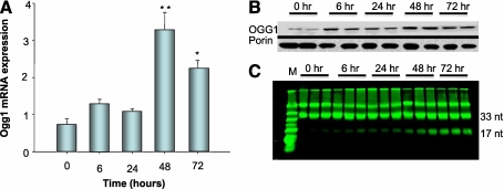
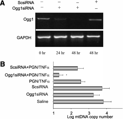
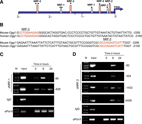
Methods: Using a well-characterized mouse model of Staphylococcus aureus sepsis, we analyzed molecular markers for mitochondrial biogenesis and OGG1 translocation into liver mitochondria as well as OGG1 mRNA expression at 0, 24, 48, and 72 hours after infection. The effects of OGG1 RNA silencing on mtDNA content were determined in control, tumor necrosis factor-α, and peptidoglycan-exposed rat hepatoma cells. Based on in situ analysis of the OGG1 promoter region, chromatin immunoprecipitation assays were performed for nuclear respiratory factor (NRF)-1 and NRF-2α GA-binding protein (GABP) binding to the promoter of OGG1.

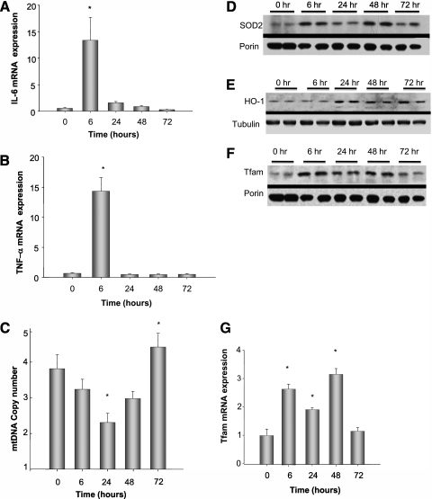
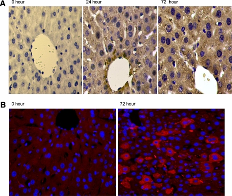
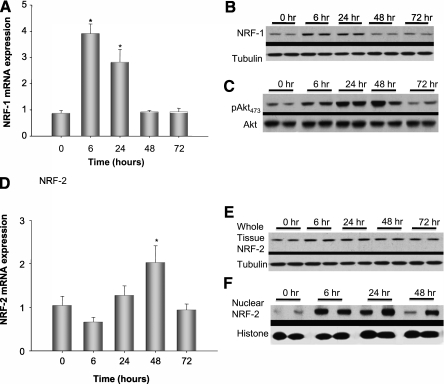
Measurements and main results: Mice infected with 10(7) cfu S. aureus intraperitoneally demonstrated hepatic oxidative mtDNA damage and significantly lower hepatic mtDNA content as well as increased mitochondrial OGG1 protein and enzyme activity compared with control mice. The infection also caused increases in hepatic OGG1 transcript levels and NRF-1 and NRF-2α transcript and protein levels. A bioinformatics analysis of the Ogg1 gene locus identified several promoter sites containing NRF-1 and NRF-2α DNA binding motifs, and chromatin immunoprecipitation assays confirmed in situ binding of both transcription factors to the Ogg1 promoter within 24 hours of infection.

Conclusions: These studies identify OGG1 as an early mitochondrial response protein during sepsis under regulation by the NRF-1 and NRF-2α transcription factors that regulate mitochondrial biogenesis.


Figures

Figure 1.
Figure 1.
IL-6, tumor necrosis factor (TNF)-α mRNA expression, mitochondrial (mt)DNA copy number, antioxidant enzyme expression, and markers of mtDNA replication during Staphylococcus aureus peritonitis in mice. (A, B) Pooled analysis of three mice per time point of IL-6 and TNF-α mRNA expression by real-time reverse transcriptase–polymerase chain reaction in liver tissue from S. aureus–infected mice. Cytokine levels increased significantly by 6 hours and returned toward baseline by 24 hours. (C) mtDNA copy number amount significantly decreased to its lowest amount 24 hours after infection and significantly increased above baseline at 72 hours after infection, *P < 0.05 by analysis of variance follow-up by Tukey analysis. (D) Representative Western blot of the mitochondrial protein fraction from two mice at each time point for superoxide dismutase (SOD2) compared with porin. SOD2 increased at 6 hours after infection and remained elevated for 72 hours. (E) Representative Western blot of whole cell lysate for HO-1 expression compared with tubulin. HO-1 increased at 24 hours after infection. (G) Mitochondrial transcription factor-A (Tfam) mRNA levels in liver tissue from S. aureus–infected mice. Tfam levels increase significantly at 6 hours and return to baseline by 72 hours after infection, *P < 0.05 by analysis of variance. (F) Representative Western analysis of the mitochondrial fraction for Tfam protein demonstrating an increase in mitochondrial Tfam at 6 hours and remaining elevated at least for 48 hours in the mitochondrial fraction.
Figure 2.
Figure 2.
Hepatic 8-hydroxyguanine (8-OHdG) and 8-oxoguanine DNA glycosylase (OGG1) staining in live S. aureus peritonitis in mice. (A) Left to right, hepatic 8-OHdG staining in C57Bl6 mice at time 0, 24, and 72 hours after intraperitoneal implantation of fibrin clot containing Staphylococcus aureus. 8-OHdG staining is brown and appears primarily as punctuate granules within the cytoplasm, suggesting mitochondrial DNA staining after infection. (B) Hepatic immunochemistry for OGG1 in mice infected with S. aureus. OGG1 by fluorescence immunohistochemistry (red staining) is increased in the liver at 72 hours after infection compared with control level. In this setting the bulk of the OGG1 is found in the cytoplasm.
Figure 3.
Figure 3.
Analysis of hepatic Ogg1 in control and S. aureus–infected mice. (A) Pooled analysis of three mice at each time point of real-time reverse transcriptase–polymerase chain reaction for Ogg1 expression in liver tissues and indicates that transcript levels reached a peak 48 hours after infection and remained elevated at 72 hours; *P < 0.05 by analysis of variance with follow-up by Tukey analysis. (B) Representative Western blot from two mice for mitochondrial 8-oxoguanine DNA glycosylase (OGG1) protein relative to porin. OGG1 protein increased within the mitochondrial fraction compared with porin within 6 hours of infection and remained elevated at 72 hours. Western analysis is shown for two mice at each time point; however, pooled data from three mice were used for statistical significance, which was present beginning 6 hours after infection (data not shown). (C) OGG1 excision assay of the liver mitochondrial fraction from S. aureus–infected mice against fluorescein isothiocyanate–labeled 8-hydroxyguanine oligonucleotides. The samples were done in triplicate and show increased excision activity as demonstrated by increased 17mer nucleotide beginning at 6 hours and remaining elevated for at least 72 hours.
Figure 4.
Figure 4.
Markers of hepatic mitochondrial biogenesis by real-time reverse transcriptase–polymerase chain reaction and Western blot analysis. (A) Pooled analysis of three mice at each time point for nuclear respiratory factor (NRF)-1 transcript levels by real-time reverse transcriptase–polymerase chain reaction. NRF-1 mRNA increased significantly by 6 hours and decreased by 48 hours after infection, *P < 0.05 by analysis of variance (ANOVA) and Tukey analysis. (B) Representative Western analysis from two mice per time point for NRF-1 relative to tubulin. NRF-1 protein, similar to mRNA expression, increased at 6 hours and decreased to baseline by 48 hours after infection. (C) Shows that Akt Ser473 phosphorylation by Western analysis is significantly increased by 6 hours after Staphylococcus aureus infection and sustained for at least 48 hours after infection. (D) Analysis of liver NRF-2 mRNA expression from the infected mice, *P < 0.05 by ANOVA. (E) Representative Western blot from two mice per time point for hepatic NRF-2. NRF-2 remained elevated even at 48 hours after infection. (F) Representative Western blot of NRF-2 from liver nuclear isolates. The increase in nuclear NRF-2 at 24 hours was more pronounced than in whole cell lysates. Three mice were used for each time point and statistical significance determined by ANOVA (data not shown). Representative Western blots from two mice are shown at each time point.
Figure 5.
Figure 5.
Ogg1 silencing in rat hepatoma cells. (A) Semiquantitative mRNA evaluation of rat hepatoma cells transfected with 8-oxoguanine DNA glycosylase (OGG1) small interfering (si)RNA demonstrating decreased OGG1 mRNA levels compared with a scrambled siRNA control. OGG1 improves mitochondrial (mt)DNA copy number on exposure to the Toll-like receptor agonist, peptidoglycan (PGN), and tumor necrosis factor (TNF)-α. (B) Decreased mtDNA copy number in rat hepatoma cells transfected with siOGG1 and stimulated with peptidoglycan and TNF-α, *P < 0.05. GAPDH = glyceraldehyde 3-phosphate dehydrogenase.
Figure 6.
Figure 6.
In silico and chromatin immunoprecipitation analysis of Ogg1 promoter site for nuclear respiratory factor (NRF)-1 and NRF-2α. NRF-1 and NRF-2α consensus site binding motifs were found for Ogg1 at 3 kb from the transcription start site. (A, B) Indicate the NRF-2α site within the Ogg1 promoter that is conserved between mice and humans. (C, D) Chromatin immunoprecipitation assays of liver nuclei at time 0, 24, and 48 hours for NRF-1 and NRF-2α binding on the Ogg1 promoter after Staphylococcus aureus infection.

Similar articles

Cited by

References

    1. Angus DC, Linde-Zwirble WT, Lidicker J, Clermont G, Carcillo J, Pinsky MR. Epidemiology of severe sepsis in the United States: analysis of incidence, outcome, and associated costs of care. Crit Care Med 2001;29:1303–1310. - PubMed
    1. Hamilton BE, Minino AM, Martin JA, Kochanek KD, Strobino DM, Guyer B. Annual summary of vital statistics: 2005. Pediatrics 2007;119:345–360. - PubMed
    1. Shigenaga MK, Hagen TM, Ames BN. Oxidative damage and mitochondrial decay in aging. Proc Natl Acad Sci USA 1994;91:10771–10778. - PMC - PubMed
    1. Raha S, Robinson BH. Mitochondria, oxygen free radicals, disease and ageing. Trends Biochem Sci 2000;25:502–508. - PubMed
    1. Taylor DE, Kantrow SP, Piantadosi CA. Mitochondrial respiration after sepsis and prolonged hypoxia. Am J Physiol 1998;275:L139–L144. - PubMed

Publication types