. 2010 Nov;30(21):5033-42.
doi: 10.1128/MCB.00922-10. Epub 2010 Aug 23.

Protein misfolding induces hypoxic preconditioning via a subset of the unfolded protein response machinery

Affiliations

Protein misfolding induces hypoxic preconditioning via a subset of the unfolded protein response machinery

Xianrong R Mao et al. Mol Cell Biol. 2010 Nov.

Abstract

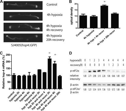
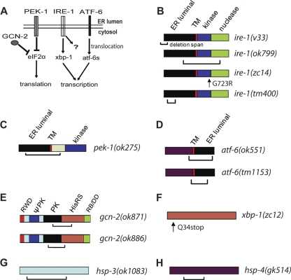
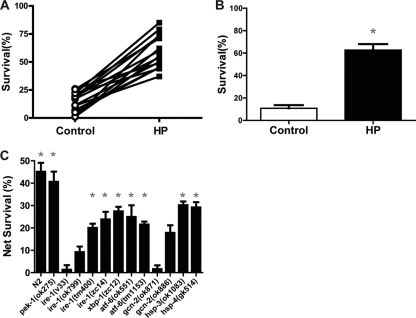
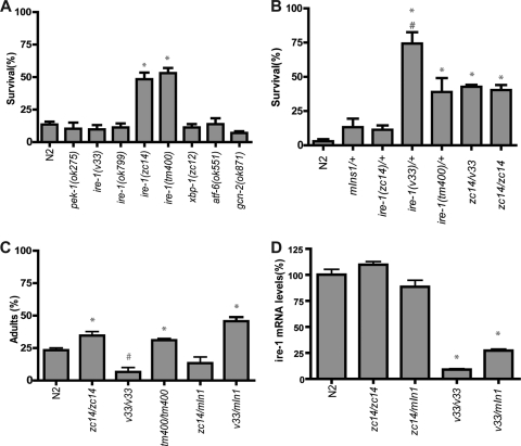
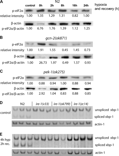
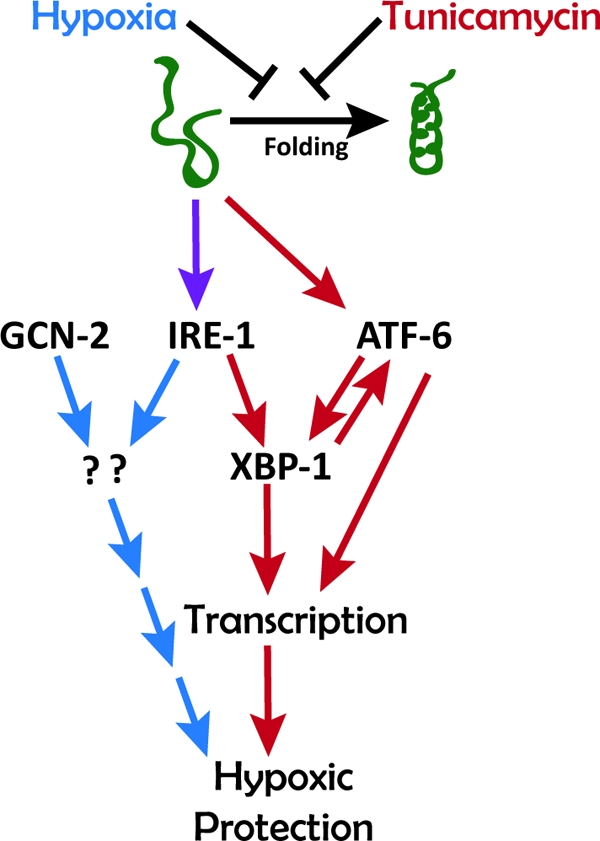
Prolonged cellular hypoxia results in energy failure and ultimately cell death. However, less-severe hypoxia can induce a cytoprotective response termed hypoxic preconditioning (HP). The unfolded protein response pathway (UPR) has been known for some time to respond to hypoxia and regulate hypoxic sensitivity; however, the role of the UPR, if any, in HP essentially has been unexplored. We have shown previously that a sublethal hypoxic exposure of the nematode Caenorhabditis elegans induces a protein chaperone component of the UPR (L. L. Anderson, X. Mao, B. A. Scott, and C. M. Crowder, Science 323:630-633, 2009). Here, we show that HP induces the UPR and that the pharmacological induction of misfolded proteins is itself sufficient to stimulate a delayed protective response to hypoxic injury that requires the UPR pathway proteins IRE-1, XBP-1, and ATF-6. HP also required IRE-1 but not XBP-1 or ATF-6; instead, GCN-2, which is known to suppress translation and induce an adaptive transcriptional response under conditions of UPR activation or amino acid deprivation, was required for HP. The phosphorylation of the translation factor eIF2α, an established mechanism of GCN-2-mediated translational suppression, was not necessary for HP. These data suggest a model where hypoxia-induced misfolded proteins trigger the activation of IRE-1, which along with GCN-2 controls an adaptive response that is essential to HP.


Figures

FIG. 1.
FIG. 1.
Hypoxia and HP activate the UPR. HP activates an hsp-4 promoter-GFP fusion reporter (A and B) and the endogenous hsp-4 gene (C). (B) After HP, the level of GFP increased significantly after a 4-h recovery (*, P < 0.001, two-tailed t test), and it returned to the control level after 16 more hours. (C) The level of hsp-4 mRNA was measured by qRT-PCR and was significantly elevated after a 2- and 4-h recovery from 4 h of hypoxia (P < 0.05, two-tailed t test), while hypoxia alone (up to 8 h) or a shorter HP incubation had no effect. (D) Hypoxia induced eIF2α phosphorylation. The level of phosphorylated eIF2α increased after 1 h of hypoxia and remained high under hypoxic conditions but rapidly returned to baseline during normoxic recovery. Relative band intensities normalized to no hypoxia are given. β-Actin levels decreased relative to total protein during the hypoxic incubation, thus the p-eIF2α/β-actin ratio increased greatly. The 0-h hypoxia/0 recovery and the 4-h hypoxia/0 recovery conditions were repeated for a total of four trials, and the relative p-eIF2α induction (1.96 ± 0.29) was statistically significant (P < 0.01, paired t test).
FIG. 2.
FIG. 2.
Tunicamycin-induced hypoxic protection. (A) Tunicamycin (Tm) pretreatment induced protection from subsequent hypoxic injury. Worms were treated with the indicated concentrations of Tm for 4 h before being recovered for 16 h. After recovery, Tm-pretreated worms were challenged with hypoxia for 22 h, and survival was scored after another 24-h recovery. Values are means ± standard deviations (SD) from three trials (* P < 0.001, paired t test, Tm versus buffer control). (C) Time course of Tm-induced hypoxia protection. The experiment was performed as described above with 10 μg/ml Tm or buffer only with various recovery times prior to the 22-h hypoxic exposure. The control value is for animals receiving no pretreatment as opposed to buffer pretreatment. Values are means ± SD from three trials (*, P < 0.01, paired t test, Tm versus buffer). (D) Wild-type (N2) or mutant animals were tested for Tm (10 μg/ml)-induced hypoxia protection. Animals were exposed for 4 h to Tm or buffer control and then recovered for 20 h prior to a 22-h hypoxic exposure, and then they were scored 24 h later for survival. Net survival (Tm survival − buffer survival) was calculated for each genotype. Tm induced significant hypoxic protection compared to that by buffer (P < 0.01, paired t test) in all strains except for the ire-1, atf-6, and xbp-1 mutants. Each bar represents the means ± SD from a minimum of three independent trials with at least 30 animals/trial. *, P < 0.01, paired t test, Tm versus buffer.
FIG. 3.
FIG. 3.
UPR pathway and mutants. (A) Schematic of UPR pathways. PEK-1, IRE-1, and ATF-6 are activated in the presence of unfolded proteins in the ER lumen. These pathways can promote adaptation to unfolded proteins via translational suppression or through a transcriptional response. GCN-2 functions along with activated PEK-1 to suppress translation. (B) ire-1 mutations. ire-1(v33) has an N-terminal 878-bp deletion resulting in a frameshift and stop and is a presumptive null mutation (48). ire-1(ok799) has a 2,093-bp deletion and 409-bp insertion and also should represent a null mutation (50). ire-1(zc14) has a missense mutation in a conserved residue in the kinase domain (8). ire-1(tm400) has a 600-bp deletion and 1-bp insertion that ends in an intron (see Wormbase.org and Materials and Methods). The mutant product is unclear. (C) pek-1(ok275) has a 2,073-bp deletion that produces a frameshift and stop codon (48). (D) Proteolysis of ATF-6 produces ATF-6s with only the maroon domain that is truncated by both mutations. ok551 has a 1,900-bp deletion (49); tm1153 has a 643-bp frameshift deletion (Wormbase.org). (E) gcn-2(ok871) has a 1,481-bp in-frame deletion starting and ending in exons (33); gcn-2(ok886) has a 1,179-bp in-frame deletion that starts and ends in exons (33). (F) xbp-1(zc12) has an early stop (8). (G) hsp-3(ok1083) has a 1,422-bp deletion that starts and ends in exons, causing frameshift (22). (H) hsp-4(gk514) has a 752-bp deletion that starts and ends in exons, causing frameshift (46). TM, transmembrane domain. The RWD domain was named after three major RWD-containing proteins: RING finger-containing proteins, WD-repeat-containing proteins, and yeast DEAD (DEXD)-like helicases. ΨPK, degenerate kinase domain; PK, kinase domain; HisRS, histidyl-tRNA synthetase; RB/DD, ribosome-binding and dimerization domain.
FIG. 4.
FIG. 4.
UPR components required for hypoxic preconditioning (HP). (A and B) Wild-type (N2) animals were exposed to hypoxia (HP) or normoxia (control) incubations for 4 h and then allowed to recover for 20 h prior to a 20-h hypoxic incubation. Survival was scored after another 24-h recovery. (C) Survival from HP in the wild type and UPR mutants. Net survival (survival of HP-treated animals − survival of control animals) is plotted for each genotype. Each bar represents the means ± standard deviations from a minimum of three independent trials with at least 30 animals/trial. *, P < 0.01, paired t test, HP versus control.
FIG. 5.
FIG. 5.
Effect of UPR mutants on hypoxic and tunicamycin sensitivity. (A) Animals with the indicated alleles were exposed to hypoxia for 20 h without any pretreatment, and survival was scored after a 24-h recovery (*, P < 0.0001 versus N2 by unpaired t test). (B) Animals with different ire-1 genetic backgrounds were tested for hypoxic survival without any pretreatment. (*, P < 0.001 versus N2; #, P < 0.05 versus zc14/zc14 or zc14/v33, unpaired t test). (C) Animals with different ire-1 genetic backgrounds were tested for sensitivity to Tm toxicity. Eggs were laid on the plates with 1 μg/ml Tm. After 3 days, the percentage of adult worms was scored (*, P < 0.01 for results greater than those for N2; #, P < 0.01 for results less than those for N2, unpaired t test). (D) The levels of ire-1 mRNA from the wild type and mutants were determined by quantitative RT-PCR by using a primer pair annealed 5′ of ire-1 cDNA. {*, P < 0.01, versus N2, zc14/zc14, or zc14/mIn1[dpy-10(e128) mIs14(p-myo-2::GFP)], unpaired t test}.
FIG. 6.
FIG. 6.
eIF2α phosphorylation and XBP-1 splicing after HP. (A to C) N2, pek-1(ok275), and gcn-2(ok871) animals were treated with hypoxia for 4 h and recovered under normoxia. Protein samples from various recovery time points were subjected to Western blotting and were probed by an antibody against p-eIF2α (ser51). The same blots were stripped and reprobed with a β-actin antibody. Intensity values for the p-eIF2α bands normalized to the control are indicated along with the ratio of the normalized intensities of the p-eIF2α bands to β-actin. Four independent trials of the control and 0-h recovery time point gave normalized p-eIF2α intensities of 1.96 ± 0.29 for N2, 1.80 ± 0.07 for gcn-2(ok871), and 1.14 ± 0.15 for pek-1(ok275). P values for the change in intensities (paired t test) were the following: for N2, 0.00531; for gcn-2(ok871), 0.00028; and for pek-1(ok275), 0.399. (D and E) The unspliced and spliced forms of xbp-1 mRNA were amplified by RT-PCR in N2 and ire-1 mutant animals under control conditions (D) and after a 2-h recovery from a 4-h HP incubation (E). Spliced xbp-1 was undetectable in all three ire-1 mutant alleles under both conditions.
FIG. 7.
FIG. 7.
Working model for the role of the UPR in HP and TmP in C. elegans. Both hypoxia and tunicamycin inhibit protein folding and thereby activate signaling through IRE-1 and ATF-6 pathways. IRE-1 is required for both HP and TmP. GCN-2 is required for HP only, and ATF-6 is required for TmP only. The mechanisms downstream of IRE-1 and GCN-2 to induce HP are unknown.

Similar articles

Cited by

References

    1. Anderson, L. L., X. Mao, B. A. Scott, and C. M. Crowder. 2009. Survival from hypoxia in C. elegans by inactivation of aminoacyl-tRNA synthetases. Science 323:630-633. - PMC - PubMed
    1. Azfer, A., J. Niu, L. M. Rogers, F. M. Adamski, and P. E. Kolattukudy. 2006. Activation of endoplasmic reticulum stress response during the development of ischemic heart disease. Am. J. Physiol. Heart Circ. Physiol. 291:H1411-H1420. - PMC - PubMed
    1. Bernales, S., K. L. McDonald, and P. Walter. 2006. Autophagy counterbalances endoplasmic reticulum expansion during the unfolded protein response. PLoS Biol. 4:e423. - PMC - PubMed
    1. Bi, M., C. Naczki, M. Koritzinsky, D. Fels, J. Blais, N. Hu, H. Harding, I. Novoa, M. Varia, J. Raleigh, D. Scheuner, R. J. Kaufman, J. Bell, D. Ron, B. G. Wouters, and C. Koumenis. 2005. ER stress-regulated translation increases tolerance to extreme hypoxia and promotes tumor growth. EMBO J. 24:3470-3481. - PMC - PubMed
    1. Bischof, L. J., C. Y. Kao, F. C. Los, M. R. Gonzalez, Z. Shen, S. P. Briggs, F. G. van der Goot, and R. V. Aroian. 2008. Activation of the unfolded protein response is required for defenses against bacterial pore-forming toxin in vivo. PLoS Pathog. 4:e1000176. - PMC - PubMed

Publication types

MeSH terms