. 2010 Aug 30;207(9):1951-66.
doi: 10.1084/jem.20100098. Epub 2010 Aug 23.

CXCR3-dependent accumulation and activation of perivascular macrophages is necessary for homeostatic arterial remodeling to hemodynamic stresses

Affiliations

CXCR3-dependent accumulation and activation of perivascular macrophages is necessary for homeostatic arterial remodeling to hemodynamic stresses

Jing Zhou et al. J Exp Med. .

Abstract

Sustained changes in blood flow modulate the size of conduit arteries through structural alterations of the vessel wall that are dependent on the transient accumulation and activation of perivascular macrophages. The leukocytic infiltrate appears to be confined to the adventitia, is responsible for medial remodeling, and resolves once hemodynamic stresses have normalized without obvious intimal changes. We report that inward remodeling of the mouse common carotid artery after ligation of the ipsilateral external carotid artery is dependent on the chemokine receptor CXCR3. Wild-type myeloid cells restored flow-mediated vascular remodeling in CXCR3-deficient recipients, adventitia-infiltrating macrophages of Gr1(low) resident phenotype expressed CXCR3, the perivascular accumulation of macrophages was dependent on CXCR3 signaling, and the CXCR3 ligand IP-10 was sufficient to recruit monocytes to the adventitia. CXCR3 also contributed to selective features of macrophage activation required for extracellular matrix turnover, such as production of the transglutaminase factor XIII A subunit. Human adventitial macrophages displaying a CD14(+)/CD16(+) resident phenotype, but not circulating monocytes, expressed CXCR3, and such cells were more frequent at sites of disturbed flow. Our observations reveal a CXCR3-dependent accumulation and activation of perivascular macrophages as a necessary step in homeostatic arterial remodeling triggered by hemodynamic stress in mice and possibly in humans as well.


Figures

Figure 1.
Figure 1.
CXCR3 is necessary for flow-mediated inward vascular remodeling. (a) The left external carotid artery was ligated, and morphometric assessment at the midpoint of the left and right common carotid arteries was performed after 2 wk. (b) Representative photomicrographs of hematoxylin and eosin–stained transverse sections of right (unligated side) and left (ligated side) common carotid arteries in WT and CXCR3−/− mice. Bar, 200 µm. The vessel walls at twofold higher magnification are depicted in the insets. (c and d) Common carotid artery size (external elastic lamina or EEL perimeter; c) and wall (medial) thickness (d) was determined by image analysis software in WT, CXCR3−/−, IL-1R−/−, IL-6−/−, TNFR1/R2−/−, and iNOS−/− mice. Data are means ± SE (n = 9–10 pooled from six operative sessions). *, P < 0.05; **, P < 0.001, KO versus WT.
Figure 2.
Figure 2.
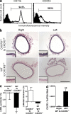
WT myeloid cells restore flow-mediated inward vascular remodeling in CXCR3−/− mice. (a) Bone marrow myeloid cells were negatively selected using density centrifugation and immunomagnetic beads and analyzed by FACS for CD11b and CXCR3 expression. (b) Representative photomicrographs of hematoxylin and eosin–stained transverse sections of the right (unligated side) and left (ligated side) common carotid arteries from CXCR3−/− animals that received 2.7 × 10⁶ bone marrow monocytes i.v. from CXCR3−/− mice or from WT mice 12 h before ligation of the left external carotid artery. Bar, 200 µm. The vessel walls at twofold higher magnification are depicted in the insets. (c) Common carotid artery size (external elastic lamina or EEL perimeter) and wall (medial) thickness was determined by image analysis software in these animals. Data are means ± SE (n = 9–10 pooled from two operative sessions). *, P < 0.05; **, P < 0.001, WT to CXCR3−/− versus CXCR3−/− to CXCR3−/−. (d) CXCR3 transcripts were quantified by real-time PCR and normalized to GAPDH from left and right common carotid arteries of CXCR3−/− mice at 3 d after adoptive transfer of WT bone marrow myeloid cells and left external carotid artery ligation. Data are means ± SE (n = 8 from one operative session). **, P < 0.001, left versus right.
Figure 3.
Figure 3.
CXCR3 is expressed by artery-associated macrophages. (a) CXCR3 transcripts were quantified by real-time PCR and normalized to GAPDH from left and right common carotid arteries of WT mice at 6 h (n = 10), 3 d (n = 9), and 10 d (n = 4) after left external carotid artery ligation. Data are means ± SE (pooled from three operative sessions). *, P < 0.05, left versus right. (b and c) CXCR3 (b) and IP-10 (c) immunohistochemical reactivity (brown, arrowheads) in left common carotid arteries at 3 d after outflow reduction. Bars, 100 µm. Absent immunostaining in right common carotid arteries is shown in insets. (d) F4/80 (green) and CXCR3 (red) immunofluorescence signal in left common carotid artery at 3 d after outflow reduction. The overlapping dual signal (orange) is marked by arrowheads. Bar, 50 µm. Irrelevant isotype-matched antibody reactivity is depicted in the inset. (e) Cells were isolated from common carotid arteries at 3 d after partial outflow ligation using CD11b antibody-coated magnetic beads and analyzed by flow cytometry for CXCR3 and F4/80 expression. (f) CXCR3 expression by common carotid artery, blood, spleen, and bone marrow (BM) CD11b-selected F4/80-expressing cells. Data are means ± SE (n = 4 pooled from four independent experiments).
Figure 4.
Figure 4.
CXCR3 expression by Gr1-defined populations of blood monocytes and artery macrophages. Cells were isolated from blood (a) or left common carotid arteries (pooled from 22 mice; b) at 3 d after partial outflow ligation using CD11b antibody-coated magnetic beads and analyzed by flow cytometry for control IgG reactivity or F4/80 and Gr1 expression, which delineated three discrete populations of myeloid cells. Their frequency is shown, calculated as the percentage of total cells. The Gr1-defined cell populations were further analyzed for CXCR3 expression (open histograms) or irrelevant isotype-matched antibody reactivity (shaded histograms), and the mean fluorescence intensity is shown. Data are representative of three independent experiments.
Figure 5.
Figure 5.
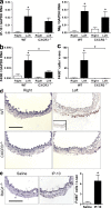
CXCR3 is necessary and sufficient for perivascular macrophage accumulation. (a and b) Transcripts for IP-10 and Mig at 6 h (a) and F4/80 at 3 d (b) were quantified by real-time PCR and normalized to GAPDH from right and left common carotid arteries of WT and CXCR3−/− mice after left external carotid artery ligation (n = 8–10 pooled from four operative sessions). (c) The number of F4/80+ cells per vessel cross section (x-sec) were counted from immunohistochemical analyses of common carotid arteries at 3 d after outflow reduction (n = 8 pooled from two operative sessions). (d) Representative photomicrographs from c are shown. Bar, 100 µm. Irrelevant isotype-matched antibody reactivity is depicted in the inset. (e) F4/80+ cells per vessel cross section were also counted from common carotid arteries infiltrated with saline or IP-10 at 300 ng/µl for 3 d in RAG1−/− mice (n = 6 pooled from two operative sessions). Bar, 100 µm. Data are means ± SE. *, P < 0.05, left versus right, CXCR3−/− versus WT, or IP-10 versus saline.
Figure 6.
Figure 6.
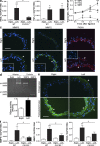
Production of macrophage mediators of vascular inflammation and remodeling after decreased flow is CXCR3 dependent. (a) Transcripts for iNOS and TNF were quantified by real-time PCR and normalized to GAPDH from right and left common carotid arteries of WT and CXCR3−/− mice at 3 d after left external carotid artery ligation (n = 9 pooled from two operative sessions). (b) Transcripts for MMP9, MMP2, and FXIIIA in left common carotid arteries of WT mice at 0 h, 6 h, 3 d, and 7 d after operation (n = 9–10 pooled from four operative sessions). (c) Immunofluorescence analyses of MMP9, MMP2, and FXIIIA expression in common carotid arteries at 3 d after outflow reduction, orientation with lumen below and adventitia above. Arrowheads mark immunofluorescence signal. Bar, 50 µm. Irrelevant isotype-matched antibody reactivity is depicted in the insets. (d) Collagenase and transglutaminase (TGase) activity in lysates of common carotid arteries at 3 d after outflow reduction. Standards of purified MMP9 and MMP2 indicate pro- and active MMP bands in gel zymogram. n = 3 from one operative session in transglutaminase assay. (e) In situ collagenase and transglutaminase activity in common carotid arteries at 3 d after outflow reduction. Enzyme activity is colored green and is marked by arrowheads. Bar, 50 µm. (f) Transcripts for MMP9 at 6 h, MMP2 at 7 d, and FXIIIA at 7 d after operation in common carotid arteries of WT and CXCR3−/− mice (n = 7–10 pooled from two operative sessions). Data are means ± SE. *, P < 0.05; **, P < 0.01, left versus right, CXCR3−/− versus WT, or after ligation versus 0 h.
Figure 7.
Figure 7.
FXIIIA production is limited to CXCR3-expressing macrophages and FXIIIA is frequently coexpressed with MMP9 in situ. (a) Perivascular macrophages were isolated with anti-CD11b–coated magnetic beads from common carotid arteries 3 d after ipsilateral external carotid artery ligation. CXCR3 and CXCR3+ F4/80-expressing macrophages were sorted by flow cytometry, RNA was extracted from an equal number of cells, and FXIIIA, MMP9, and TNF transcripts were measured by real-time PCR (n = 5–7 pooled from three independent experiments). Data are means ± SE. **, P < 0.001, CXCR3+ versus CXCR3. (b) Alternatively, CD11b-selected CD3/B220/CD11c adventitial macrophages were isolated by immunomagnetic beads and flow cytometry from 18 remodeling arteries (generated in one operative session) at 3 d after operation, treated with PMA and ionomycin for 5 h, cytospins were prepared, and the expression of FXIIIA and MMP9 was analyzed by immunofluorescence (IF) microscopy and image analysis software from 134 images. (c) Representative photomicrographs are shown of predominantly MMP9-expressing (i and iv), predominantly FXIIIA-expressing (ii and v), and dual FXIIIA-MMP9-expressing (iii and vi) small (top) and large (bottom) macrophages. (d) Similar analysis was performed on cytospins of untreated adventitial macrophages. Bar, 15 µm. Irrelevant isotype-matched antibody reactivity is depicted in the insets. Data are representative of two independent experiments.
Figure 8.
Figure 8.
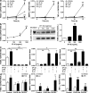
IP-10 induces FXIIIA expression in cultured macrophages. (a) Bone marrow monocytes were matured in culture in the presence of M-CSF–enriched medium for 7–10 d before treatment with IP-10 at 300 ng/ml, LPS at 10 ng/ml, or IL-4 at 10 ng/ml for various times, and FXIIIA, MMP9, and TNF transcripts were measured (n = 4 pooled from four independent experiments). (b) The cells were also treated with IP-10 at various concentrations. FXIIIA transcripts were measured by quantitative PCR (n = 3 pooled from three independent experiments) at 72 h. FXIIIA protein was assessed by immunoblotting of cell lysates at 96 h, purified human FXIIIA (75 kD) was included at 100 ng/ml, β-actin expression was used as loading controls, and blots are representative of three independent experiments. Transglutaminase activity was determined in macrophage lysates after IP-10 treatment for 96 h (n = 2 pooled from two independent experiments). (c) Quantitative PCR was also performed after treatment with single or combined proinflammatory factors at 72 h (n = 6–8 pooled from three independent experiments). (d) FXIIIA, MMP9, and TNF transcripts were measured in macrophages from WT mice after treatment with IP-10, IL-4, or LPS in the presence of CXCR3 blocking antibody (Ab) at 10 µg/ml versus irrelevant isotype-matched IgG or in macrophages from CXCR3−/− mice and compared with that of WT mice (n = 6–12 pooled from three independent experiments). Data are means ± SE. *, P < 0.05; **, P < 0.001, treated versus untreated, or single versus combined treatment.
Figure 9.
Figure 9.
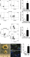
Human perivascular macrophages express CXCR3. (a) CD11b+ cells were isolated from blood or aortic adventitia using magnetic beads, analyzed by flow cytometry for control IgG reactivity or CD14 and CXCR3 expression, and a representative dot plot and combined results (n = 4) are shown. CXCR3 expression by perivascular macrophages was also compared between the nonaneurysmal (n = 5) and aneurysmal aortas (n = 3). (b) CD11b+/CD14+ adventitial cells were further analyzed for the CD16 resident macrophage marker in addition to CXCR3 and CD163 expression, and a representative dot plot and combined results (n = 7) are shown. (c) Distal (sinus) and mid-common carotid arteries (n = 3) were analyzed for CD68 (green) and CXCR3 (red) expression by immunofluorescence, and the frequency of CXCR3+/CD68+ cells (double-positive cells marked by arrowheads) were expressed as the percentage of total CD68+ cells. Bar, 25 µm. Data are means ± SE (each replicate is derived from a different donor and each specimen was processed independently). *, P < 0.05.

Similar articles

Cited by

References

    1. Auffray C., Fogg D., Garfa M., Elain G., Join-Lambert O., Kayal S., Sarnacki S., Cumano A., Lauvau G., Geissmann F. 2007. Monitoring of blood vessels and tissues by a population of monocytes with patrolling behavior. Science. 317:666–670 10.1126/science.1142883 - DOI - PubMed
    1. Bakker E.N., Pistea A., Spaan J.A., Rolf T., de Vries C.J., van Rooijen N., Candi E., VanBavel E. 2006. Flow-dependent remodeling of small arteries in mice deficient for tissue-type transglutaminase: possible compensation by macrophage-derived factor XIII. Circ. Res. 99:86–92 10.1161/01.RES.0000229657.83816.a7 - DOI - PubMed
    1. Bakker E.N., Matlung H.L., Bonta P., de Vries C.J., van Rooijen N., Vanbavel E. 2008. Blood flow-dependent arterial remodelling is facilitated by inflammation but directed by vascular tone. Cardiovasc. Res. 78:341–348 10.1093/cvr/cvn050 - DOI - PubMed
    1. Bassiouny H.S., Song R.H., Hong X.F., Singh A., Kocharyan H., Glagov S. 1998. Flow regulation of 72-kD collagenase IV (MMP-2) after experimental arterial injury. Circulation. 98:157–163 - PubMed
    1. Baumbach G.L., Heistad D.D. 1989. Remodeling of cerebral arterioles in chronic hypertension. Hypertension. 13:968–972 - PubMed

Publication types

MeSH terms