. 2010 Aug 30;207(9):1853-62.
doi: 10.1084/jem.20100239. Epub 2010 Aug 23.

PAD4 is essential for antibacterial innate immunity mediated by neutrophil extracellular traps

Affiliations

PAD4 is essential for antibacterial innate immunity mediated by neutrophil extracellular traps

Pingxin Li et al. J Exp Med. .

Abstract

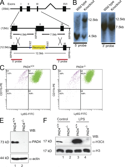
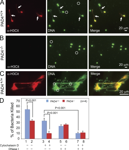
Neutrophils trap and kill bacteria by forming highly decondensed chromatin structures, termed neutrophil extracellular traps (NETs). We previously reported that histone hypercitrullination catalyzed by peptidylarginine deiminase 4 (PAD4) correlates with chromatin decondensation during NET formation. However, the role of PAD4 in NET-mediated bacterial trapping and killing has not been tested. Here, we use PAD4 knockout mice to show that PAD4 is essential for NET-mediated antibacterial function. Unlike PAD4(+/+) neutrophils, PAD4(-/-) neutrophils cannot form NETs after stimulation with chemokines or incubation with bacteria, and are deficient in bacterial killing by NETs. In a mouse infectious disease model of necrotizing fasciitis, PAD4(-/-) mice are more susceptible to bacterial infection than PAD4(+/+) mice due to a lack of NET formation. Moreover, we found that citrullination decreased the bacterial killing activity of histones and nucleosomes, which suggests that PAD4 mainly plays a role in chromatin decondensation to form NETs instead of increasing histone-mediated bacterial killing. Our results define a role for histone hypercitrullination in innate immunity during bacterial infection.


Figures

Figure 1.
Figure 1.
PAD4 is essential for histone citrullination in mouse neutrophils. (A) Schematic illustration of the replacement of exon II in the PAD4 gene with the FRT site–flanked neomycin cassette. A Hind III restriction site adjacent to exon II was also replaced. (B) Replacement of exon II leads to an appearance of a 12.5-kb fragment in addition to the 4.5-kb fragment detected by the 5′ probe (left) or the 7.5-kb fragment detected by the 3′ probe (right) in the Southern blot after digestion of ES cell genomic DNA with Hind III (representative results from three independent experiments are shown). (C and D) Flow cytometry analyses of peripheral blood neutrophils in PAD4+/+ (C) and PAD4−/− (D) mice using antibodies against neutrophil surface markers CD11b and Ly6G. Peripheral blood neutrophils were purified from five PAD4+/+ or five PAD4−/− paired mouse siblings in each experiment. Representative results from three independent experiments are shown. (E) Western blotting of PAD4 protein in PAD4+/+ and PAD4−/− neutrophils. Mouse PAD4 was detected as an ∼73-kD protein in SDS-PAGE (five mice per genotype, representative results of two independent experiments). (F) Western blotting of histone citrullination in PAD4+/+ and PAD4−/− neutrophils were performed before and after LPS treatment. General H3 antibody was used to ensure equal loading (five mice per genotype, three independent experiments).
Figure 2.
Figure 2.
PAD4 is required for chromatin decondensation and NET formation after LPS and H2O2 treatment. (A) Histone citrullination and nuclear morphology in untreated neutrophils. A small number (∼3.69%) of PAD4+/+ neutrophils showed robust histone H3 citrullination (H3Cit) staining before stimulation. DNA dye Hoechst staining was pseudo-colored green. Decondensed chromatin was not observed before stimulation. Histone H3Cit or chromatin decondensation were not observed in PAD4−/− neutrophils before stimulation. (B) LPS treatment induced histone citrullination and chromatin structural changes in PAD4+/+ neutrophils. DNA was pseudo-colored green. Notice the swelling nucleus (top) and chromatin elongation (middle). In contrast, LPS treatment did not induce histone citrullination or chromatin structural changes in PAD4−/− neutrophils (bottom). (C) Histone citrullination and neutrophil elastase staining colocalized with decondensed chromatin stained by DNA dye in PAD4+/+ neutrophils after LPS treatment. (D) H2O2 treatment induced histone citrullination and chromatin structural changes in PAD4+/+ neutrophils but not in PAD4−/− neutrophils. For assays in A–D, peripheral blood neutrophils were purified from five PAD4+/+ or five PAD4−/− paired mouse siblings in each experiment, and at least three independent experiments for each treatment condition were performed.
Figure 3.
Figure 3.
PAD4 is required for bacterial killing mediated by NETs. (A and B) Preincubation of neutrophils with IL-8 followed by incubation with S. flexneri bacteria induced histone citrullination and NET formation in PAD4+/+ (A) but not in PAD4−/− neutrophils (B). Arrows in A denote decondensed chromatin forming NETs. Circles in A and B highlight bacteria stained by the DNA dye. (C) Higher magnification images showing histone H3 citrullination and DNA staining of decondensed chromatin associated with bacteria. Circles indicate bacteria stained by DNA dye. (D) Percentages of S. flexneri bacteria killed by PAD4+/+ or PAD4−/− neutrophils, or neutrophils treated with cytochalasin D, DNase I, or both cytochalasin D and DNase I before incubation with bacteria. P-values (n = 4) were determined by a Student’s t test. Error bars indicate standard deviation. For assays in A–D, peripheral blood neutrophils were purified from five PAD4+/+ or five PAD4−/− paired mouse siblings in each experiment, and at least three independent experiments were performed.
Figure 4.
Figure 4.
Antagonism of PAD4-mediated NET formation and bacterial extracellular DNase-mediated NET destruction. (A) Genomic DNA (untreated in lane 1) was incubated with cell culture supernatant from wild-type M1 GAS (lane 2) or M1 ΔSda1 GAS (lane 3). DNA degradation by DNase Sda1 was observed (lane 2; representative results of three independent experiments). (B) Upon incubation of M1 GAS with PAD4+/+ neutrophils, histone H3 citrullination was detected but NETs were rarely observed by immunostaining (top). In contrast, histone H3 citrullination or NETs were not detected after incubation of M1 GAS with PAD4−/− neutrophils (bottom). (C) Both histone H3 citrullination and NETs were detected after incubation of M1 ΔSda1 GAS with PAD4+/+ neutrophils (top, arrows denote NETs). In contrast, histone H3 citrullination or NETs were not detected after incubation of M1 ΔSda1 GAS with PAD4−/− neutrophils (bottom). (D) Percentages of PAD4+/+ neutrophils with H3 citrullination staining or with both H3 citrullination and NET formation after incubation with M1 GAS or M1 ΔSda1 GAS were analyzed. The presence of Sda1 decreased NET formation by ∼4.2-fold (P < 0.003 by a Student’s t test). All NETs were positive for the H3 citrullination antibody staining. Error bars indicate standard deviation. (E) Higher-magnification images show NETs formed in PAD4+/+ neutrophils after incubation with M1 ΔSda1 GAS (arrows denote NETs). For assays in B-E, peripheral blood neutrophils were purified from five PAD4+/+ or five PAD4−/− paired mouse siblings in each experiment, and three independent experiments were performed.
Figure 5.
Figure 5.
PAD4 is important in immune defense against GAS in a mouse model of necrotizing fasciitis. (A) In necrotizing fasciitis assays, M1 GAS but not M1 ΔSda1 GAS induced large lesions in PAD4+/+ mice (two representative PAD4+/+ mice are shown on the left panels). In contrast, both M1 GAS and M1 ΔSda1 GAS formed large lesions in PAD4−/− mice (right). Arrows indicate lesion sites. (B) Lesion size was measured using ImageJ. The size of lesion formed by M1 ΔSda1 GAS in PAD4−/− mice increased ∼4.03-fold compared with PAD4+/+ mice (P < 0.001 by a Student’s t test). (C) The number of bacteria recovered from the lesion formation site was analyzed by colony formation assays. The number of M1 ΔSda1 GAS bacteria recovered from PAD4−/− mice increased ∼3.97-fold compared with PAD4+/+ mice (P < 0.001 by a Student’s t test). For A-C, three independent experiments were performed, with two pairs of PAD4+/+ and PAD4−/− mouse siblings used for each experiment. (D) Representative photomicrographs of skin lesions from PAD4+/+ and PAD4−/− mice infected with M1 GAS and M1 ΔSda1 GAS. Arrows indicate neutrophilic infiltrates and the star highlights intact epithelium that is absent in the other sections. Magnification is 100×. Three independent experiments, with one pair of PAD4+/+ and PAD4−/− mouse siblings, were formed for each experiment. (E) GST-PAD4 expressed and purified from Escherichia coli was used to treat histone H3 and mononucleosomes. The citrullination of histone H3 was tested by using histone H3 citrullination antibody (H3Cit) in Western blotting. Histone H3 was probed to show the amount of histone H3 in each lane (representative results of three independent experiments). (F) Analyses of the percentages of S. flexneri bacteria killed by histone H3, citrullinated histone H3, mononucleosomes, and citrullinated mononucleosomes at concentrations indicated. Standard deviations (indicated by error bars) are calculated from three independent repeat experiments.

References

    1. Bianchi M., Hakkim A., Brinkmann V., Siler U., Seger R.A., Zychlinsky A., Reichenbach J. 2009. Restoration of NET formation by gene therapy in CGD controls aspergillosis. Blood. 114:2619–2622 - PMC - PubMed
    1. Brinkmann V., Zychlinsky A. 2007. Beneficial suicide: why neutrophils die to make NETs. Nat. Rev. Microbiol. 5:577–582 10.1038/nrmicro1710 - DOI - PubMed
    1. Brinkmann V., Reichard U., Goosmann C., Fauler B., Uhlemann Y., Weiss D.S., Weinrauch Y., Zychlinsky A. 2004. Neutrophil extracellular traps kill bacteria. Science. 303:1532–1535 10.1126/science.1092385 - DOI - PubMed
    1. Brown D.T., Izard T., Misteli T. 2006. Mapping the interaction surface of linker histone H1(0) with the nucleosome of native chromatin in vivo. Nat. Struct. Mol. Biol. 13:250–255 10.1038/nsmb1050 - DOI - PMC - PubMed
    1. Buchanan J.T., Simpson A.J., Aziz R.K., Liu G.Y., Kristian S.A., Kotb M., Feramisco J., Nizet V. 2006. DNase expression allows the pathogen group A Streptococcus to escape killing in neutrophil extracellular traps. Curr. Biol. 16:396–400 10.1016/j.cub.2005.12.039 - DOI - PubMed

Publication types