. 2010 Aug 23;190(4):575-86.
doi: 10.1083/jcb.201002124.

A chromatin-bound kinase, ERK8, protects genomic integrity by inhibiting HDM2-mediated degradation of the DNA clamp PCNA

Affiliations

A chromatin-bound kinase, ERK8, protects genomic integrity by inhibiting HDM2-mediated degradation of the DNA clamp PCNA

Angela L Groehler et al. J Cell Biol. .

Abstract

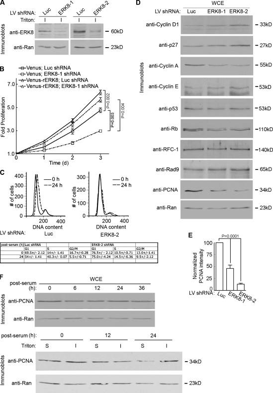
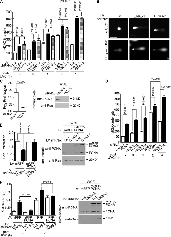
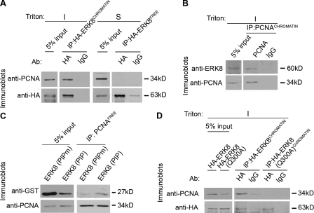
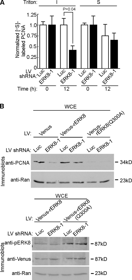
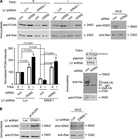
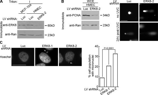
Proliferating cell nuclear antigen (PCNA) acts as a scaffold, coordinator, and stimulator of numerous processes required for faithful transmission of genetic information. Maintaining PCNA levels above a critical threshold is essential, but little is known about PCNA protein turnover. We now show that ERK8 (extracellular signal-regulated kinase 8) is required for PCNA protein stability. ERK8 contains a conserved PCNA-interacting protein (PIP) box. Chromatin-bound ERK8 (ERK8(CHROMATIN)) interacts via this motif with PCNA(CHROMATIN), which acts as a platform for numerous proteins involved in DNA metabolism. Silencing ERK8 decreases PCNA levels and increases DNA damage. Ectopic expression of PCNA blocks DNA damage induced by ERK8 loss. ERK8 prevents HDM2-mediated PCNA destruction by inhibiting the association of PCNA with HDM2. This regulation is physiologically relevant as ERK8 activity is inhibited in transformed mammary cells. Our results reveal an unanticipated mechanism to control PCNA levels in normal cycling mammary epithelial cells and implicate ERK8 in the regulation of genomic stability.


Figures

Figure 1.
Figure 1.
ERK8 regulates proliferation of MCF-10A cells. (A) Analysis of endogenous ERK8 knockdown in MCF-10A cells transduced with shRNA to luciferase (Luc) or one of two different ERK8 targeting sequences for 5 d. The detergent-insoluble fraction (I) of the transduced lysates was normalized for Ran expression and immunoblotted for ERK8. (B) Scatter plot showing the rate of proliferation of transduced MCF-10A cells. Analysis was started 2 d after transduction. Mean is shown (n = 5, quadruplicates), and error bars indicate SEM. (C) A representative flow cytometric analysis of cell cycle progression in MCF-10A cells transduced as in A. Transduced cells were identified by GFP fluorescence, and DNA was stained with DRAQ5. The times indicated are sampling times after the cells were arrested by serum and growth factor depletion, followed by release into the cell cycle by the addition of complete media. The table summarizes the results (mean ± range [n = 2]) generated by ModFit LT (Verity Software House). (D) Analysis of whole cell extracts (WCE) of MCF-10A cells transduced as in A. The lysates were normalized for Ran expression and immunoblotted for regulators of the cell cycle and DNA damage. (E) Quantitation of the relative amount of PCNA in comparison to Ran and normalized to the control, which was transduced with luciferase shRNA. Mean is shown (n = 6), and error bars indicate SEM. (F) Cell cycle analysis of PCNA in whole cell extracts and detergent-insoluble (I) and -soluble (S) fractions. MCF-10A cells were arrested and released into the cell cycle as in C. LV, lentivirus.
Figure 2.
Figure 2.
ERK8 regulates DNA repair by a PCNA-dependent mechanism. (A) Analysis of DNA damage by γ-H2AX immunofluorescence in ERK8 knockdown cells. MCF-10A cells transduced for 5 d were treated with or without (−) 20 J/m2 UVC. The times indicated refer to the length of time after irradiation. At the indicated time, the cells were treated with detergent, fixed, and immunostained with an anti–γ-H2AX antibody and an anti–mouse fluorescent secondary antibody. Nuclei were stained with DRAQ5. The intensity of γ-H2AX staining was determined and normalized to the levels obtained in the control cells, which were transduced with luciferase (Luc) shRNA and not irradiated. Mean is shown (n = 2, duplicates, ≥35 cells/condition), and error bars indicate SEM. (B) Analysis of DNA damage by comet assay of cells transduced as in A, without irradiation or 2 h after UVC treatment. DNA was visualized by staining with Sybr green (Trevigen, Inc.). Bar, 50 µm. (C) Rate of proliferation of MCF-10A cells transfected with control or PCNA-specific siRNA. The rate of proliferation was determined over 48 h, starting at 2 d after transfection. Mean is shown (n = 4, sextuplicate), and error bars indicate SEM. The right panel shows the extent of PCNA knockdown 4 d after transfection in lysates normalized for Ran. (D) Analysis of DNA damage by γ-H2AX immunofluorescence in PCNA knockdown cells. Cells transfected as in C were treated as in A, and the intensity of γ-H2AX staining was determined and normalized to the levels obtained in the control cells, which were transfected with control siRNA and not irradiated. Mean is shown (n = 2, duplicates, ≥40 cells/condition), and error bars indicate SEM. (E) Rescue of proliferation by ectopic expression of PCNA in ERK8 knockdown cells. MCF-10A cells were transduced with mRFP or mRFP-PCNA and then transduced a second time with control or ERK8-specific shRNA. The rate of proliferation was determined over 48 h, starting at 4 d after transduction. Mean is shown (n = 4, quadruplicate), and error bars indicate SEM. The right panel shows the level of mRFP-PCNA (∼62 kD) in comparison with endogenous PCNA (∼34 kD). (F) Comet assay of MCF-10A cells transduced as in E. Transduced cells were treated as in B, and tail length was measured. Mean is shown (n = 2, duplicates), and error bars indicate SEM. The right panel shows the level of mRFP-PCNA (∼62 kD) in comparison with endogenous PCNA (∼34 kD). LV, lentivirus; WCE, whole cell extract.
Figure 3.
Figure 3.
ERK8 interacts with PCNA via a PIP box. (A and B) Chromatin was immunoprecipitated with antibodies against ectopically expressed HA-tagged ERK8 (A) or endogenous PCNA (B), treated with DNase I, and analyzed by immunoblotting. A fraction of the input is shown for comparison to the level of associated PCNA or ERK8. (C) GST–ERK8 (PIP) fusion proteins were immunoprecipitated with antibodies to PCNA. The soluble fraction of MCF-10A lysates was incubated with GST-PIP box fusions containing the wild-type (QALQHPYVQRFH) or mutant ERK8 PIP (QALAHPYVQRFH) box. PCNA was immunoprecipitated and electrophoresized, and associating proteins were analyzed by immunoblotting. A fraction of the input is shown for comparison to the level of associated GST–ERK8 (PIP). (D) Chromatin was immunoprecipitated with antibodies (Ab) against ectopically expressed HA-tagged wild type or ERK8(Q300A) mutant, treated with DNase I, and analyzed by immunoblotting. A fraction of the input is shown for comparison to the level of associated PCNA. I, insoluble fraction; IP, immunoprecipitation; S, soluble fraction.
Figure 4.
Figure 4.
The association of ERK8 with PCNA inhibits PCNA protein turnover. (A) Time course of PCNA degradation by [35S]Met pulse-chase labeling. MCF-10A cells transduced for 5 d were depleted of intracellular Met, labeled with [35S]Met for 4 h, washed, and incubated in excess cold Met for 0 or 12 h. Lysates were separated into insoluble (I) and soluble (S) fractions, and each fraction was immunoprecipitated with an anti-PCNA antibody. The amount of [35S]Met labeling was divided by the relative level of immunoprecipitated PCNA and then normalized to the control, which was transduced with luciferase (Luc) shRNA and analyzed at 0 h after the addition of excess, cold Met. Mean is shown (n = 3, duplicates), and error bars indicate SEM. (B) Rescue of PCNA levels by ectopic expression of resistant ERK8 (rERK8). MCF-10A cells were transduced with resistant ERK8 constructs or Venus control, followed by a second transduction with control or ERK8-specific shRNA. Lysates were taken 5 d after transduction, normalized for Ran expression, and immunoblotted for PCNA. The bottom panel shows the expression levels and activation of the Venus-ERK8(Q300A) in comparison with wild type. IP, immunoprecipitation; LV, lentivirus; WCE, whole cell extract.
Figure 5.
Figure 5.
ERK8 regulates the association of PCNA with HDM2. (A) Silencing HDM2 enhances PCNA levels in the absence or presence of ERK8. MCF-10A cells were transduced with control (con) or ERK8-specific shRNA, followed by transfection with control or HDM2 siRNA. Lysates were taken 5 d after transduction and divided into insoluble (I) and soluble (S) fractions, normalized for Ran levels, and immunoblotted for PCNA. The top right panel shows the extent of HDM2 knockdown in lysates normalized for Ran levels. Quantitation of the relative amount of PCNA in comparison to Ran and normalized to the appropriate control, which was transduced with luciferase (Luc) shRNA and transfected with control siRNA. Mean is shown (n = 3), and error bars indicate SEM. Silencing HDM2 decreased levels of ubiquitinated PCNA. MCF-10A cells were transduced with ERK8-specific shRNA followed by transfection with a plasmid encoding His6-Ub and control or HDM2 siRNA. PCNA was immunoprecipitated 5 d after transduction, and the normalized PCNA immunoprecipitates were analyzed for ubiquitination. The IgG band is from the immunoprecipitating antibody. (B) Loss of ERK8 increases the association of HDM2 with PCNA. MCF-10A cells were transduced with control or ERK8-specific shRNA for 5 d. PCNA was immunoprecipitated, and the normalized PCNA immunoprecipitates were analyzed for HDM2. The right panel shows the level of HDM2 expression in lysates normalized for Ran. IP, immunoprecipitation; LV, lentivirus; WCE, whole cell extract.
Figure 6.
Figure 6.
ERK8 activity is regulated by autoinhibition. (A) Representative images of wild-type and mutant ERK8CHROMATIN. MCF-10A cells were transduced with Venus-ERK8 constructs. Transduced cells were treated with detergent and a high salt wash and fixed. Venus-ERK8 constructs were detected by direct fluorescence. The right panel shows immunoblots of lysates normalized for Venus expression. The black line indicates that intervening lanes were removed. WCE, whole cell extract. Bar, 50 µm. (B) Graph of the proliferation rate of transduced MCF-10A cells between day 2 and 4 after transduction. Mean is shown (n = 7, quadruplicates), and error bars indicate SEM. (C) Relative activity of wild type and ERK8 (P390A, P398A) as detected by pERK8. Transduced MCF-10A lysates were divided into insoluble (I) and soluble (S) fractions and normalized to the levels of active ERK1/2, and total expression of Venus-tagged constructs and pERK8 was determined. Active ERK1/2 levels were detected by dual phosphorylation of its T-E-Y motif. (D) The percentage of the total nuclei that stained for chromatin-bound Venus-ERK8 constructs after a detergent and high salt wash extraction. The total number of nuclei was determined by staining with DRAQ5. Venus-ERK8 constructs were detected by direct fluorescence. Mean is shown (n = 2, ≥50 cells/condition), and error bars indicate SEM. LV, lentivirus.
Figure 7.
Figure 7.
ERK8 is critical for genomic integrity in mammary epithelial cells. (A) Analysis of endogenous ERK8 knockdown in HME cells (HMEC) transduced with control or ERK8 shRNA for 5 d. The detergent-insoluble fraction (I) of the transduced lysates was normalized for Ran expression. (B) Analysis of DNA damage by comet assay of transduced HME cells treated without irradiation or 2 h after UVC treatment. The left panel shows lysates from transduced HME cells that were normalized for Ran expression and immunoblotted. (C) Analysis of DNA morphology by Hoechst staining of transduced MCF-10A cells kept in long-term culture (∼2 wk). Arrows indicate micronuclei. The percentage of the total cell population that contained micronuclei was determined. Mean is shown (≥500 cells), and error bars indicate SEM. Luc, luciferase; LV, lentivirus. Bars: (B) 50 µm; (C) 10 µm.
Figure 8.
Figure 8.
Breast cancer cells do not have active ERK8. (A) Analysis of endogenous ERK8 levels in breast cancer cell lines. Whole cell extracts (WCE) from different breast cell lines were normalized for Ran. For comparison, MCF-10A cells were transduced with luciferase (Luc) or ERK8 shRNA, and the insoluble (I) fraction was analyzed 5 d after transduction. (B) Lysates from the various breast lines were transduced with Venus-ERK8, normalized to expression of Venus-ERK8, and immunoblotted for active ERK8 (pERK8). (A and B) The black lines indicate that intervening lanes were removed. (C) Loss of ERK8 does not decrease PCNA levels in MCF-7 cells. MCF-7 cells were transduced with luciferase or ERK8 shRNA for 5 d. Lysates were normalized to Ran and immunoblotted. (D) Model of ERK8 regulation of PCNA levels. Active ERK8 preferentially binds to the chromatin through its binding partners. ERK8CHROMATIN and PCNACHROMATIN interact via the ERK8 PIP box. Loss of ERK8 binding to PCNA via silencing or mutation of the PIP domain results in the increased recruitment of HDM2, which enhances PCNA turnover. Autoinhibition is alleviated by mutating Pro390 and Pro398. However, these mutations interfere with chromatin binding. LV, lentivirus.

Comment in

  • HDM2 ERKs PCNA.
    Nguyen HD, Bielinsky AK. Nguyen HD, et al. J Cell Biol. 2010 Aug 23;190(4):487-9. doi: 10.1083/jcb.201007096. J Cell Biol. 2010. PMID: 20733049 Free PMC article.

Similar articles

Cited by

References

    1. Abe M.K., Saelzler M.P., Espinosa R., III, Kahle K.T., Hershenson M.B., Le Beau M.M., Rosner M.R. 2002. ERK8, a new member of the mitogen-activated protein kinase family. J. Biol. Chem. 277:16733–16743 10.1074/jbc.M112483200 - DOI - PubMed
    1. Andersen P.L., Xu F., Xiao W. 2008. Eukaryotic DNA damage tolerance and translesion synthesis through covalent modifications of PCNA. Cell Res. 18:162–173 10.1038/cr.2007.114 - DOI - PubMed
    1. Avkin S., Sevilya Z., Toube L., Geacintov N., Chaney S.G., Oren M., Livneh Z. 2006. p53 and p21 regulate error-prone DNA repair to yield a lower mutation load. Mol. Cell. 22:407–413 10.1016/j.molcel.2006.03.022 - DOI - PubMed
    1. Azam N., Vairapandi M., Zhang W., Hoffman B., Liebermann D.A. 2001. Interaction of CR6 (GADD45gamma) with proliferating cell nuclear antigen impedes negative growth control. J. Biol. Chem. 276:2766–2774 10.1074/jbc.M005626200 - DOI - PubMed
    1. Banks D., Wu M., Higa L.A., Gavrilova N., Quan J., Ye T., Kobayashi R., Sun H., Zhang H. 2006. L2DTL/CDT2 and PCNA interact with p53 and regulate p53 polyubiquitination and protein stability through MDM2 and CUL4A/DDB1 complexes. Cell Cycle. 5:1719–1729 - PubMed

Publication types

MeSH terms