. 2010 Oct;3(5):426-35.
doi: 10.1161/CIRCGENETICS.109.934281. Epub 2010 Aug 23.

Dynamic microRNA expression programs during cardiac differentiation of human embryonic stem cells: role for miR-499

Affiliations

Dynamic microRNA expression programs during cardiac differentiation of human embryonic stem cells: role for miR-499

Kitchener D Wilson et al. Circ Cardiovasc Genet. 2010 Oct.

Abstract

Background: MicroRNAs (miRNAs) are a newly discovered endogenous class of small, noncoding RNAs that play important posttranscriptional regulatory roles by targeting messenger RNAs for cleavage or translational repression. Human embryonic stem cells are known to express miRNAs that are often undetectable in adult organs, and a growing body of evidence has implicated miRNAs as important arbiters of heart development and disease.

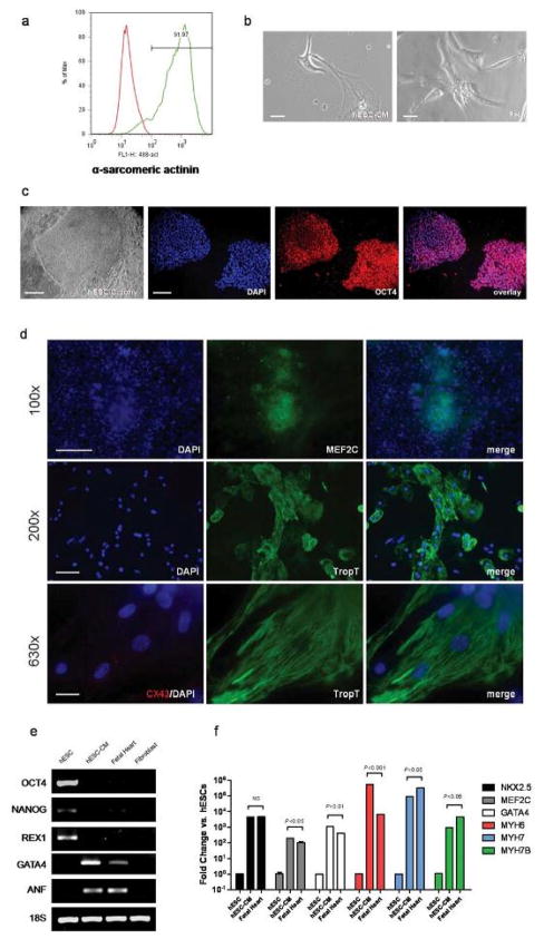
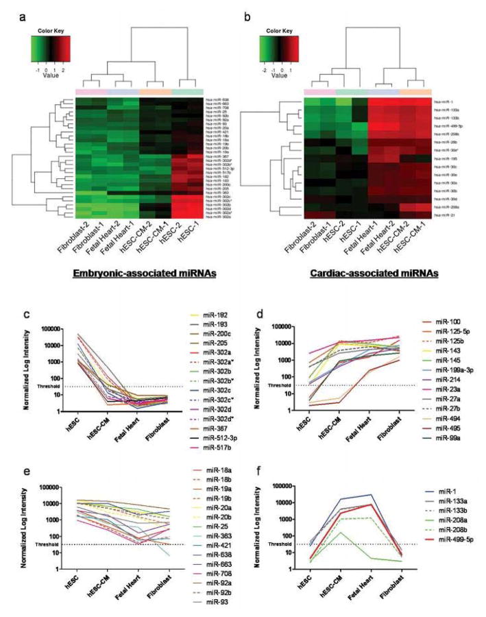
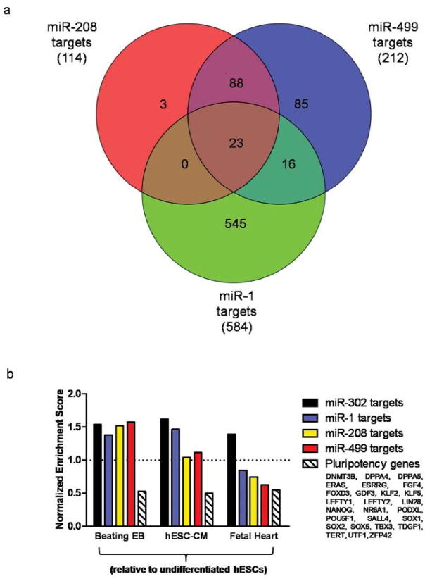
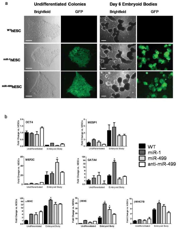
Methods and results: To better understand the transition between the human embryonic and cardiac "miRNA-omes," we report here the first miRNA profiling study of cardiomyocytes derived from human embryonic stem cells. Analyzing 711 unique miRNAs, we have identified several interesting miRNAs, including miR-1, -133, and -208, that have been previously reported to be involved in cardiac development and disease and that show surprising patterns of expression across our samples. We also identified novel miRNAs, such as miR-499, that are strongly associated with cardiac differentiation and that share many predicted targets with miR-208. Overexpression of miR-499 and -1 resulted in upregulation of important cardiac myosin heavy-chain genes in embryoid bodies; miR-499 overexpression also caused upregulation of the cardiac transcription factor MEF2C.

Conclusions: Taken together, our data give significant insight into the regulatory networks that govern human embryonic stem cell differentiation and highlight the ability of miRNAs to perturb, and even control, the genes that are involved in cardiac specification of human embryonic stem cells.


Conflict of interest statement

Conflict of Interest Disclosures None.

Figures

Figure 1
Figure 1. Differentiation of hESCs to cardiomyocytes that express lineage-specific genes
(a) Flow cytometry of differentiated hESC-CMs shows greater than 90% of cells are positive for the cardiac marker α-sarcomeric actinin compared to isotype control. (b) hESC-CMs spontaneously beat in culture and exhibited a muscle-like morphology that was similar to 20-week left ventricular fetal heart (FH) cells. (Scale bars, 40 μm) (c) Undifferentiated hESCs immunostain positive for the stem cell marker OCT4 (red). (Scale bars, 100 μm) (d) hESC-CMs express cardiac specific markers MEF2C, cardiac Troponin-T (TropT), and Connexin 43 (CX43) (Scale bars for 100X, 200X, and 630X are 200 μm, 60 μm, and 20 μm, respectively). The same hESC-CM cluster shown at 100X magnification can be seen beating in Video S1. (e) RT-PCR analysis reveals differences in expression of embryonic (OCT4, NANOG, REX1) and cardiac markers (ANF, GATA4) in hESCs, hESC-CMs, fetal heart tissue, and fetal fibroblasts. (f) qPCR of NKX2.5, MEF2C and GATA4 (early cardiac genes) expression showed significant upregulation in hESC-CMs and fetal heart relative to hESCs. Three cardiac myosin heavy chains were also upregulated: αMHC (MYH6), βMHC (MYH7), and βMHC7b (MYH7B). Error bars represent one standard deviation from the mean of biological triplicate experiments. (NS = Not Significant).
Figure 2
Figure 2
(a–b) Heatmaps of the miRNA microarray data, showing the normalized log2 miRNA expression level for each miRNA (rows) in each sample (columns), according to the indicated color scale. (a) miRNAs more highly expressed in hESCs. (b) miRNAs primarily expressed in cardiac samples. (c–f) Microarray signal intensity plots of selected miRNAs. Signal values are the mean of each pair of biological duplicate experiments; signal intensities below “threshold” may not be quantitative. (c) miRNAs expressed highly in hESCs but not detectable in fetal cell types. (d) miRNAs with increased expression during differentiation and in fetal cells. (e) miRNAs with decreasing expression during differentiation. (f) Selected miRNAs with high expression in cardiac cells (hESC-CM and/or fetal heart) but near or below threshold of detection in hESCs and fibroblasts.
Figure 3
Figure 3. Target analysis of miR-499, miR-1, and miR-208
(a) Venn diagram of TargetScan predicted gene targets. (b) Gene Set Enrichment Analysis (GSEA) of the predicted targets for the three miRNAs, as well as the miR-302 cluster and a custom set of 26 pluripotentcy-related genes, in relation to published microarray data for hESCs, beating EBs, hESC-CMs and fetal heart (GSE13834). The Normalized Enrichment Score indicates degree of gene set enrichment in the indicated cell type relative to hESCs; >1 defines upregulation, <1 defines downregulation. During cardiac differentiation, the gene targets of miR-1, −208, and −499 are increasingly less enriched (relative to hESCs) due to higher expression of these miRNAs in cardiac cell types. In contrast, the targets of the hESC-specific miR-302 cluster are consistently highly enriched in all cell types relative to hESCs, whereas pluripotency genes are consistently less enriched, as expected. Gene sets were considered significant with Q-value <0.25 (see Materials and Methods).
Figure 4
Figure 4. Over-expression of miR-1 and miR-499 during hESC differentiation
(a) miR-1 and miR-499 transduced hESCs are positive for GFP (scale bars, 100 μm), and remain positive after differentiation to day 6 embryoid bodies (scale bars, 400 μm); wild-type (WT) hESCs remain negative. (b) qPCR shows different patterns of gene expression induced by the two miRNAs, as well as knockdown of miR-499 (anti-miR-499). Error bars represent one standard deviation from the mean of biological quadruplicate experiments. (*P<0.05 vs. WTEB).
Figure 5
Figure 5. Electrophysiological changes in hESC-CMs after over-expression of miR-1 and miR-499
(a) Beating rates of spontaneously-contracting hESC-derived EB clusters after miR-1 and miR-499 transduction (N=6). (b) Left: Representative If tracings recorded from isolated single hESC-CMs with or without miR-1 transduction. Right: Bar graph summarizing the If current. *P<0.05.

Comment in

Similar articles

Cited by

References

    1. van Laake LW, Passier R, Doevendans PA, Mummery CL. Human embryonic stem cell-derived cardiomyocytes and cardiac repair in rodents. Circulation Research. 2008;102:1008–10. - PubMed
    1. Cao F, Wagner RA, Wilson KD, Xie X, Fu J-D, Drukker M, Lee A, Li RA, Gambhir SS, Weissman IL, Robbins RC, Wu JC. Transcriptional and functional profiling of human embryonic stem cell-derived cardiomyocytes. PloS ONE. 2008;3:e3474. - PMC - PubMed
    1. Laflamme MA, Chen KY, Naumova AV, Muskheli V, Fugate JA, Dupras SK, Reinecke H, Xu C, Hassanipour M, Police S, O’Sullivan C, Collins L, Chen Y, Minami E, Gill EA, Ueno S, Yuan C, Gold J, Murry CE. Cardiomyocytes derived from human embryonic stem cells in pro-survival factors enhance function of infarcted rat hearts. Nature Biotechnology. 2007;25:1015–24. - PubMed
    1. Bartel DP. MicroRNAs: genomics, biogenesis, mechanism, and function. Cell. 2004;116:281–97. - PubMed
    1. Morin RD, O’Connor MD, Griffith M, Kuchenbauer F, Delaney A, Prabhu A-L, Zhao Y, McDonald H, Zeng T, Hirst M, Eaves CJ, Marra MA. Application of massively parallel sequencing to microRNA profiling and discovery in human embryonic stem cells. Genome Research. 2008;18:610–21. - PMC - PubMed

Publication types

MeSH terms