. 2010 Sep 21;107(38):16715-20.
doi: 10.1073/pnas.1011190107. Epub 2010 Aug 23.

Physical interaction between VIVID and white collar complex regulates photoadaptation in Neurospora

Affiliations

Physical interaction between VIVID and white collar complex regulates photoadaptation in Neurospora

Chen-Hui Chen et al. Proc Natl Acad Sci U S A. .

Abstract

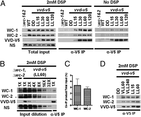
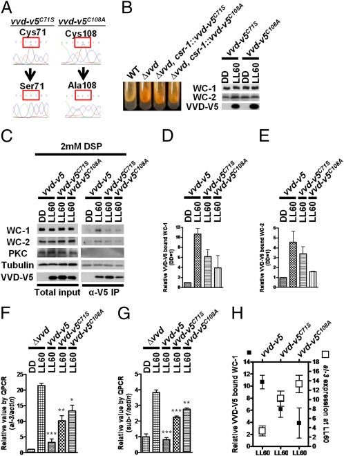
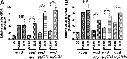
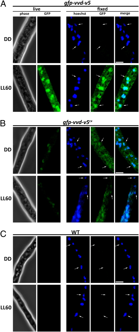
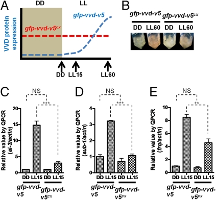
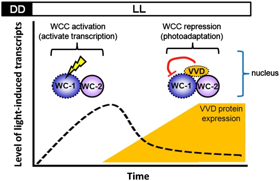
Photoadaptation, the ability to attenuate a light response on prolonged light exposure while remaining sensitive to escalating changes in light intensity, is essential for organisms to decipher time information appropriately, yet the underlying molecular mechanisms are poorly understood. In Neurospora crassa, VIVID (VVD), a small LOV domain containing blue-light photoreceptor protein, affects photoadaptation for most if not all light-responsive genes. We report that there is a physical interaction between VVD and the white collar complex (WCC), the primary blue-light photoreceptor and the transcription factor complex that initiates light-regulated transcriptional responses in Neurospora. Using two previously characterized VVD mutants, we show that the level of interaction is correlated with the level of WCC repression in constant light and that even light-insensitive VVD is sufficient partly to regulate photoadaptation in vivo. We provide evidence that a functional GFP-VVD fusion protein accumulates in the nucleus on light induction but that nuclear localization of VVD does not require light. Constitutively expressed VVD alone is sufficient to change the dynamics of photoadaptation. Thus, our results demonstrate a direct molecular connection between two of the most essential light signaling components in Neurospora, VVD and WCC, illuminating a previously uncharacterized process for light-sensitive eukaryotic cells.


Conflict of interest statement

The authors declare no conflict of interest.

Figures

Fig. 1.
Fig. 1.
Physical interaction between VVD-V5 and WCC revealed by co-IP after DSP treatment. (A) Anti-V5 co-IP assay with or without DSP treatment following a time course up to 2 h. (B) Relative amount of WC-1 and WC-2 interacting with VVD as determined by the anti-V5 co-IP assay. (C) Densitometric measurements of WC-1 and WC-2 coimmunoprecipitated with the VVD-V5 (n = 5). The data were quantified by comparison with the diluted inputs with the closest image intensity, as shown in B. (D) Anti-V5 co-IP assay with DSP treatment following a very low light (VLL, 3 μmol·m2·s) to LL (20 μmol·m2·s) transfer. NS, nonspecific band on the WC-1 blot as a loading control. Asterisks in A and B highlight a nonspecific band close in size to the WC-2 signal. In all figures, LL indicates a constant white light stimulus with a photon flux of 20 μmol·m2·s.
Fig. 2.
Fig. 2.
The level of interaction between VVD and the WCC is correlated with the level of WCC repression in constant light. (A) Sequence confirmation of the vvd-v5C71S and vvd-v5C108A alleles. (B) Carotenoid accumulation as a measure of photoadaptation defects in the vvd-v5C71S and vvd-v5C108A strains. Photoadaptation limits the expression of genes whose products are involved in the carotenogenesis pathway. (Right) Light induction of the VVD-V5 protein in either the vvd-v5C71S or the vvd-v5C108A strain. (C) Anti-V5 co-IP assay with the vvd-5, vvd-v5C71S, and vvd-v5C108A strains. PKC and tubulin are controls as described in the text. Densitometric measurements of WC-1 (D) and WC-2 (E) coimmunoprecipitated with VVD-V5 (n = 3). The data were normalized to the background signals in DD. Photoadaptation defects were determined by measuring light induction of al-3 (F) and sub-1 (G) at LL60 using RT quantitative PCR analysis (n = 3, mean values ± SE). Asterisks indicate statistical significance when compared with the Δvvd strain at LL60 as determined by an unpaired t test. ***P < 0.001; **P < 0.01; *P < 0.05. (H) Relative amount of WC-1 coimmunoprecipitated with VVD-V5 (filled squares) vs. al-3 expression at LL60 (open squares) in respective strains (n = 3). LL indicates constant white light stimulus with a photon flux of 20 μmol·m2·s.
Fig. 3.
Fig. 3.
Light-insensitive VVD is sufficient to regulate photoadaptation in vivo. Photoadaptation defects were determined by measuring induction of al-3 (A) and sub-1 (B) following a very low light (VLL, 3 μmol·m2·s) to LL (20 μmol·m2·s) transfer using RT quantitative PCR (QPCR) analysis (n = 3, mean values ± SE). Asterisks indicate statistical significance when compared with the level of expression at VLL240 as determined by an unpaired t test. ***P < 0.001; **P < 0.01. NS, difference between the two groups is not significant.
Fig. 4.
Fig. 4.
GFP-VVD accumulates in the nucleus on light induction in live cells, and the nuclear localization of GFP-VVD does not require light activation. (A) Localization of GFP-VVD-V5 before and after light treatment. Mycelia samples for live-cell imaging were collected before (DD) and after light treatment for 60 min. For fixed-cell images, the mycelia were fixed in paraformaldehyde for 7 min and then incubated with Hoechst dye for another 30 min before imaging. (B) Localization of GFP-VVD-V5cx (constitutively expressed) before and after light treatment. (C) WT strain under the same light conditions. LL indicates constant white light (photon flux of 20 μmol·m2·s). White arrows highlight representative nuclei. (Scale bar: 5 μm.) All images were acquired using identical light exposure settings, deconvolved, and displayed using identical linear contrast enhancement.
Fig. 5.
Fig. 5.
Constitutively expressed VVD is sufficient to change the dynamics of photoadaptation. (A) Schematic depicting the light induction kinetics of GFP-VVD-V5 in either the gfp-vvd-v5 or gfp-vvd-v5cx strain (constitutively expressed). (B) Photoadaptation phenotype of the gfp-vvd-v5 and gfp-vvd-v5cx strains at LL60. Photoadaptation kinetics were determined by measuring light induction of al-3 (C), sub-1 (D), and frq (E) at LL15 using RT quantitative PCR (QPCR) analysis (n = 3, mean values ± SE). Asterisks indicate statistical significance as determined by an unpaired t test. ***P < 0.001; **P < 0.01. NS, difference between two groups is not significant. LL indicates constant white light stimulus with a photon flux of 20 μmol·m2·s.
Fig. 6.
Fig. 6.
Photoadaptation in Neurospora. After light activation, the WCC transiently binds to the promoter of light-responsive genes to activate transcription, including vvd. The induced VVD protein accumulates in the nucleus and physically interacts with WCC to regulate photoadaptation by repressing WCC activity in constant light. The kinetics of photoadaptation are predominantly regulated by the amount of VVD protein in the system. Components capable of sensing light directly through a chromophore are marked with dashed lines.

Similar articles

Cited by

References

    1. Herrera-Estrella A, Horwitz BA. Looking through the eyes of fungi: Molecular genetics of photoreception. Mol Microbiol. 2007;64:5–15. - PubMed
    1. Heintzen C, Liu Y. The Neurospora crassa circadian clock. Adv Genet. 2007;58:25–66. - PubMed
    1. Corrochano LM. Fungal photoreceptors: Sensory molecules for fungal development and behaviour. Photochem Photobiol Sci. 2007;6:725–736. - PubMed
    1. Chen CH, Loros JJ. Neurospora sees the light: Light signaling components in a model system. Commun Integr Biol. 2009;2:448–451. - PMC - PubMed
    1. Bahn YS, et al. Sensing the environment: Lessons from fungi. Nat Rev Microbiol. 2007;5:57–69. - PubMed

Publication types

MeSH terms

LinkOut - more resources