. 2011 Mar;68(5):847-62.
doi: 10.1007/s00018-010-0496-2. Epub 2010 Aug 25.

Serine residue 115 of MAPK-activated protein kinase MK5 is crucial for its PKA-regulated nuclear export and biological function

Affiliations

Serine residue 115 of MAPK-activated protein kinase MK5 is crucial for its PKA-regulated nuclear export and biological function

Sergiy Kostenko et al. Cell Mol Life Sci. 2011 Mar.

Abstract

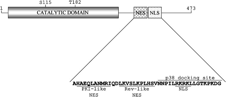
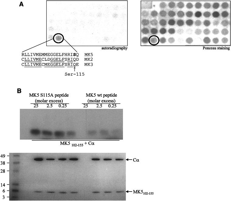
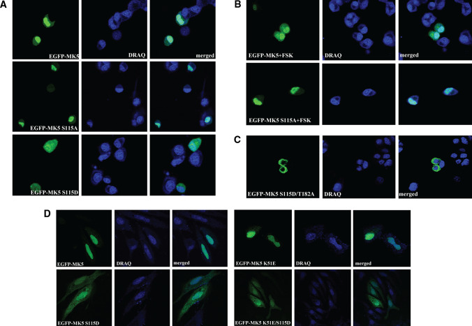
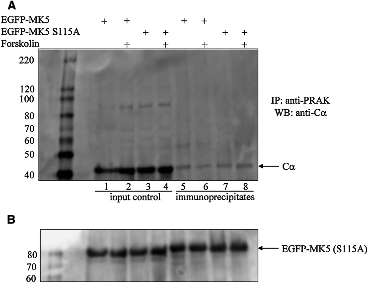
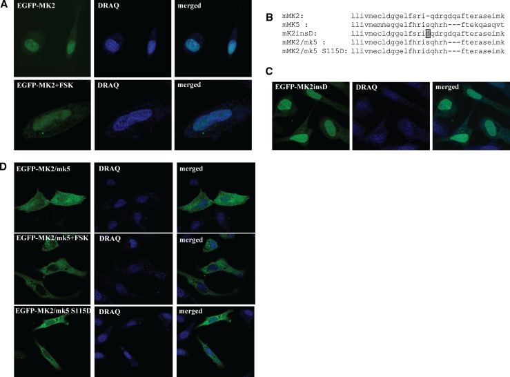
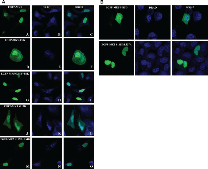
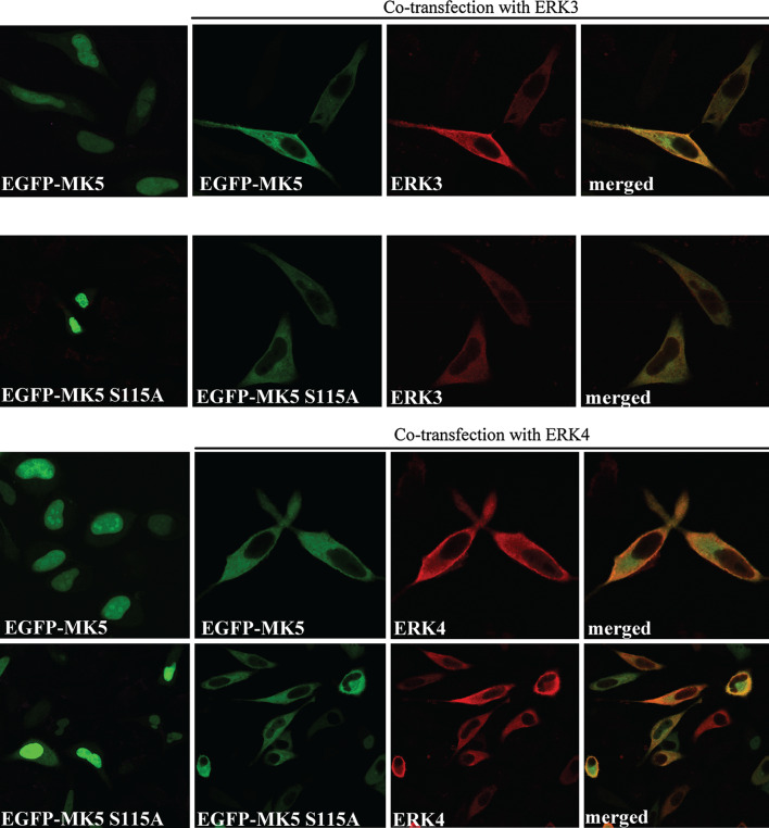
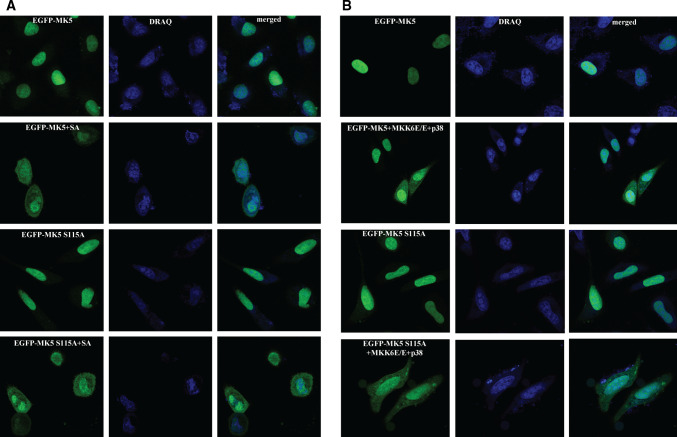
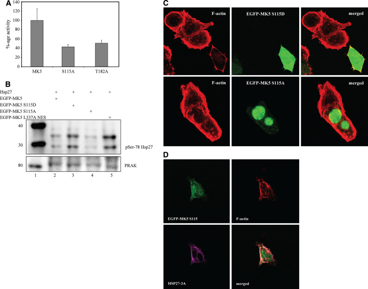
The mitogen-activated protein kinase-activated protein kinase-5 (MK5) resides predominantly in the nucleus of resting cells, but p38(MAPK), extracellular signal-regulated kinases-3 and -4 (ERK3 and ERK4), and protein kinase A (PKA) induce nucleocytoplasmic redistribution of MK5. The mechanism by which PKA causes nuclear export remains unsolved. In the study reported here we demonstrated that Ser-115 is an in vitro PKA phosphoacceptor site, and that PKA, but not p38(MAPK), ERK3 or ERK4, is unable to redistribute MK5 S115A to the cytoplasm. However, the phospho-mimicking MK5 S115D mutant resides in the cytoplasm in untreated cells. While p38(MAPK), ERK3 and ERK4 fail to trigger nuclear export of the kinase dead T182A and K51E MK5 mutants, S115D/T182A and K51E/S115D mutants were able to enter the cytoplasm of resting cells. Finally, we demonstrated that mutations in Ser-115 affect the biological properties of MK5. Taken together, our results suggest that Ser-115 plays an essential role in PKA-regulated nuclear export of MK5, and that it also may regulate the biological functions of MK5.


Figures

Fig. 1
Fig. 1
Schematic representation of the structure of MK5. The protein consists of 473 amino acids. The catalytic domain and the NES and NLS are shown. The phosphoacceptor site in the activation loop (T182) is indicated, as well as the serine residue (S115) that was identified in this work as a PKA site. The primary sequence of the NES and NLS are given. The p38 docking site, which overlaps with the NLS, is indicated
Fig. 2
Fig. 2
PKA phosphorylates MK5 at serine residue 115 in vitro. a In vitro kinase assay of a peptide array membrane that was spotted with three amino acid overlapping oligopeptides 20 amino acids long that spanned the complete MK5 protein. The autoradiographic exposure is shown on the left and the phosphorylated peptide is encircled. The right panel represents the Ponceau-stained membrane used for the peptide kinase assay. The peptide spot corresponding to the phosphorylated peptide is encircled. The sequence of the oligopeptide that was vividly phosphorylated by PKA is given with serine residue 115 indicated in bold. Comparison of the corresponding sequences of MK2 and MK3 reveals that S115 is not conserved in MK2 and MK3. The identical amino acids in the three proteins are underlined. b In vitro kinase assay on recombinant MK5102–155 fragment. GST-MK5102–155 fusion protein was purified from E. coli and the MK5 moiety was released by thrombin. The MK5 fragment was incubated for 30 min at 30°C with Cα in the presence of [γ-32P]ATP. Decreasing amounts (25, 2.5, and 0.25 M excess) of wild-type or S115A mutant competitor peptide encompassing amino acid residues 107–116 were added. Proteins were separated by polyacrylamide gel electrophoresis and phosphorylation was visualized by autoradiography. Bottom panel Coomassie blue staining ensured equal loading of the proteins in each sample. The molecular masses (in kilodaltons) of the protein marker are indicated
Fig. 3
Fig. 3
Phosphomimicking mutation of Ser-115 affects the subcellular distribution of EGFP-MK5 fusion protein in resting cells. a PC12 cells were transfected with expression plasmids for EGFP-tagged MK5 (top panels), EGFP-tagged MK5 S115A (middle panels), or EGFP-tagged MK5 S115D (bottom panels) and left untreated. b PC12 cells were transfected with expression plasmids for EGFP-MK5 or EGFP-MK5 S115A fusion proteins. Cells were exposed 24 h after transfection to 10 μM forskolin for 30 min and were then fixed. c PC12 cells were transfected with expression plasmids for EGFP-MK5 S115D/T182A and left untreated. d Cells transfected with plasmids encoding EGFP-tagged wild-type MK5, mutant MK5 S115D, MK5 K51E, or MK5 K51E/S115D. The subcellular location of ectopically expressed MK5 variants was monitored by EGFP fluorescence (green channel), while the cell nuclei were visualized by DRAQ5 staining (blue channel). On average 50 cells expressing EGFP-MK5 were analysed and representative images are shown. Similar results were obtained in independent experiments
Fig. 4
Fig. 4
Substitution of Ser-115 into Ala does not abrogate the interaction between the catalytic subunit of PKA (Cα) and MK5. HEK293 cells were transfected with expression plasmids for EGFP-MK5 or EGFP-MK5 S115A and then either left untreated or stimulated with 10 μM forskolin for 30 min. EGFP-MK5 fusion proteins were immunoprecipitated (IP) from cell lysates using anti-PRAK antibodies. These antibodies are directed against the human homologue of MK5 and crossreact with MK5. The immunoprecipitates were then examined for the presence of Cα by western blot (WB) with anti-Cα antibodies (lanes 58). The presence of Cα in the immunocomplexes is indicated by the arrow. Total cell lysates (input control) are shown in lanes 14. Bottom panel: to ensure equal loading and expression of EGFP-MK5 and EGFP-MK5 S115A, membranes were stripped and probed with anti-PRAK antibodies. The position of EGFP-MK5 (EGFP-MK5 S115A) is indicated by the arrow. The molecular mass (in kilodaltons) of the protein marker is given
Fig. 5
Fig. 5
Subcellular localization of MK2 and MK2/mk5 hybrids. a EGFP-MK2 resides predominantly in the nucleus of untreated cells and this subcellular localization is not changed upon activation of the cAMP/PKA pathway by forskolin. Cells were treated with 50 μM forskolin (FSK) for 30 min. b Alignment of the region spanning Ser-115 in MK5 with the corresponding sequence in MK2. The insertion of Asp (d) is highlighted in grey. c Insertion of a phosphomimicking Asp in MK2 that corresponds to Ser-115 in MK5 does not affect the subcellular localization of this MK2insD mutant. d Swapping the MK2 domain with the MK5 motif encompassing Ser-115 changes the subcellular localization of the MK2 from almost exclusively nuclear to both cytoplasmic and nuclear for the MK2/mk5 hybrid protein (top panels). Forskolin triggers nuclear exclusion of MK2/mk5 (middle panels). The MK2/mk5 S115D mutant resides predominantly in the cytoplasm of untreated cells (bottom panels). The EGFP fusion proteins were visualized as described in the legend for Fig. 3. On average 50 cells expressing EGFP-MK5 were analysed and representative images are shown. Similar results were obtained in independent experiments
Fig. 6
Fig. 6
Both PKA-induced nucleocytoplasmic redistribution of MK5 and cytoplasmic residence of MK5 S115D are CRM1-dependent processes. a HeLa cells were transfected with EGFP-MK5 (panels AI) or EGFP-MK5 S115D (panels JO) and either pretreated with 5 ng/ml leptomycin B (LMB) for 1 h (panels GI and MO) or left untreated. Cells were then exposed to 10 μM forskolin (FSK) for 30 min (panels DI and MO) or left untreated (panels AC and JO). b HeLa cells were transfected with EGFP-MK5 S115D or EGFP-MK5 S115D/L337A expression plasmids. The latter contains a mutation in the NES which renders this motif nonfunctional [19]. The subcellular localization of the EGFP-MK5 fusion proteins was monitored as described in the legend of Fig. 3. On average 50 cells expressing EGFP-MK5 were analysed and representative images are shown. Similar results were obtained in independent experiments
Fig. 7
Fig. 7
Overexpression of ERK3 and ERK4 induces nuclear export of wild-type MK5 and the S115A mutant. HeLa cells were cotransfected with either expression plasmids for EGFP-MK5 or EGFP-MK5 S115A alone, or cotransfected with myc-tagged ERK4 (upper panels) or myc-tagged ERK3 expression plasmids (lower panels). The subcellular location of ectopic expressed EGFP-MK5 (or EGFP-MK5 S115A) was monitored by EGFP fluorescence (green channel) and the subcellular distribution of ERK3 (or ERK4) is visualized as the red channel using Alexa Fluor 647-conjugated Myc-tag mouse monoclonal antibody. Merged images show cytoplasmic colocalization of MK5 and ERK3 or ERK4. On average 50 cells expressing EGFP-MK5 were analysed and representative images are shown. Similar results were obtained in independent experiments
Fig. 8
Fig. 8
Ser-115 is not implicated in p38MAPK-induced nuclear export of MK5. a HeLa cells transfected with EGFP-MK5 or EGFP-MK5 S115A plasmids were left untreated or exposed to sodium arsenite (250 μM for 120 min). b As in a, but cells were contransfected with plasmids encoding activated MKK6 (MKK6 E/E) and p38MAPK. The subcellular localization of the ectopically expressed MK5 variants was visualized as described in the legend for Fig. 3. On average 50 cells expressing EGFP-MK5 were analysed and representative images are shown. Similar results were obtained in independent experiments
Fig. 9
Fig. 9
Mutations in Ser-115 affect the biological properties of MK5. a HEK293 cells were cotransfected with the luciferase plasmid G5-E1b-LUC, a plasmid encoding GAL4-p53 fusion protein, and expression vectors for the fusion proteins EGFP-MK5 wild-type (MK5), EGFP-MK5 S115A (S115A) or kinase dead EGFP-MK5 T182A (T182A). Luciferase activity was determined in three independent parallel experiments and the p53 transcriptional activity in the presence of wild-type MK5 was arbitrarily set as 100% and the activity in the presence of the MK5 S115A or MK5 T182A was related to this. The experiment was repeated three times and similar results were obtained. b HEK293 cells were transfected with an expression plasmid for Hsp27 and with plasmids encoding either EGFP-MK5 (lane 1), EGFP-MK5 S115D (lane 2), EGFP-MK5 S115A (lane 3), or EGFP-MK5 L337A NES (lane 4). The last of these is a constitutive active MK5 that is exclusively localized in the cytoplasm. Phosphorylation of Hsp27 was monitored by western blotting using a specific antibody that recognized the relevant phosphoSer-78 site (top panel). The membrane was stripped and degree of equality in the expression of the EGFP-MK5 variants was assayed by western blotting with an anti-PRAK antibody (bottom panel). c PC12 cells were transfected with plasmids encoding EGFP-MK5 S115D (top panel) or EGFP-MK5 S115A (bottom panel) and F-actin was visualized using Alexa Fluor 594 Phalloidin. The expression of the EGFP-tagged MK5 was visualized by EGFP fluorescence (green channel). The superimposed images demonstrate that the architecture of F-actin is changed in cells expressing EGFP-MK5 S115D, but not in nontransfected cells nor in cells expressing EGFP-MK5 S115A. d PC12 cells were cotransfected with expression plasmids for EGFP-MK5 S115D and Hsp27-3A. Hsp27-3A was visualized using anti-Flag antibody and Alexa 647-coupled secondary antibody (magenta). MK5 S115D and F-actin were stained as described above

Similar articles

Cited by

References

    1. Johnson GL, Lapadat R. Mitogen-activated protein kinase pathways mediated by ERK, JNK, and p38 protein kinases. Science. 2002;298:1911–1912. doi: 10.1126/science.1072682. - DOI - PubMed
    1. Roux PP, Blenis J. ERK and p38 MAPK-activated protein kinases: a family of protein kinases with diverse biological functions. Microbiol Mol Biol Rev. 2004;68:320–344. doi: 10.1128/MMBR.68.2.320-344.2004. - DOI - PMC - PubMed
    1. Imajo M, Tsuchiya Y, Nishida E. Regulatory mechanisms and functions of MAP kinase signaling pathways. IUBMB Life. 2006;58:312–317. doi: 10.1080/15216540600746393. - DOI - PubMed
    1. Cuenda A, Rousseau S. p38 MAP-kinases pathway regulation, function and role in human diseases. Biochim Biophys Acta. 2007;1773:1358–1375. doi: 10.1016/j.bbamcr.2007.03.010. - DOI - PubMed
    1. Cuevas B, Abell A, Johnson G. Role of mitogen-activated protein kinase kinase kinases in signal integration. Oncogene. 2007;26:3159–3171. doi: 10.1038/sj.onc.1210409. - DOI - PubMed

Publication types

MeSH terms

Substances