. 2010 Sep;48(1):31-47.
doi: 10.1007/s10858-010-9436-6. Epub 2010 Aug 24.

Conformational dependence of 13C shielding and coupling constants for methionine methyl groups

Affiliations

Conformational dependence of 13C shielding and coupling constants for methionine methyl groups

Glenn L Butterfoss et al. J Biomol NMR. 2010 Sep.

Abstract

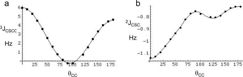
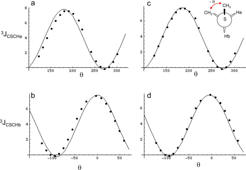
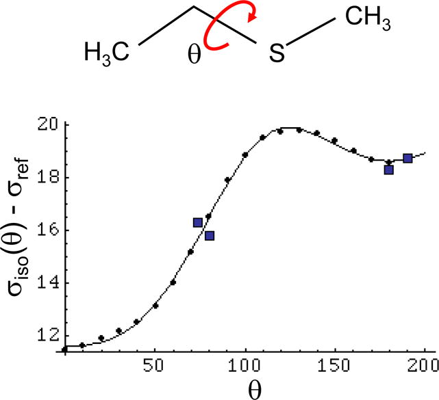
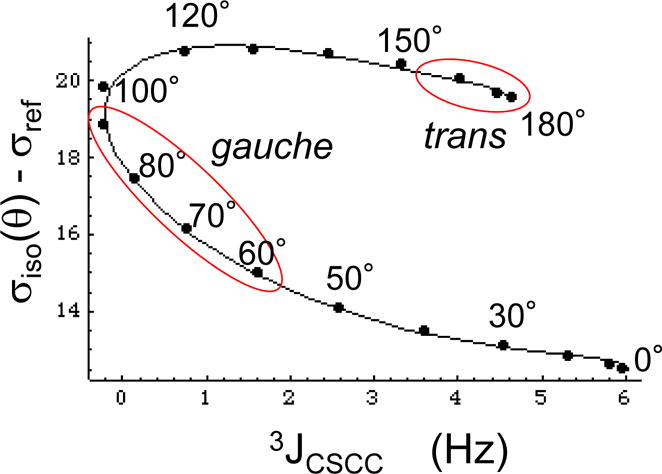
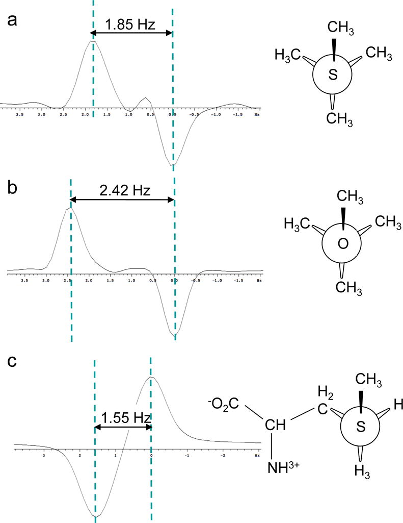
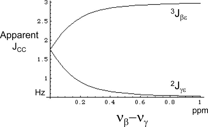
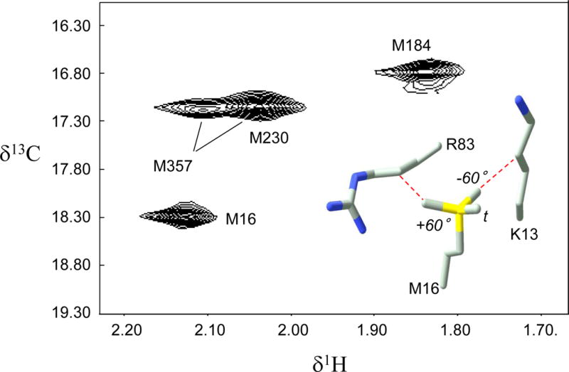
Methionine residues fulfill a broad range of roles in protein function related to conformational plasticity, ligand binding, and sensing/mediating the effects of oxidative stress. A high degree of internal mobility, intrinsic detection sensitivity of the methyl group, and low copy number have made methionine labeling a popular approach for NMR investigation of selectively labeled protein macromolecules. However, selective labeling approaches are subject to more limited information content. In order to optimize the information available from such studies, we have performed DFT calculations on model systems to evaluate the conformational dependence of (3)J (CSCC), (3)J (CSCH), and the isotropic shielding, sigma(iso). Results have been compared with experimental data reported in the literature, as well as data obtained on [methyl-(13)C]methionine and on model compounds. These studies indicate that relative to oxygen, the presence of the sulfur atom in the coupling pathway results in a significantly smaller coupling constant, (3)J (CSCC)/(3)J (COCC) approximately 0.7. It is further demonstrated that the (3)J (CSCH) coupling constant depends primarily on the subtended CSCH dihedral angle, and secondarily on the CSCC dihedral angle. Comparison of theoretical shielding calculations with the experimental shift range of the methyl group for methionine residues in proteins supports the conclusion that the intra-residue conformationally-dependent shift perturbation is the dominant determinant of delta(13)Cepsilon. Analysis of calmodulin data based on these calculations indicates that several residues adopt non-standard rotamers characterized by very large approximately 100 degrees chi(3) values. The utility of the delta(13)Cepsilon as a basis for estimating the gauche/trans ratio for chi(3) is evaluated, and physical and technical factors that limit the accuracy of both the NMR and crystallographic analyses are discussed.


Figures

Figure 1
Figure 1
Conformational dependence of scalar coupling constants in ethylmethyl sulfide. a) The filled circles (③) correspond to the calculated 3JCSCC values, and the smooth curve to the fitted values, Eq. 3; b) the filled circles (③) correspond to the calculated 2JCSC values, and the smooth curve to the fitted values, Eq. 4.
Figure 2
Figure 2
Conformational dependence of 3JCSCH. Panels a and b show the calculated coupling constants at discrete values represnted by the closed circles, and the Karplus relations that best fit the data for 3JCSCHa and 3JCSCHb. Panels c and d were fit using equation 6, where the - value corresponds to Ha and the + value to Hb. 360° was added to negative values of θ in order to make the data cover a continuous range, however the resulting Karplus relations are not altered by this procedure.
Figure 3
Figure 3
Isotropic shielding of the S-CH3 carbon. Theoretical determination of the isotropic shielding for the S-CH3 group of methyl ethyl sulfide as a function of dihedral angle. The discrete points correspond to the calculated values, and the smooth curve is the Fourier series approximation given in Equation 10. The value of σref = 178.8 ppm was chosen to give the best agreement with the solid state NMR values obtained by Diaz et al. (1986) for methionine (Table 1), which are indicated by squares.
Figure 4
Figure 4
Parametric plot of the theoretical relationship between shielding and vicinal coupling as a function of the dihedral angle θ. The figure shows a parametric plot of 3JCSCC vs. σiso – σref determined for the model compound methyl ethyl sulfide. The points correspond to the values calculated for EMS in 10° increments, while the smooth curves are obtained from Equations 3 and 10. Generously defined trans and gauche parameter ranges are indicated by red ovals. For this calculation, we set σref = 179.9 ppm, as discussed in the text.
Figure 5
Figure 5
3JCSCC measurements on S-CH3 groups obtained using the INADEQUATE pulse sequence. A) methyl t-butyl sulfide; b) methyl t-butyl ether; c) [methyl-13C]methionine. The first two samples used 10% dissolved acetone-d6 for the lock, and the last was measured in D2O.
Figure 6
Figure 6
Calculated vs. experimental δ1H shift values for the methionine methyl resonances of calmodulin complexed with the M13 peptide. Panel a used the 2BBM structure for the SHIFTX calculations; panel b represents an average of 15 SHIFTX calculations for Amber-generated structures covering the period from 14 to 30 ns; panel c corresponds to the best of these calculations obtained for the 22 ns simulation. The correlation coefficients were determined to be r=0.41, r=0.83, and r=0.95, respectively.
Figure 7
Figure 7
Effect of high order interaction on the apparent 3JCC and 2JCC coupling constants. The apparent coupling constants between Cε and Cβ, and between Cε and Cγ are plotted as a function of the chemical shift difference between Cβ and Cγ, Δβγ = νβ − νγ (expressed in ppm for Ho = 14.1 T). Values used for the simulation are: 1JCβCγ = 33 Hz, 3JCβCε = 3.0 Hz, and 2JCγCε = 0.5 Hz. At zero shift difference, the virtual coupling effect is complete and the Cε resonance appears as a triplet with apparent coupling constant = ½(3JCβCε + 2JCγCε).
Figure 8
Figure 8
Behavior of the methionine resonances of [methyl-13C]methionine in UvrB from B. caldotenax. Panel a shows the 1H-13C HSQC spectrum with assigned methionine resonances; panel b shows the electron density near M551 of chain B, the originally selected methionine rotamer (green and yellow), and the revised conformation (purple); panel c shows the electron density for M457, the original conformation (green and yellow), and the optimal single rotamer (purple). The inadequacy of the original conformation is indicated by the region in green. For M457, the best fitting single rotamer (purple) provides a poorer fit to the density than a mixture of rotamers (not shown). The red and green polygons correspond to regions of excessive and insufficient electron density predicted by the original conformational selection.
Figure 9
Figure 9
1H-13C spectrum of [methyl-13C]methionine66 HIV-1 reverse transcriptase (RT), where the subscript indicates labeling in the p66 subunit of the RT heterodimer. Assignments are from Zheng et al.29. The insert shows M16 and two nearby residues, K13 and R83, that constrain the conformation of M16. The three possible staggered conformations for M16 χ3 are also shown.

References

    1. Anbanandam A, Bieber Urbauer RJ, Bartlett RK, Smallwood HS, Squier TC, Urbauer JL. Mediating molecular recognition by methionine oxidation: conformational switching by oxidation of methionine in the carboxyl-terminal domain of calmodulin. Biochemistry. 2005;44:9486–9496. - PubMed
    1. Babu YS, Bugg CE, Cook WJ. Structure of calmodulin refined at 2.2 A resolution. J Mol Biol. 1988;204:191–204. - PubMed
    1. Barfield M, Marshall JL, Canada ED. Nuclear Spin-Spin Coupling Via Nonbonded Interactions .2. Gamma-Substituent Effects for Vicinal Coupling-Constants Involving C-13. Journal of the American Chemical Society. 1980;102:7–12.
    1. Batchelor JG, Feeney J, Roberts GCK. C-13 Nmr Protonation Shifts of Amines, Carboxylic-Acids and Amino-Acids. Journal of Magnetic Resonance. 1975;20:19–38.
    1. Batra VK, Beard WA, Shock DD, Krahn JM, Pedersen LC, Wilson SH. Magnesium-induced assembly of a complete DNA polymerase catalytic complex. Structure. 2006;14:757–766. - PMC - PubMed

Publication types

LinkOut - more resources