

doi: 10.1002/ajmg.a.33657.
De novo ACTA2 mutation causes a novel syndrome of multisystemic smooth muscle dysfunction
Affiliations
- PMID: 20734336
- PMCID: PMC3573757
- DOI: 10.1002/ajmg.a.33657
Item in Clipboard
De novo ACTA2 mutation causes a novel syndrome of multisystemic smooth muscle dysfunction
Am J Med Genet A.
2010 Oct.
Abstract
Smooth muscle cells (SMCs) contract to perform many physiological functions, including regulation of blood flow and pressure in arteries, contraction of the pupils, peristalsis of the gut, and voiding of the bladder. SMC lineage in these organs is characterized by cellular expression of the SMC isoform of α-actin, encoded by the ACTA2 gene. We report here on a unique and de novo mutation in ACTA2, R179H, that causes a syndrome characterized by dysfunction of SMCs throughout the body, leading to aortic and cerebrovascular disease, fixed dilated pupils, hypotonic bladder, malrotation, and hypoperistalsis of the gut and pulmonary hypertension.
Copyright © 2010 Wiley-Liss, Inc.
Figures
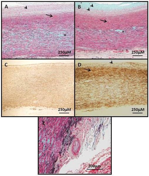
Panel A and B show the aortic pathology of the ascending aneurysms associated with ACTA2 R179H mutations (patients D and C, respectively). Movat staining of the aortas shows significant fibroproliferative lesions in the intima (arrowheads) characterized by SMCs (red) and extracellular matrix accumulation, including collagens (yellow) and proteoglycans (blue). In the medial layer of the aorta, there is subintimal proliferation of SMCs (arrows) and medial degeneration with proteoglycan accumulation and loss of elastic fibers (black), which is most apparent in the middle of medial layer (asterisks). Panel C is an age-matched control aorta and panel D is an ascending aneurysm (patient C) immunostained with α-actin to identify SMCs. These panels confirm intimal SMC hyperplasia (arrowheads) and increased SMCs in the subintimal medial layer (arrows) in the patient compared with the control. Panel E shows an artery in the aortic vasa vasorum with intimal fibrocellular proliferation (the thin black line in the artery is the internal elastic lamina, which demarcates the intimal from the medial layer). Magnification is indicated on the images.
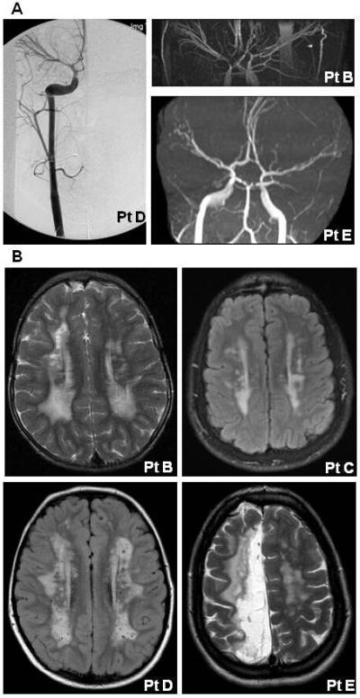
Panel A shows angiogram and MRAs images from three patients with the ACTA2 R179H mutation. These images illustrate the bilateral ectasia of internal carotid arteries from the cavernous to the clinoidal segments and stenosis of the carotid terminus in these patients. Panel B shows MRIs from four patients illustrating increased T2 signal intensity in the periventricular white matter bilaterally. In addition, MRI of patient E shows white matter changes consistent with an ischemic stroke in the distribution of the anterior cerebral artery.
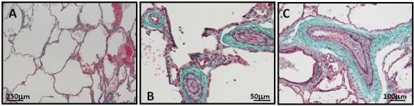
Panel A illustrates the emphysematous changes in the lungs in patient D (modified Trichrome staining elastin purple and collagen blue). Panel B and C show the increased thickness of the medial layer and subintimal proliferation of SMCs in the pulmonary vasculature of the same patient (dark purple lines surrounding the artery lumen are the internal and external elastic laminas, demarcating the intimal and medial layers and the medial and adventitial layers, respectively).
References
-
- Adès LC, Davies R, Haan EA, Holman KJ, Watson KC, Sreetharan D, Cao SN, Milewicz DM, Bateman JF, Chiodo AA, Eccles M, McNoe L, Harbord M. Aortic dissection, patent ductus arteriosus, iris hypoplasia and brachytelephalangy in a male adolescent. Clin Dysmorphol. 1999;8:269–276. - PubMed
-
- Fatigati V, Murphy RA. Actin and tropomyosin variants in smooth muscles. Dependence on tissue type. J Biol Chem. 1984;259:14383–14388. - PubMed
-
- Gerthoffer WT. Actin cytoskeletal dynamics in smooth muscle contraction. Can J Physiol Pharmacol. 2005;83:851–856. - PubMed
-
- Graf MH, Jungherr A. Clinicopathologic reports, case reports, and small case series: congenital mydriasis, failure of accommodation, and patent ductus arteriosus. Arch Ophthalmol. 2002;120:509–510. - PubMed
Publication types
MeSH terms
Substances
Grants and funding
LinkOut - more resources
Full Text Sources
Medical
Molecular Biology Databases
Miscellaneous
