. 2011 Feb;26(2):331-40.
doi: 10.1002/jbmr.226.

The biological function of DMP-1 in osteocyte maturation is mediated by its 57-kDa C-terminal fragment

Affiliations

The biological function of DMP-1 in osteocyte maturation is mediated by its 57-kDa C-terminal fragment

Yongbo Lu et al. J Bone Miner Res. 2011 Feb.

Abstract

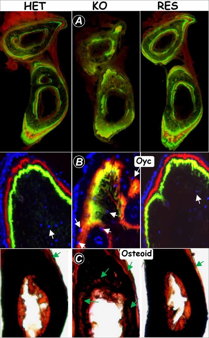
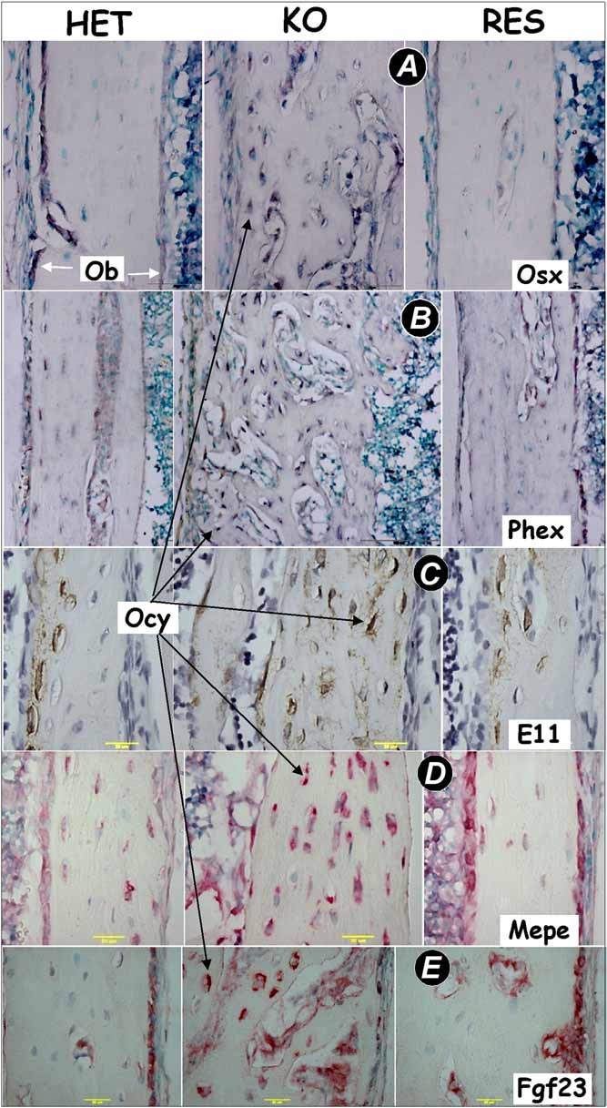
Dentin matrix protein 1 (DMP-1) is a key molecule in controlling osteocyte formation and phosphate homeostasis. Based on observations that full-length DMP-1 is not found in bone, but only cleaved fragments of 37 and 57 kDa are present, and in view of the finding that mutations in the 57-kDa fragment result in disease, we hypothesized that the 57-kDa C-terminal fragment is the functional domain of DMP-1. To test this hypothesis, a 3.6-kb type I collagen promoter was used to express this 57-kDa C-terminal fragment for comparison with full-length DMP-1 in Dmp1 null osteoblasts/osteocytes. Not only did expression of the full-length DMP-1 in bone cells fully rescue the skeletal abnormalities of Dmp1 null mice, but the 57-kDa fragment also had similar results. This included rescue of growth plate defects, osteomalacia, abnormal osteocyte maturation, and the abnormal osteocyte lacunocanalicular system. In addition, the abnormal fibroblast growth factor 23 (FGF-23) expression in osteocytes, elevated circulating FGF-23 levels, and hypophosphatemia were rescued. These results show that the 57-kDa C-terminal fragment is the functional domain of DMP-1 that controls osteocyte maturation and phosphate metabolism.


Figures

Fig. 1
Fig. 1
Reexpression patterns of full-length DMP-1 in Dmp1 null (KO) mice driven by the 3.6-kb murine Col1a1 promoter. DMP-1 expression in Dmp1 heterozygous (HET) control mice (left panels), Dmp1 null mice (middle panels), and Col1a1 full-length DMP-1 in the Dmp1 null background (RES, right panels) was analyzed in tibias from 4-week-old mice by in situ hybridization (a, signal in red color), immunohistochemistry (b, signal in brown color), and immunogold labeling and transmission electron microscopy (c). The assays showed that expression of endogenous DMP-1 in HET mice was found predominantly in the osteocytes (Oyc, black arrows) embedded in the bone matrix. No expression of endogenous DMP-1 was detected in Dmp1 null mice. Note that in situ hybridization showed that the transgene was highly expressed in osteoblasts (Ob, black arrows) but that the protein is present in both osteoblasts and osteocytes. Immunogold staining showed that the endogenous DMP-1 protein was localized to the lamina limitans at the edges of the canaliculi in the bone matrix in HET mice. As expected, there was no DMP-1 protein present in the Dmp1 null mouse bone (middle panels in a, b, and c). However, the transgenic DMP-1 protein was localized predominantly in both the osteoblast layer and in lamellae closely adjacent to the osteocytes (RES). Note that in d, the endogenous Dmp1 mRNA was weakly expressed in chondrocytes (left panel), but the transgenic Dmp1 mRNA was not detected in chondrocytes (right panel).
Fig. 2
Fig. 2
Rescue of Dmp1 null bone abnormalities with the full-length Dmp1 transgene at the ages of 10 days and 5 weeks. (A) Representative µCT images of tibias from the HET, KO, and rescued mice (RES) at the age of 10 days. The whole-mount view is shown in the left panel, the sagittal sections of metaphyses are shown in the middle panel, and the cross sections of midshaft are shown in the right panel; the quantified data are shown in the lower panels. (Data are mean ± SEM; n = 4; ***p < .001). (B) Whole-mount views of 5-week-old tibias from the HET, KO, and rescued (RES) mice are shown in the left panel; the quantified data are shown in the right panel. (Data are mean ± SEM; n = 4; ***p < .001).
Fig. 3
Fig. 3
Rescue of Dmp1 null bone defects in mineralization with the full-length Dmp1 transgene. (A) The double fluorochrome labels displayed an identical bone-formation rate in HET (left panel) and RES (right panel) mice in contrast to the diffuse label in KO mice (middle panel) at 2 months of age. Diffuse fluorochrome labels in the Dmp1 null mice (middle panel) were fully restored in the rescue group (right panel). (B) Confocal microscopic images of fluorochrome labeling, counterstained with DAPI for visualization of osteocyte nuclei (blue, white arrows), showed that osteocytes are separated from the mineralization front in HET control mice (left panel) and RES mice (right panel). Diffuse fluorochrome labeling in the Dmp1 null mice (middle panel) was replaced with sharp lines in the RES group (right panel). (C) von Kossa staining showed restored mineralization in the RES mice, whereas abundant osteoid (red color, arrows) was present in the KO tibias at the age of 2 months.
Fig. 4
Fig. 4
Rescue of the malformed osteocyte lacunocanalicular network and growth plate defects. (A) Visualization of the disorganized osteocyte canalicular system in 7-week-old Dmp1 null mice (middle panel) with procion red injection compared with the well-organized control osteocytes (left panel) and the rescued osteocytes (right panel) using confocal microscopy at 565-nm excitation and 610-nm emission. (B) SEM images of the acid-etched, resin-casted osteocyte canalicular system from the 7-week-old HET control mice (left panel), the Dmp1 null mice (middle panel), and the rescued mice(right panel). Note that the poorly formed osteocyte canalicular system in Dmp1 null mice was fully rescued by the full-length Dmp1 transgene. (C) Safranin-O staining of the tibia growth plate, showing that Dmp1 null mice displayed about a threefold increase in the hypertrophic zone at the age of 10 days and formation of multiple chondrocyte clusters with a loss of continuity at 7 weeks of age (middle panels). All these morphological changes were completely rescued by the full-length Dmp1 transgene in the Dmp1 null background (right panels) compared with the HET controls (left panels).
Fig. 5
Fig. 5
Targeted expression of DMP-1 rescued the gene expression patterns in the Dmp1 null cortical bone. In situ hybridization (A, Osx, osterix; B, Phex, phosphate-regulating gene with homologies to endopeptidases on the X chromosome; D, MEPE, matrix extracellular phosphoglycoprotein; E, Fgf23, fibroblast growth factor 23) and immunohistochemistry (C, E11) assays were performed on the HET control tibias (left panels), the Dmp1 null tibias (middle panels), and the RES (right panels) tibias at the age of 10 days. All these genes in Dmp1 null cortical bone were increased (mainly in osteocytes) and were restored to control levels by targeted expression of the full-length Dmp1. Signal in dark purple color (A, B), in brown color (C), and in red color (D, E).
Fig. 6
Fig. 6
Targeted expression of the full-length DMP-1 or the 57-kD C-terminal fragment in Dmp1 KO mice rescued the skeletal abnormalities, serum FGF-23, Pi homeostasis, and malformed osteocyte lacunocanalicular network. (A) Western blot data obtained from HET, KO, and rescued long bones documenting expression of the 57-kDa fragment in 2-month-old Dmp1 KO mice. (B) µCT images showing similar restoration of the morphology of the femur by targeted expression of either the full-length DMP-1 or the 57-kDa fragment in the Dmp1 KO background at 7 weeks of age. (C) Restoration of serum levels of FGF-23 (upper panel), Pi (phosphorus, middle panel), and Ca (calcium, lower panel) to normal levels in both the full-length and the 57-kDa rescued mice at 7 weeks of age; Data are mean ± SEM; n = 4 to 6; **p < .01; ***p < .001. (D) The double fluorochrome labels showed a similar bone-formation rate in HET mice (left panel) and the 57-kDa RES mice (right panel) in contrast to the diffuse label in the KO mice (middle panel) at 7 weeks of age. Immunohistochemistry (E, sclerostin) and in situ hybridization (F, type I collagen; G, FGF-23) assays were performed on HET control tibias (left panels), Dmp1 KO tibias (middle panels), and the 57-kDa RES tibias (right panels) at the age of 10 days. Sclerostin, a marker for mature osteocytes, in Dmp1 KO osteocytes was largely undetectable, whereas ColI and Fgf23 mRNAs were increased dramatically in the Dmp1 KO osteocytes. All these changes were restored to control levels by targeted expression of the 57-kDa C-terminal fragment. Signal in brown color (E) and in red color (F, G).
Fig. 7
Fig. 7
A working model of DMP-1 function in osteocyte maturation, mineralization, and phosphate homeostasis. We propose that the full-length DMP-1, secreted mainly from osteocytes (Oyc), is cleaved by BMP-1/subtilisin-like proprotein convertase into a 37-kDa N-terminal fragment and a 57-kDa C-terminal fragment.(16,24) The function of the former is not known, whereas the latter (57-kDa C-terminal) (1) likely accelerates osteocyte maturation through downregulation of osteoblast-expressed genes, such as Osx and Col1, and mineralization, and (2), FGF-23, which is normally secreted from osteoblasts (Ob) but sharply increased in osteocytes in diseases such as Phex mutations or Dmp1 mutations or these knockout mouse models, decreases phosphorus (Pi) reabsorption in kidney, leading to hypophosphatemic rickets and osteomalacia.

References

    1. George A, Sabsay B, Simonian PA, Veis A. Characterization of a novel dentin matrix acidic phosphoproteimplications for induction of biomineralization. J Biol Chem. 1993;268:12624–12630. - PubMed
    1. Toyosawa S, Shintani S, Fujiwara T, et al. Dentin matrix protein 1 is predominantly expressed in chicken and rat osteocytes but not in osteoblasts. J Bone Miner Res. 2001;16:2017–2026. - PubMed
    1. Fen JQ, Zhang J, Dallas SL, et al. Dentin matrix protein 1, a target molecule for Cbfa1 in bone, is a unique bone marker gene. J Bone Miner Res. 2002;17:1822–1831. 2001. - PubMed
    1. Ye L, Mishina Y, Chen D, et al. DMP-1-deficient mice display severe defects in cartilage formation responsible for a chondrodysplasia-like phenotype. J Biol Chem. 2005;280:6197–6203. - PMC - PubMed
    1. Ye L, MacDougall M, Zhang S, et al. Deletion of dentin matrix protein 1 leads to a partial failure of maturation of predentin into dentin, hypomineralization, and expanded cavities of pulp and root canal during postnatal tooth development. J Biol Chem. 2004;279:19141–19148. - PubMed

Publication types

MeSH terms