. 2010 Nov 15;432(1):65-76.
doi: 10.1042/BJ20100584.

Lipin proteins form homo- and hetero-oligomers

Affiliations

Lipin proteins form homo- and hetero-oligomers

Guang-Hui Liu et al. Biochem J. .

Abstract

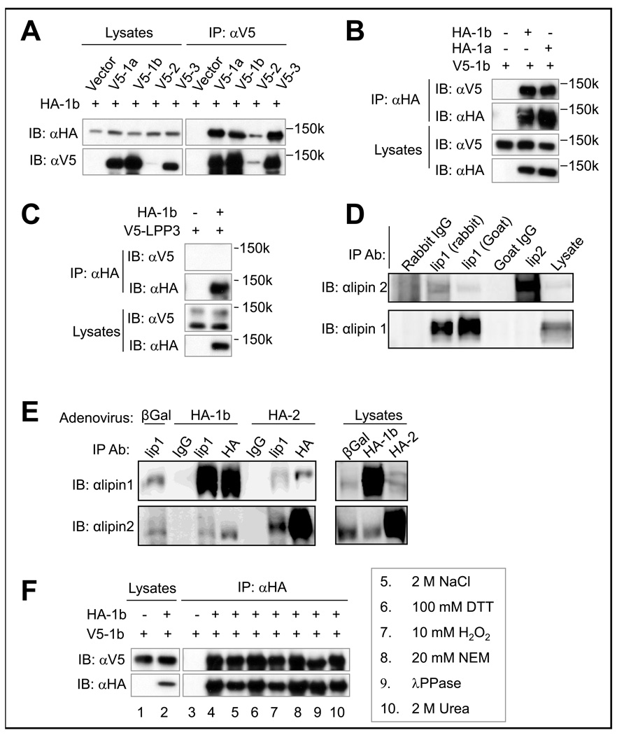
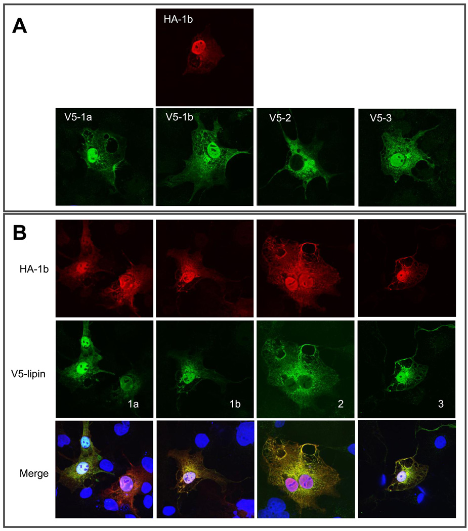
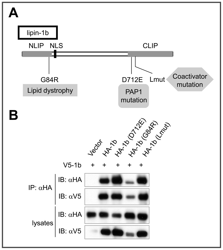
Lipin family members (lipin 1, 2 and 3) are bi-functional proteins that dephosphorylate PA (phosphatidic acid) to produce DAG (diacylglycerol) and act in the nucleus to regulate gene expression. Although other components of the triacylglycerol synthesis pathway can form oligomeric complexes, it is unknown whether lipin proteins also exist as oligomers. In the present study, using various approaches, we revealed that lipin 1 formed stable homo-oligomers with itself and hetero-oligomers with lipin 2/3. Both the N- and C-terminal regions of lipin 1 mediate its oligomerization in a head-to-head/tail-to-tail manner. We also show that lipin 1 subcellular localization can be influenced through oligomerization, and the individual lipin 1 monomers in the oligomer function independently in catalysing dephosphorylation of PA. The present study provides evidence that lipin proteins function as oligomeric complexes and that the three mammalian lipin isoforms can form combinatorial units.


Figures

Figure 1
Figure 1. Lipin 1 coimmunoprecipitates with itself and lipin 2, and 3, but not LPP3
(A) COS-7 cells were transfected with constructs for expression of HA-lipin 1b and various V5-tagged lipin expression vectors as indicated. 36 hours after transfection, cell lysates were subjected to immunoprecipitation with an anti-V5 antibody, followed by immunoblotting with anti-HA or anti-V5 antibodies. (B) COS-7 cells were cotransfected with vectors for expression of V5-lipin 1b and HA-tagged lipin 1a or lipin 1b as indicated. 36 hours after transfection, lysates were subjected to immunoprecipitation with anti-HA antibody, followed by immunoblotting with anti-HA or anti-V5 antibodies. (C) COS-7 cells were cotransfected with vectors for expression of V5-LPP3 and HA-tagged lipin 1b. 36 hours after transfection, lysates were subjected to immunoprecipitation with anti-HA antibody, followed by immunoblotting with anti-HA or anti-V5 antibodies. (D) 3T3-L1 adipocytes were homogenized in buffer A containing 10 mM Triton X-100 and equal amounts of lysates were used for immunoprecipitation with rabbit or goat non-immune IgG, lipin 1 (LAb2 or Santa Cruz C-15), or lipin 2 antibodies, followed by immunoblotting with anti-lipin 1 (LAb1) or 2 antibodies. (E) 3T3-L1 adipocytes were infected for two days with adenovirus vectors expressing HA-tagged lipin 1 or 2. Cells were homogenized and immunoprecipitations were performed as in 1D, but with anti-lipin 1 (LAb2) or HA (12CA5) antibodies. (F) COS-7 cells were transfected with vectors for expression of V5-lipin 1b, with or without HA-lipin 1b, and after 36 hours cell lysates were prepared and were subjected to immunoprecipitation with anti-HA antibody. After 3 washes with lysis buffer, the immunoprecipitates were washed another time with either Tris buffer (50 mM Tris-HCl pH 7.5, 50 mM NaCl) (lanes 4), or Tris buffer containing indicated materials (lanes 5–10). The beads were subjected to another round of washing with lysis buffer and then subjected to immunoblot analysis.
Figure 2
Figure 2. Mapping the lipin 1b domains in mediating its oligomerization
(A) Schematic representation of the V5-tagged lipin 1b truncation mutant constructs used in co-IP and GST-pull down experiments. Their abilities to interact with HA-lipin 1b or GST-fused lipin 1b fragments are indicated as “+” (positive), “−” (negative) or “N/D” (not determined). NLIP, CLIP, and NLS (nuclear localization sequence) are shown. (B) COS-7 cells were co-transfected with HA-lipin 1b and various V5-tagged lipin 1b fragments as indicated. 36 hours after transfection, the lysates were subjected to immunoprecipitation with anti-V5 antibody, followed by immunoblotting with anti-HA or anti-V5 antibody. (C) In vitro interaction between GST-lipin 1b fragments and immunopurified V5-tagged lipin 1b fragments.
Figure 3
Figure 3. Colocalization of lipin 1 and lipin 1, 2, and 3 in cells
(A) COS-7 cells were transfected with vectors for expression of V5-tagged lipin 1a, 1b, 2 or 3, or HA-tagged lipin 1b. 36 hours after transfection, the cells were fixed and subjected to immunostaining with anti-HA or anti-V5 antibody. (B) COS-7 cells were cotransfected with HA-tagged lipin 1b and V5-tagged lipin 1a, 1b, 2 or 3. 36 hours after transfection, the cells were subjected to immunostaining with anti-HA and anti-V5 antibody. Red: HA-lipin 1b; Green: V5-tagged lipin isoforms; Blue: Hoechst 33342.
Figure 4
Figure 4. FRET analysis of fluorescent-tagged lipin 1
(A) Co-localization of Venus-lipin 1b and Cerulean-lipin 1b. Images merged in Image J. (B) Single cell example of uncorrected emission spectra from co-transfected Venus-lipin 1b and Cerulean-lipin 1b (Co-transfection) at 514 or 458nm, and single transfected Venus-lipin 1b (Acceptor alone) or Cerulean-lipin 1b (Donor Alone) excited at 458nm. (C) (Left) Intensity map showing E% of a representative cell where spectral bleed through of both donor and acceptor have been removed by linear unmixing and a computer algorithm (PFRET). (Right) Graph of E% versus acceptor intensity of individual cell shown on left. (D) Decay curve for cells transfected with either Cerulean-lipin 1b (Cer-lip) alone or Cerulean-lipin 1b and Venus-lipin 1b (Cer-lip + Ven-lip). (E) Average decay time (nsec) per cell for Cerulean-lipin 1b (Cer-lip) and Cerulean-lipin 1b + Venus-lipin 1b (Cer-lip + Ven-lip), * indicates statistical significance (P < 0.00001).
Figure 5
Figure 5. Forced plasma membrane-targeting of lipin 1 via a CAAX domain can recruit soluble lipin 1 to the plasma membrane
COS-7 cells were transfected with plasmids for expression of CAAX-tagged lipin 1b and/or myc-tagged lipin 1b. 48 hours after transfection cells were fixed and subjected to immunostaining with anti-HA and/or anti-Myc antibodies. Transfections indicated to the left, and immunostaining shown at the top. Red: HA-lipin 1b-CAAX; Green: Myc-lipin 1b, Blue: DAPI.
Figure 6
Figure 6. Lipin 1 exists as an oligomer in 3T3-L1 adipocytes
(A) Column fractions from Superose 6 gel filtration chromatography of 3T3-L1 adipocte cytosolic extract immunoblotted for lipin 1 and 2. Bottom panel is same extracts incubated with 10mM TX-100 before loading on column. (B) Lipin 1 immunoreactivity (arbitrary units) in column fractions versus Mg2+-dependent PAP activity (nmol/min/ml).
Figure 7
Figure 7. Recombinant lipin 1 forms dimers and tetramers
(A) Column fractions from Superose 6 gel filtration chromatography of purified recombinant lipin 1b were collected and analyzed for PAP activity. (B) Immunoblotting of column fractions. (C) HEK293A cells transiently expressing V5-lipin 1b or V5-lipin 1b (D712E) were homogenized in digitonin-containing lysis buffer, the lysates separated by BN-PAGE and subjected to immunoblotting with anti-V5 antibody.
Figure 8
Figure 8. Co-immunoprecipitation between lipin 1b and various lipin 1b mutants
(A) Schematic representation of the functional effects of the lipin 1b point mutant constructs used in co-IP experiments. (B) COS-7 cells were co-transfected with V5-lipin 1b and various HA-tagged lipin 1b mutants as indicated. 36 hours after transfection, the lysates were subjected to immunoprecipitation with anti-HA antibody, followed by immunoblotting with anti-HA or anti-V5 antibody.
Figure 9
Figure 9. Individual lipin 1b monomers in oligomers dephosphorylate PA independently
(A and B) HEK-293T cells were transfected with 0.5 µg of plasmids for expression of myc-lipin 1b (+), and either 0.5 µg (+), or 0.25, 0.5 or 1.0 µg of plasmids for expression of HA-lipin 1b (D712E). The cells were lysed and after immunoprecipitating HA-lipin 1b (D712E) the immunoprecipitate was split into separate fractions, with one fraction used for Western blotting (A) and the other fraction divided into multiple aliquots and measured for PAP activity in the presence and absence of Mg2+ (B). (C) Simplified model depicting the putative dimeric arrangement and activity of individual lipin 1b monomers in a lipin 1b dimer. Black: catalytically inactive monomer; Gray: catalytically active monomer.

References

    1. Han GS, Wu WI, Carman GM. The Saccharomyces cerevisiae Lipin homolog is a Mg2+-dependent phosphatidate phosphatase enzyme. J. Biol. Chem. 2006;281:9210–9218. - PMC - PubMed
    1. Peterfy M, Phan J, Xu P, Reue K. Lipodystrophy in the fld mouse results from mutation of a new gene encoding a nuclear protein, lipin. Nat. Genet. 2001;27:121–124. - PubMed
    1. Harris TE, Huffman TA, Chi A, Shabanowitz J, Hunt DF, Kumar A, Lawrence JC., Jr. Insulin controls subcellular localization and multisite phosphorylation of the phosphatidic acid phosphatase, lipin 1. J. Biol. Chem. 2007;282:277–286. - PubMed
    1. Huffman TA, Mothe-Satney I, Lawrence JC., Jr. Insulin-stimulated phosphorylation of lipin mediated by the mammalian target of rapamycin. Proc. Natl. Acad. Sci. U. S. A. 2002;99:1047–1052. - PMC - PubMed
    1. Peterfy M, Phan J, Reue K. Alternatively spliced lipin isoforms exhibit distinct expression pattern, subcellular localization, and role in adipogenesis. J. Biol. Chem. 2005;280:32883–32889. - PubMed

Publication types

MeSH terms