. 2010 Sep;161(2):308-20.
doi: 10.1111/j.1476-5381.2010.00744.x.

GPR55-dependent and -independent ion signalling in response to lysophosphatidylinositol in endothelial cells

Affiliations

GPR55-dependent and -independent ion signalling in response to lysophosphatidylinositol in endothelial cells

Alexander Bondarenko et al. Br J Pharmacol. 2010 Sep.

Abstract

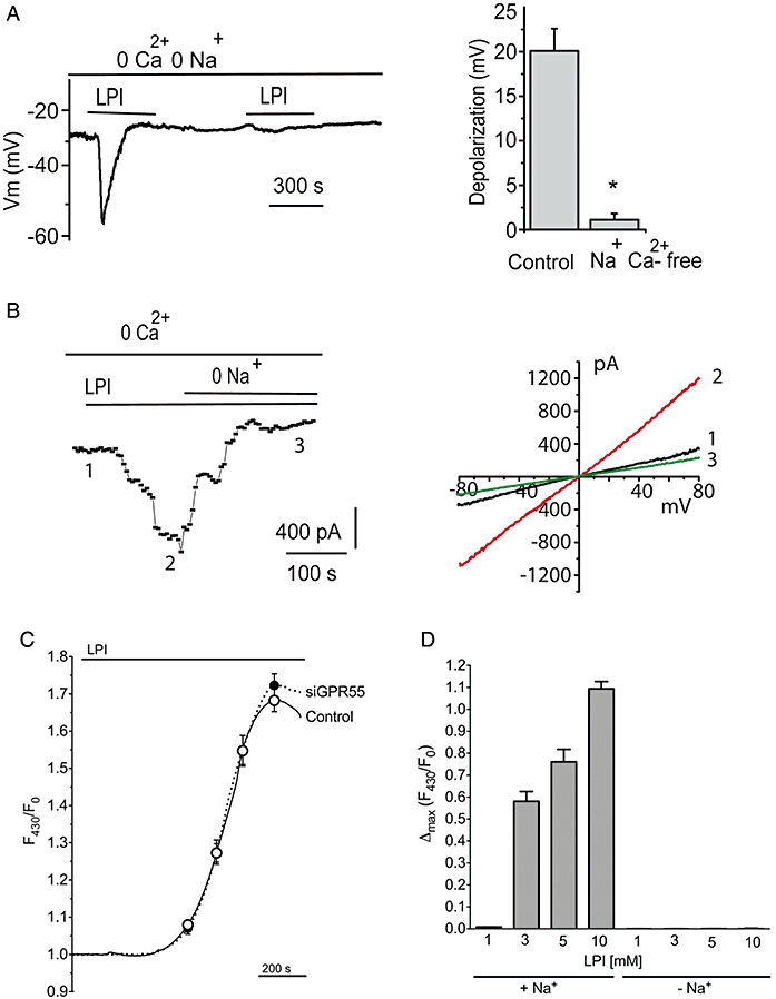
Background and purpose: The glycerol-based lysophospholipid lysophosphatidylinositol (LPI) is an endogenous agonist of the G-protein-coupled receptor 55 (GPR55) exhibiting cannabinoid receptor-like properties in endothelial cells. To estimate the contribution of GPR55 to the physiological effects of LPI, the GPR55-dependent and -independent electrical responses in this cell type were investigated.

Experimental approach: Applying small interference RNA-mediated knock-down and transient overexpression, GPR55-dependent and -independent effects of LPI on cytosolic free Ca(2+) concentration, membrane potential and transmembrane ion currents were studied in EA.hy296 cells.

Key results: In a GPR55-dependent, GDPbetaS and U73122-sensitive manner, LPI induced rapid and transient intracellular Ca(2+) release that was associated with activation of charybdotoxin-sensitive, large conductance, Ca(2+)-activated, K(+) channels (BK(Ca)) and temporary membrane hyperpolarization. Following these initial electrical reactions, LPI elicited GPR55-independent long-lasting Na(+) loading and a non-selective inward current causing sustained membrane depolarization that depended on extracellular Ca(2+) and Na(+) and was partially inhibited by Ni(2+) and La(3+). This inward current was due to the activation of a voltage-independent non-selective cation current. The Ni(2+) and La(3+)-insensitive depolarization with LPI was prevented by inhibition of the Na/K-ATPase by ouabain.

Conclusions and implications: LPI elicited a biphasic response in endothelial cells of which the immediate Ca(2+) signalling depends on GPR55 while the subsequent depolarization is due to Na(+) loading via non-selective cation channels and an inhibition of the Na/K-ATPase. Thus, LPI is a potent signalling molecule that affects endothelial functions by modulating several cellular electrical responses that are only partially linked to GPR55.


Figures

Figure 1
Figure 1
Effect of LPI on free intracellular Ca2+ and membrane potential of endothelial cells. Representative effect of 5 µM LPI on free intracellular Ca2+ in the presence of 2 mM extracellular Ca2+ (n = 32) (A) and in nominally Ca2+-free solution (n = 27) (B). Concentration-response correlation of LPI on cytosolic Ca2+ concentration measured at the initial transient peak (Peak Phase) and the subsequent plateau phase (Plateau Phase) (1 µM, n = 9; 3 µM, n = 9; 5 µM, n = 15; 10 µM, n = 14) (C). Representative biphasic effect of LPI (5 µM) on membrane potential in the presence of extracellular Ca2+ (n = 9) (D). Concentration-response correlation of LPI in terms of initial membrane hyperpolarization and subsequent depolarization (1 µM, n = 17; 3 µM, n = 7; 10 µM, n = 7) (E). Representative changes in endothelial membrane potential evoked by repetitive stimulations with 5 µM LPI (n = 5) (F). Representative membrane currents evoked by repetitive stimulations by LPI (5 µM) at −40 mV holding potential (n = 3) (G).
Figure 2
Figure 2
The GPR55 blocker rimonabant prevented only the initial Ca2+ transients and membrane hyperpolarization evoked by LPI. Cells were pre-treated with 1 µM rimonabant as shown. This GPR55 blocker abolished the transient elevation of the cytosolic-free Ca2+ concentration in response to 5 µM LPI (A). Typical membrane potential recording showing the inhibitory effect of rimonabant (1 µM) of a LPI (5 µM)-evoked hyperpolarization (B). Statistical evaluation of LPI (5 µM)-induced biphasic changes in endothelial cell membrane potential under control conditions (LPI, n = 9) and in the presence of 1 µM rimonabant (n = 14) (C). Rimonabant (1 µM) prevented the development of the initial outward current (1st phase) but failed to prevent the sustained inward current (2nd phase) in response to 5 µM LPI (n = 3) (D). *P < 0.005 versus control.
Figure 3
Figure 3
The effects of LPI on initial Ca2+ signalling and membrane hyperpolarization essentially depend on GPR55 expression. Intracellular Ca2+ signalling in response to 5 µM LPI was recorded in cells transiently transfected with the respective controls (same vector encoding nuclear GFP) or a GPR55 encoding vector (Control: n = 17; GPR55ox: n = 24) (A). In addition, cells were transfected with siRNA control (AllStar®) or siRNA against GPR55 (Control: n = 12; siRNA GPR55: n = 19) (B). In conventional whole cell configuration, the LPI- (5 µM) induced hyperpolarization was measured in cells transfected either siRNA control (n = 4), siRNA against GPR55 (n = 5) or a vector encoding GPR55 (n = 6) (C). *P < 0.005 versus control.
Figure 4
Figure 4
LPI-induced hyperpolarization but not depolarization is sensitive to inhibition of phospholipase C and to GDPβS. In conventional whole cell configuration, the effect of the inhibitor of phospholipase C U73122 (10 µM) (Control: n = 5; U73122: n = 6) (A) or 1 mM GDPβS in the pipette (Control: n = 7; GDPβS: n = 12) (B) on LPI- (3 µM) evoked initial hyperpolarization and subsequent depolarization was evaluated. *P < 0.005 versus control. The effect of 10 µM LPI in the bath in the inside-out configuration (PS#4) in the presence of 100 nM free Ca2+ (EB#4) on plasma membrane currents at +70 and −70 mV membrane potentials (left) and the respective voltage-current relationship before (1) and after (2) LPI application (right) (n = 6) were recorded (C).
Figure 5
Figure 5
The initial transient response to LPI is due to a stimulation of outwardly rectifying, Ca2+-dependent, charybdotoxin-sensitive, K+ channels. Representative biphasic changes in membrane currents evoked by 5 µM LPI at a holding portential of −40 mV (n = 14) (A). Voltage ramps of the outward rectifying membrane currents under resting conditions (control, n = 5) and upon cell stimulation with 5 µM (LPI (n = 8) (B). Representative time course of the effect of LPI on single BKCa channels in the cell-attached mode at a holding potential (Vhold) of 20 mV (n = 7) (C). Representative membrane potential recording showing a lack of hyperpolarization in response to 5 µM LPI in the presence of 100 nM charybdotoxin (n = 5) (D). Statistical evaluation of LPI (5 µM)-induced membrane hyperpolarization in endothelial cells under control conditions (LPI, n = 4) and in the presence of 100 nM charybdotoxin (100 nM, n = 5) (E). *P < 0.005 versus control.
Figure 6
Figure 6
LPI-evoked membrane currents that account for membrane depolarization. Representative tracings of membrane currents induced by 5 µM LPI measured at holding potentials of −75 and +75 mV (n = 7). In the whole-cell mode, 1 s voltage ramps from −80 mV to +80 mV were applied every 5 s from a holding potential of −40 mV (A). Current-voltage relationship constructed from current responses to voltage ramps before and after application of 5 µM LPI at points specified in A (B). Statistical evaluation of the effects of 5 µM LPI on endothelial cell membrane potential under control conditions (n = 7) and in the presence of 100 µM DIDS (n = 3) (C).
Figure 7
Figure 7
Dependency of the LPI-evoked electrical responses on extracellular Ca2+. Original tracing of membrane potential fluctuations in response to 5 µM LPI in the absence of extracellular Ca2+ (n = 16) (A). Representative effect of re-adding Ca2+ in the continued presence of 5 µM LPI on membrane potential (n = 3) (B). Original tracing of the effect of 5 µM LPI on endothelial membrane potential in the presence of 2 mM Ni2+ (n = 7) (C). Effect of 2 mM Ni2+ on the LPI-induced inward current measured at −75mV (n = 10) (D). Effect of 100 µM La3+ on LPI-evoked depolarization (n = 4) (E). Statistical evaluation of the effects of different ionic conditions on the depolarization evoked by 5 µM LPI in endothelial cells: Control (n = 14), in the absence of extracellular Ca2+ (0 Ca2+, n = 5), in the presence of 2 mM Ni2+ (Ni2+, n = 10) or 100 µM La3+ (La3+, n = 4), and on Ca2+ re-addition (Ca2+, n = 3) (F). *P < 0.05 versus control.
Figure 8
Figure 8
The LPI-evoked electrical responses depend on extracellular Na+ and LPI-triggered, slow cytosolic Na+ accumulation. Representative membrane potential recording showing LPI-evoked depolarization (n = 6) in the absence of Na+ and Ca2+ in the bath (left panel) and the corresponding statistical evaluation of LPI (5 µM)-evoked depolarization under control conditions (Control) and in the absence of Na+ and Ca2+ (right panel) (A). Representative recording of the inhibitory effect of Na+ withdrawal on LPI-evoked inward current (left panel) measured at a holding potential of −40 mV (n = 3). Endothelial cells were dialyzed with a Cs+- containing pipette solution and superfused with a Ca2+- and K+-free solution. Corresponding current-voltage relationship obtained from current responses to voltage ramps before and after the application of 5 µM LPI at time points indicated (left panel) (B). The effect of 5 µM LPI on intracellular Na+ was recorded in CoroNa™ Green-loaded cells that were transiently transfected with either siRNA control (Control: n = 17) or siRNA against GPR55 (n = 10) (C). Concentration-response correlation of LPI on the accumulation of cytosolic Na+ in a conventional buffer (EB#1) (left; n = 15–30) or in the absence of extracellular Na+ (right; n = 8–15) (D). *P < 0.005 versus control.
Figure 9
Figure 9
LPI-evoked sustained depolarization is partially due to inhibition of the Na+/K+-ATPase. Representative tracings of the endothelial cell membrane potential showing the inhibitory effect of 5 µM LPI on the hyperpolarization induced by re-adding K+ to K+-free solution (A) and the inhibitory effect of 250 µM ouabain on LPI-induced depolarization (B). Statistical evaluation of results from panels (A) and (B), showing the mean of the maximal membrane depolarization in response to 5 µM LPI in the presence of 100 µM La3+ (n = 4), 250 µM ouabain (n = 9), or 100 µM La3+ and 250 µM ouabain (n = 3) (C). *P < 0.05 versus control.

References

    1. Alexander SPH, Mathie A, Peters JA. Guide to receptors and channels (GRAC) Br J Pharmacol. (4th edn) 2009;158(Suppl 1):S1–S254. - PMC - PubMed
    1. Beny JL, Pacicca C. Bidirectional electrical communication between smooth muscle and endothelial cells in the pig coronary artery. Am J Physiol. 1994;266(4 Pt 2):H1465–H1472. - PubMed
    1. Bondarenko A. Sodium-calcium exchanger contributes to membrane hyperpolarization of intact endothelial cells from rat aorta during acetylcholine stimulation. Br J Pharmacol. 2004;143:9–18. - PMC - PubMed
    1. Bondarenko A, Sagach V. Na+-K+-ATPase is involved in the sustained ACh-induced hyperpolarization of endothelial cells from rat aorta. Br J Pharmacol. 2006;149:958–965. - PMC - PubMed
    1. Chen GF, Suzuki H. Calcium dependency of the endothelium-dependent hyperpolarization in smooth muscle cells of the rabbit carotid artery. J Physiol (Lond) 1990;421:521–534. - PMC - PubMed

Publication types

MeSH terms