. 2010 Oct;78(1):187-202.
doi: 10.1111/j.1365-2958.2010.07324.x. Epub 2010 Feb 8.

Regulated maturation of malaria merozoite surface protein-1 is essential for parasite growth

Affiliations
Free PMC article

Regulated maturation of malaria merozoite surface protein-1 is essential for parasite growth

Matthew A Child et al. Mol Microbiol. 2010 Oct.
Free PMC article

Abstract

The malaria parasite Plasmodium falciparum invades erythrocytes where it replicates to produce invasive merozoites, which eventually egress to repeat the cycle. Merozoite surface protein-1 (MSP1), a prime malaria vaccine candidate and one of the most abundant components of the merozoite surface, is implicated in the ligand-receptor interactions leading to invasion. MSP1 is extensively proteolytically modified, first just before egress and then during invasion. These primary and secondary processing events are mediated respectively, by two parasite subtilisin-like proteases, PfSUB1 and PfSUB2, but the function and biological importance of the processing is unknown. Here, we examine the regulation and significance of MSP1 processing. We show that primary processing is ordered, with the primary processing site closest to the C-terminal end of MSP1 being cleaved last, irrespective of polymorphisms throughout the rest of the molecule. Replacement of the secondary processing site, normally refractory to PfSUB1, with a PfSUB1-sensitive site, is deleterious to parasite growth. Our findings show that correct spatiotemporal regulation of MSP1 maturation is crucial for the function of the protein and for maintenance of the parasite asexual blood-stage life cycle.


Figures

Fig. 1
Fig. 1
Most primary processing site sequences diverge between the two MSP1 allelic types.Primary proteolytic processing of MSP1 by PfSUB1 results in the production of the MSP183, MSP130, MSP138 and MSP142 fragments (top centre). The amino acid sequences within which cleavage occurs are shown for the two major dimorphic forms of MSP1, typified by those of the 3D7 and Wellcome isolates (centre). Also shown is an additional cleavage site (called 83int, indicated by dotted lines) that occurs only in the 3D7-type MSP1. The only detected microheterogeneity in the cleavage sites is a Thr-to-Ala substitution in the P4′ position of the 3D7-type 83/30 cleavage site. The positions of the primary cleavage sites relative to the distribution of conserved (open), variable (black) and semi-conserved (grey) blocks within MSP1, numbered 1–17 according to Tanabe et al. (1987) are shown (bottom). The sequence at which PfSUB2-mediated secondary processing occurs is also shown (top right). Note that PfSUB1 cannot cleave C-terminal to a Leu residue, and the secondary processing site is completely refractory to PfSUB1 (Withers-Martinez et al., 2002; Yeoh et al., 2007; Koussis et al., 2009).
Fig. 2
Fig. 2
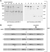
PfSUB1-mediated proteolytic processing of the 3D7 MSP1 is ordered.A. Typical time-course of digestion of native 3D7 P. falciparum-derived MSP1. Saponin-permeabilized 3D7 schizonts were incubated at 37°C in the presence of rPfSUB1. Samples taken at intervals were analysed by Western blot, probing with either the MSP183-specific mAb 89.1 (left-hand side), or the MSP142-specific monoclonal antibody X509 (right-hand side). For both blots the MSP1 precursor is indicated with a black arrow, processing intermediates are marked with an open arrow, or closed round or square-head lollipop, while terminal processing products are marked with an open round or square-head lollipop. No processing was observed in the absence of added rPfSUB1 (not shown).B. Schematic of the order of 3D7-type MSP1 primary processing, determined from the digestion assay shown in (A). Individual processed species are marked with arrow or lollipop symbols to aid comparison with the corresponding bands on the blots.
Fig. 3
Fig. 3
PfSUB1-mediated proteolytic processing of the Wellcome-type MSP1 is ordered.A. Typical time-course of digestion of native T9/94 P. falciparum-derived MSP1. Saponin-permeabilized T9/94 schizonts were incubated at 37°C in the presence of rPfSUB1. Samples taken at intervals were analysed by Western blot, probing with either the MSP183-specific mAb 89.1 (left-hand side), or a MSP142-specific polyclonal antibody (right-hand side). For both blots the MSP1 precursor is indicated with a black arrow, processing intermediates are marked with an open arrow, or closed square-head lollipop, while terminal processing products are marked with an open round or square-head lollipop. No processing was observed in the absence of added rPfSUB1 (not shown).B. Schematic of the order of Wellcome-type MSP1 primary processing, determined from the digestion assay shown in A. Individual processed species are marked with arrow or lollipop symbols to aid comparison with the corresponding bands on the blots.
Fig. 4
Fig. 4
Synthetic peptides based on the 38/42 processing sites in the 3D7 and Wellcome-type MSP1 are poor substrates for PfSUB1.RP-HPLC chromatograms showing partial digestion by rPfSUB1 of N-acetylated decapeptides based on the primary processing sites in the Wellcome-type (A–C) and 3D7-type (D) MSP1. The top chromatogram in each panel shows the elution profile of an equimolar mix of the undigested peptides, while the lower chromatogram shows the RP-HPLC elution profile after partial digestion with rPfSUB1. Identities of major peaks as determined by electrospray mass spectrometry (data not shown but see Koussis et al., 2009), are indicated. As described previously (Yeoh et al., 2007; Koussis et al., 2009), highly polar cleavage products were usually not retained by the RP-HPLC column but eluted in the column flow-through. Note that for determination of relative initial rates of cleavage, cleavage rates were compared when the faster-cleaved peptide of each pair was digested by ≤ 10%, as described in Experimental procedures. For clarity, digestion was allowed to proceed much more than 10% in most of the profiles shown in this figure.
Fig. 5
Fig. 5
Substitution of the MSP1 secondary processing site with a PfSUB1-sensitive sequence.A. Western blot time-course analysis of rPfSUB1-mediated processing of rMSP138/42 either in wild-type form (WT) or after modification of the secondary processing site by site-directed mutagenesis to convert it to a predicted PfSUB1-sensitive site (SUB2to1). The WT rMSP138/42 (80 kDa, arrowed) is converted to a stable 42 kDa terminal species (open round-head lollipop) equivalent to MSP142. The SUB2to1 mutant (arrowed) is similarly converted to the normal MSP142 species (open round-head lollipop), but also undergoes an additional processing step that converts it via an approximate 70 kDa intermediate (closed square-head lollipop) to a stable 33 kDa form (open square-head lollipop). The 2 h tracks show the results of incubation for 2 h in the absence of added rPfSUB1. The bands marked with a star are likely truncated forms of rMSP138/42 derived from breakdown of the recombinant protein during its purification and refolding.B and C. Schematic depiction of processing patterns for the WT (B) and SUB2to1 mutant rMSP138/42 protein (C). The relative position of the secondary processing site, the sequence modifications made and the region of the protein recognized by the rabbit anti-MSP142 antibodies used to probe the blots, are indicated. Individual processed species are marked with arrow or lollipop symbols to aid comparison with the corresponding bands on the blots.
Fig. 6
Fig. 6
Strategy for modification of the MSP1 secondary processing site in P. falciparum by single cross-over homologous recombination.A. Integration construct (pMSP1chimWT or pMSP1chimSUB2to1) used for targeted homologous recombination. The plasmid contains 1036 bp of targeting sequence (dark grey) derived from the 3D7 msp1 gene, fused in frame to synthetic recodonized sequence (sMSP1-19+) encoding the C-terminal 119 residues of the Wellcome-type MSP1, including its GPI anchor sequence. The position of the PfSUB2 cleavage (the secondary processing site) is arrowed. In pMSP1chimSUB2to1 this site is modified to encode a PfSUB1-sensitive sequence, but otherwise the two constructs are identical. Because the recodonized sequence shares low identity with the authentic msp1 sequence, cross-over (dotted lines) is expected to occur only within the targeting sequence. Pbdt 3′ untranslated region (UTR), UTR of the Plasmodium berghei dihydrofolate reductase gene to ensure correct transcription termination and polyadenylation of the modified gene. hdhfr-ts, human dihydrofolate reductase–thymidylate synthase cassette, providing resistance to the antifolate drug WR99210.B. The 3D7 P. falciparum genomic msp1 locus.C. Expected result of integration, which displaces the 3′ end of the msp1 gene downstream, and replaces it with a chimera of the native sequence and the recodonized Wellcome-type sequence. As the integration plasmid contains only a partial msp1 open reading frame, not preceded by a promoter, the only copy to be transcribed is the modified chimeric msp1 gene directly downstream of the endogenous promoter. The positions of epitopes recognized by mAbs X509 and 111.4 are indicated. Restriction sites used for Southern blot analysis (see Fig. 8), the predicted sizes of restriction products, and the position of hybridization of the probe used for Southern analysis are shown.
Fig. 8
Fig. 8
Transgenic P. falciparum clones stably expressing a chimeric MSP1 with an unmodified secondary processing site are phenotypically normal.A. IFA examination of transgenic clones 3D7W-E6 and 3D7W-G3 shows reactivity with the Wellcome-specific mAb 111.4, confirming stable expression of a chimeric form of MSP1. Nuclei were counterstained with DAPI. Scale bar, 5 µm.B. Southern blot analysis. Genomic DNA from parental wild-type 3D7 parasites, and clones 3D7W-E6 and 3D7W-G3, was digested with ClaI and EcoNI and analysed by Southern blot using a 480 bp PCR product amplified from the 3′ region of the 3D7 msp1 gene (see Fig. 6). The endogenous locus band is visible at 8.6 kb in the parental 3D7 digest, whereas its replacement with a 6.2 kb signal in the clone DNA digests is consistent with modification of the msp1 locus as predicted (see Fig. 6).C. Western blot confirms expression of chimeric MSP1 in the transgenic clones. Schizont extracts from clones 3D7W-E6 and 3D7W-G3, as well as from parental 3D7 and T9/94 (Wellcome-type) parasites, were fractionated on 10% SDS-PAGE gels and probed with either mAb 111.4 or mAb X509. In contrast to the parental extracts, the full-length MSP1 and MSP142 species from the transgenic clones were recognized by both mAbs. Note that mAb 111.4 recognizes a reduction-sensitive epitope, so the left-hand blot was from a gel run under non-reducing conditions, whereas the right-hand blot was from a gel run under reducing conditions.D. Growth assay comparing in vitro replication rates of parental 3D7 and transgenic clones 3D7W-E6 and 3D7W-G3. All three clones were co-synchronized to a 2 h window following 8 days (four intraerythrocytic cycles) of culture in the absence of drug pressure). Initial parasitaemias, measured by FACS, were adjusted to 0.5%, then subsequently assessed at 48 h intervals and cultures diluted as described in Experimental procedures. Individual points represent mean values from triplicate samples for all three lines and error bars represent the standard deviations of these values. There was no significant difference in growth rates of the clones.
Fig. 7
Fig. 7
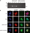
Production of transgenic P. falciparum lines expressing chimeric MSP1.A. PCR analysis to detect correct genomic integration of the homologous recombination constructs pMSP1chimWT and pMSP1chimSUB2to1. As controls, template DNA was prepared from the parental 3D7 clone, or input pMSP1chimWT plasmid DNA only (P). For test samples, genomic DNA was prepared after four rounds of drug cycling from three lines transfected with pMSP1chimWT (a–c), or three lines transfected with pMSP1chimSUB2to1 (d–f), or from one of the pMSP1chimSUB2to1-transfected line following seven drug cycles (d*). Oligonucleotide primers used in the PCR were designed to hybridize upstream of the region used as the targeting sequence in the 3D7 MSP1 locus, as well as to an allele-specific portion of the recodonized Wellcome sMSP1-19+ sequence in the transfection constructs (see Fig. 6C). A PCR product of approximately 1.2 kb can therefore only be produced if the predicted integration event has taken place. The integration-specific PCR product was detected for all three pMSP1chimWT-transfected lines but not for the pMSP1chimSUB2to1-transfected lines, except for one line examined after seven drug cycles (d*) (arrowed).B. IFA examination of the uncloned transgenic lines confirms expression of chimeric forms of MSP1. Samples of the pMSP1chimWT-transfected line (line a, drug cycle 4) and the pMSP1chimSUB2to1-transfected line (line d*, drug cycle 8) were double-probed with the 3D7 MSP1-specific mAb X509 and the Wellcome MSP1-specific mAb 111.4. Whereas the parental 3D7 and T9/94 parasite lines showed reactivity only with one or other type-specific mAb as expected, both transgenic populations contained parasites reactive with both mAbs, indicating expression of the predicted chimeric MSP1. The proportion of schizonts in the transgenic lines reactive with both antibodies was approximately 95% for the pMSP1chimWT-transfected line, and approximately 0.2% for the pMSP1chimSUB2to1-transfected line. Nuclei were counterstained with DAPI. Scale bar, 5 µm.

Similar articles

Cited by

References

    1. Bergmann-Leitner ES, Duncan EH, Mullen GE, Burge JR, Khan F, Long CA, et al. Critical evaluation of different methods for measuring the functional activity of antibodies against malaria blood stage antigens. Am J Trop Med Hyg. 2006;75:437–442. - PubMed
    1. Blackman MJ, Holder AA. Secondary processing of the Plasmodium falciparum merozoite surface protein-1 (MSP1) by a calcium-dependent membrane-bound serine protease: shedding of MSP133 as a noncovalently associated complex with other fragments of the MSP1. Mol Biochem Parasitol. 1992;50:307–315. - PubMed
    1. Blackman MJ, Heidrich HG, Donachie S, McBride JS, Holder AA. A single fragment of a malaria merozoite surface protein remains on the parasite during red cell invasion and is the target of invasion-inhibiting antibodies. J Exp Med. 1990;172:379–382. - PMC - PubMed
    1. Blackman MJ, Whittle H, Holder AA. Processing of the Plasmodium falciparum major merozoite surface protein-1: identification of a 33-kilodalton secondary processing product which is shed prior to erythrocyte invasion. Mol Biochem Parasitol. 1991;49:35–44. - PubMed
    1. Blackman MJ, Chappel JA, Shai S, Holder AA. A conserved parasite serine protease processes the Plasmodium falciparum merozoite surface protein-1. Mol Biochem Parasitol. 1993;62:103–114. - PubMed

Publication types

MeSH terms