. 2010 Aug 24:6:48.
doi: 10.1186/1744-8069-6-48.

Familial hemiplegic migraine Ca(v)2.1 channel mutation R192Q enhances ATP-gated P2X3 receptor activity of mouse sensory ganglion neurons mediating trigeminal pain

Affiliations

Familial hemiplegic migraine Ca(v)2.1 channel mutation R192Q enhances ATP-gated P2X3 receptor activity of mouse sensory ganglion neurons mediating trigeminal pain

Asha Nair et al. Mol Pain. .

Abstract

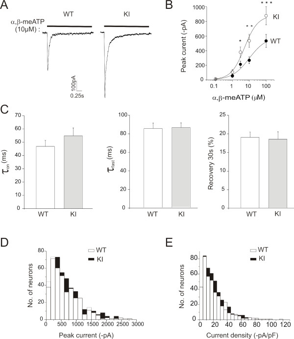
Background: The R192Q mutation of the CACNA1A gene, encoding for the α1 subunit of voltage-gated P/Q Ca2+ channels (Ca(v)2.1), is associated with familial hemiplegic migraine-1. We investigated whether this gain-of-function mutation changed the structure and function of trigeminal neuron P2X3 receptors that are thought to be important contributors to migraine pain.

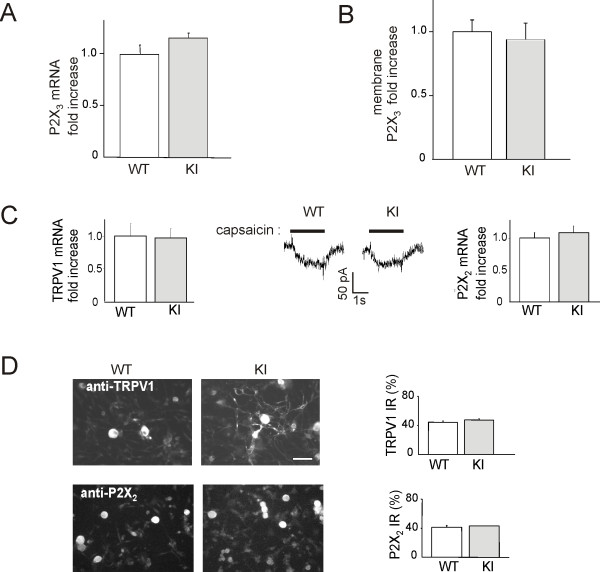
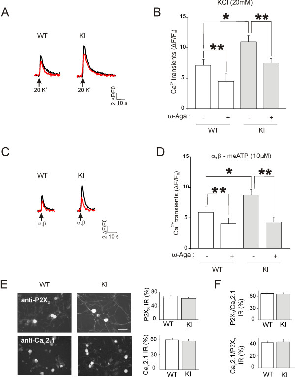
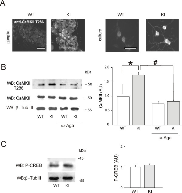
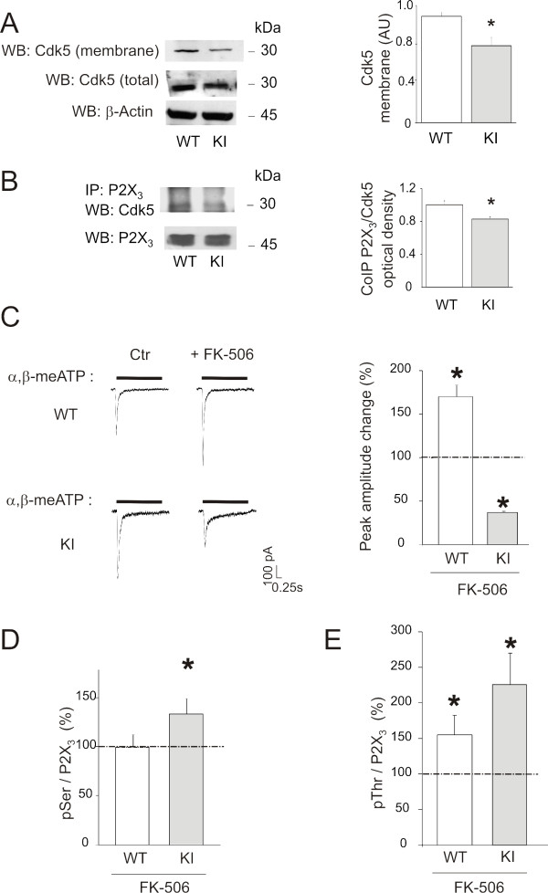
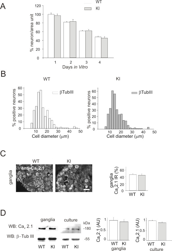
Results: Using in vitro trigeminal sensory neurons of a mouse genetic model knockin for the CACNA1A R192Q mutation, we performed patch clamp recording and intracellular Ca2+ imaging that showed how these knockin ganglion neurons generated P2X3 receptor-mediated responses significantly larger than wt neurons. These enhanced effects were reversed by the Ca(v)2.1 blocker ω-agatoxin. We, thus, explored intracellular signalling dependent on kinases and phosphatases to understand the molecular regulation of P2X3 receptors of knockin neurons. In such cells we observed strong activation of CaMKII reversed by ω-agatoxin treatment. The CaMKII inhibitor KN-93 blocked CaMKII phosphorylation and the hyperesponsive P2X3 phenotype. Although no significant difference in membrane expression of knockin receptors was found, serine phosphorylation of knockin P2X3 receptors was constitutively decreased and restored by KN-93. No change in threonine or tyrosine phosphorylation was detected. Finally, pharmacological inhibitors of the phosphatase calcineurin normalized the enhanced P2X3 receptor responses of knockin neurons and increased their serine phosphorylation.

Conclusions: The present results suggest that the CACNA1A mutation conferred a novel molecular phenotype to P2X3 receptors of trigeminal ganglion neurons via CaMKII-dependent activation of calcineurin that selectively impaired the serine phosphorylation state of such receptors, thus potentiating their effects in transducing trigeminal nociception.


Figures

Figure 1
Figure 1
Functional characterization and expression of P2X3 receptors in WT and R192Q KI trigeminal ganglion neurons. A, Representative current traces induced by α,β-meATP (10 μM, 2-s application) on WT and KI neurons. B, Dose-response curves for WT (n = 18, filled circles) and KI (n = 17, open circles) cells. * p = 0.04, ** p = 0.035, ***p = 0.03. Note larger responses of α,β-meATP-mediated currents on KI neurons with respect to WT, as the dose-effect plot is shifted upwards for KI neurons. C, Rise time (left; expressed as τon calculated on the 10-90% response rise), desensitization onset (middle; expressed as the first time constant, τfast, of current decay) and recovery from desensitization (right; expressed as% of control amplitude in a paired pulse agonist application) of P2X3 receptor currents are similar for WT and KI neurons. All responses were evoked by α,β-meATP (10 μM, 2 s). n = 154 for WT and n = 183 for KI. D, Histograms show peak amplitude distribution of α,β-meATP (10 μM)-induced P2X3 currents for WT (n = 414, open bars) and KI (n = 454, filled bars). E, Histograms show the distribution of current density (i.e., current amplitude normalized with respect to the neuronal capacitance, pA/pF) for WT (n = 414, open bars) and KI (n = 454, filled bars) neurons, indicating significantly higher α,β-meATP-evoked KI responses over a span of 15-35 pA/pF values compared to WT.
Figure 2
Figure 2
Expression of pain receptors in WT and KI neurons. A, Real-time RT-PCR experiments of mRNA extracts from trigeminal ganglia show no significant difference in P2X3 mRNA expression levels between WT and KI mice (n = 3, p > 0.05). Data were normalized with respect to β-tubulin III housekeeping mRNA content. B, Biotinylation experiments of trigeminal neurons in culture show no significant differences in P2X3 membrane protein expression levels between WT and KI mice (n = 4, p > 0.05). C, Real time RT-PCR experiments to measure mRNA expression levels of TRPV1 (left) and P2X2 (right) receptor subunits show no significant difference between trigeminal ganglia from WT and KI mice. n = 3 p > 0.05. Middle panel shows examples of TRPV1 receptor mediated responses evoked by capsaicin (1 μM) from WT or KI neuron, indicating similar amplitude and duration. D, Microphotographs show immunoreactivity of TRPV1 and P2X2 proteins in cultured trigeminal neurons from WT or KI mice. Histograms (right) represent% of immunoreactive neurons for TRPV1 (top) and P2X2 (bottom) over β-tubulin III immunoreactive neurons. n = 5, p > 0.05.
Figure 3
Figure 3
Ca2+ transients evoked by K+ depolarization or P2X3 receptors in WT and R192Q KI neurons. A, Examples of Ca2+ transients of trigeminal neurons evoked by KCl (20 mM, 2-s application) before (black trace) and after (red trace) application of ω-agatoxin (200 nM, 30 min). B, KI neurons show significant increase in KCl (20 mM, 2-s application) mediated Ca2+ transients compared to WT (*p = 0.005, n = 28 and n = 45, in WT and KI, respectively). Histograms also represent inhibition by ω-agatoxin of Ca2+ transients for WT (n = 14) and KI (n = 35) neurons. After ω-agatoxin responses of WT and KI neurons differ from their own controls (**p ≤ 0.001). C, Representative traces of α,β-meATP (10 μM, 2-s application)-evoked Ca2+ transients before (black trace) and after (red trace) application of ω-agatoxin (200 nM, 30 min). D, Histograms show larger Ca2+ transients evoked by α,β-meATP (10 μM, 2-s application) from KI (n = 26) than WT (n = 16) and neurons (*p = 0.04). Histograms also show that ω-agatoxin reduced Ca2+ transients of KI (n = 22) and WT (n = 9) neurons. **p ≤ 0.001 for each case. E, Microphotographs of immunofluorescence experiments depicting WT and KI trigeminal neurons in culture expressing P2X3 receptors or CaV2.1 channels. Bar = 50 μm. Histograms (right) show% of P2X3- (top) or CaV2.1- (bottom) immunoreactive neurons (taking as 100% the β-tubulin III immunoreactive) (n = 5, p > 0.05 for P2X3 receptors; n = 3, p > 0.05 for CaV2.1-expressing neurons). F, Histograms show% of CaV2.1-immunoreactive neurons (top; taken as 100%) which are immunopositive for P2X3 (n = 7, p > 0.05) or% of P2X3-immunoreactive neurons (bottom) which are immunopositive for CaV2.1 (n = 4, p > 0.05).
Figure 4
Figure 4
CaMKII activation of R192Q KI trigeminal neurons is reversed by ω-agatoxin. A, Microphotographs of immunofluorescence experiments with anti-phospho Thr286 CaMKII antibody (recognizing the active form of CaMKII) of WT and KI trigeminal ganglia (left panel) or trigeminal neurons in culture (right panel). Bar = 50 μm. B, Example of western immunoblots of total protein lysate from WT and KI trigeminal neurons in culture using antibodies recognizing the active form of CaMKII (phosphorylated Thr286, upper lanes) or anti-total CaMKII antibody (middle lanes). Equal loading was tested with β-tubulinIII antibodies (bottom lanes). p = 0.03, (n = 4). CaMKII activation is prevented by treatment with ω-agatoxin (200 nM, 24 h). Histograms (right) demonstrate significant increase in active (threonine-phosphorylated) CaMKII in KI neurons, an effect prevented by ω-agatoxin (n = 4, #p = 0.027). C, Western immunoblot experiments of trigeminal ganglia from WT and KI mice, using the same anti-phospho-CREB antibody (43 kDa). Gel loading quantification is obtained using anti-β-tubulinIII antibody. Histograms (right) show no significant difference in CREB phosphorylation between the two conditions. n = 3, p > 0.05.
Figure 5
Figure 5
Phosphorylation state of P2X3 receptors in KI neurons. A, Example of western blots of P2X3 receptors immunopurified from WT and KI neurons and probed with anti-phosphorylated serine antibodies. Note decreased serine phosphorylation of KI samples, a phenomenon prevented by pre-treatment with ω-agatoxin (200 nM, 24 h). B, Histograms quantify serine phosphorylation state of P2X3 receptors (*p = 0.02 vs. WT; n = 5) that is enhanced for KI receptors and prevented by ω-agatoxin (n = 3, #p = 0.027). C, Example of western blots of immunopurified P2X3 receptors from WT and KI neurons probed with anti-phosphorylated threonine (top row) or tyrosine (middle row) antibodies. Total P2X3 receptor levels are shown in bottom row. Control antibody lane (Ab) is also shown. n = 8 for threonine and n = 3 for tyrosine. D, Pretreatment of neurons with the CaMKII inhibitor KN-93 (5 μM, 90 min) restores the P2X3 serine phosphorylation state without changing total P2X3 receptor expression. Histograms (right) quantify serine phosphorylation state of P2X3 receptors, an effect blocked by KN-93 (n = 3; *p = 0.02; #p = 0.017) in KI neurons. E, Representative examples of WT and KI current traces evoked by α,β-meATP (10 μM, 2-s application) in control (Ctr) and after pre-treatment with KN-93 (5 μM, 90 min). Histograms (right) show the percent inhibition by KN-93 of peak amplitude of α,β-meATP-mediated P2X3 receptor currents in KI neurons (n = 22), that is significantly larger in KI cells (n = 17, *p = 0.039).
Figure 6
Figure 6
Cdk5 and calcineurin modulate P2X3 receptors of trigeminal neurons. A, Example of western immunoblot using anti-Cdk5 antibodies on membrane extracts from WT and KI neurons. Total Cdk5 in the soluble fraction is also shown. Actin immunoblotting ensures equal loading between WT and KI fractions (bottom row). Histograms (right) quantify average data (n = 4, p = 0.04) of membrane Cdk5. B, Example of co-immunoprecipitation experiments between P2X3 receptors and Cdk5 in WT and KI neurons showing decreased detection of Cdk5 in P2X3 immunopurified samples from KI neurons. Histograms (right) quantify lower Cdk5 signal from KI neurons (n = 3, p = 0.03). C, Example of current traces recorded from WT and KI trigeminal neurons evoked by α,β-meATP (10 μM, 2-s application) in control and after incubation with FK-506 (5 μM, 30 min). Histograms (right) quantify the effect of FK-506 on the P2X3 receptor-mediated currents (n = 18, *p = 0.035 in WT neurons and n = 20, *p = 0.027 in KI neurons). Data are normalized to the signals from WT neurons in control conditions. Note that FK-506 inhibits P2X3 current responses in KI neurons. D, Histograms of western blot signals obtained with anti-phosphorylated serine antibody applied to immunopurified P2X3 receptors from WT and KI neurons in control condition and after incubation with FK-506 (5 μM, 30 min). Note increase in serine phosphorylation after FK-506 treatment in KI neurons (n = 5, *p = 0.02). Data are normalized to the signals from WT neurons in control conditions. E, Histograms of western blot signals obtained with anti-phosphorylated threonine antibody show enhancement of threonine phosphorylation in WT (n = 5, *p = 0.046) as well as KI (n = 3, *p = 0.04) in neurons after treatment with FK-506. Data are normalized to the signals from WT neurons in control conditions.
Figure 7
Figure 7
Survival of WT and KI trigeminal neurons in culture and their expression of Cav2.1 protein. A, Survival is calculated as number of β-tubulin III positive cells per unit area after 1-4 days in culture. Data are normalized with respect to those at 1 day. n = 4, p > 0.05. B, Somatic size distribution of trigeminal neurons (β-tubulin III immunoreactive) in culture from WT and KI mice. n = 4. C, Immunocytochemical expression of CaV2.1 channels in intact trigeminal ganglia of WT and KI mice. Histograms represent% of CaV2.1 immunoreactive neurons over β-tubulinIII immunoreactive neurons in WT or KI ganglia. n = 3, p > 0.05. D, Example of western blot of protein extracts from WT and KI trigeminal ganglia or culture, probed with anti-CaV2.1 antibody. Equal loading was ensured by membrane probing with β-tubulinIII antibodies. n = 3, p > 0.05. Histograms (right) show no significant difference between these conditions.

Similar articles

Cited by

References

    1. International Headache Society. The International Classification of Headache Disorders, 2nd edition. Cephalalgia. 2004;24:S1–S160. - PubMed
    1. van den Maagdenberg AM, Haan J, Terwindt GM, Ferrari MD. Migraine: gene mutations and functional consequences. Curr Opin Neurol. 2007;20:299–305. doi: 10.1097/WCO.0b013e3281338d1f. - DOI - PubMed
    1. Ophoff RA, Terwindt GM, Vergouwe MN, van Eijk R, Oefner PJ, Hoffman SM, Lamerdin JE, Mohrenweiser HW, Bulman DE, Ferrari M, Haan J, Lindhout D, van Ommen GJ, Hofker MH, Ferrari MD, Frants RR. Familial hemiplegic migraine and episodic ataxia type-2 are caused by mutations in the Ca2+ channel gene CACNL1A4. Cell. 1996;87:543–552. doi: 10.1016/S0092-8674(00)81373-2. - DOI - PubMed
    1. Tottene A, Conti R, Fabbro A, Vecchia D, Shapovalova M, Santello M, van den Maagdenberg AM, Ferrari MD, Pietrobon D. Enhanced excitatory transmission at cortical synapses as the basis for facilitated spreading depression in CaV2.1 knockin migraine mice. Neuron. 2009;61:762–773. doi: 10.1016/j.neuron.2009.01.027. - DOI - PubMed
    1. van den Maagdenberg AM, Pietrobon D, Pizzorusso T, Kaja S, Broos LA, Cesetti T, van de Ven RC, Tottene A, van der Kaa J, Plomp JJ, Frants RR, Ferrari MD. A Cacna1a knockin migraine mouse model with increased susceptibility to cortical spreading depression. Neuron. 2004;41:701–710. doi: 10.1016/S0896-6273(04)00085-6. - DOI - PubMed

Publication types

MeSH terms