Comparative Study
. 2010 Aug 24;11(1):115.
doi: 10.1186/1465-9921-11-115.

Female sex hormones mediate the allergic lung reaction by regulating the release of inflammatory mediators and the expression of lung E-selectin in rats

Affiliations
Comparative Study

Female sex hormones mediate the allergic lung reaction by regulating the release of inflammatory mediators and the expression of lung E-selectin in rats

Ana Paula Ligeiro de Oliveira et al. Respir Res. .

Abstract

Background: Fluctuations of estradiol and progesterone levels caused by the menstrual cycle worsen asthma symptoms. Conflicting data are reported in literature regarding pro and anti-inflammatory properties of estradiol and progesterone.

Methods: Female Wistar rats were ovalbumin (OVA) sensitized 1 day after resection of the ovaries (OVx). Control group consisted of sensitized-rats with intact ovaries (Sham-OVx). Allergic challenge was performed by aerosol (OVA 1%, 15 min) two weeks later. Twenty four hours after challenge, BAL, bone marrow and total blood cells were counted. Lung tissues were used as explants, for expontaneous cytokine secretion in vitro or for immunostaining of E-selectin.

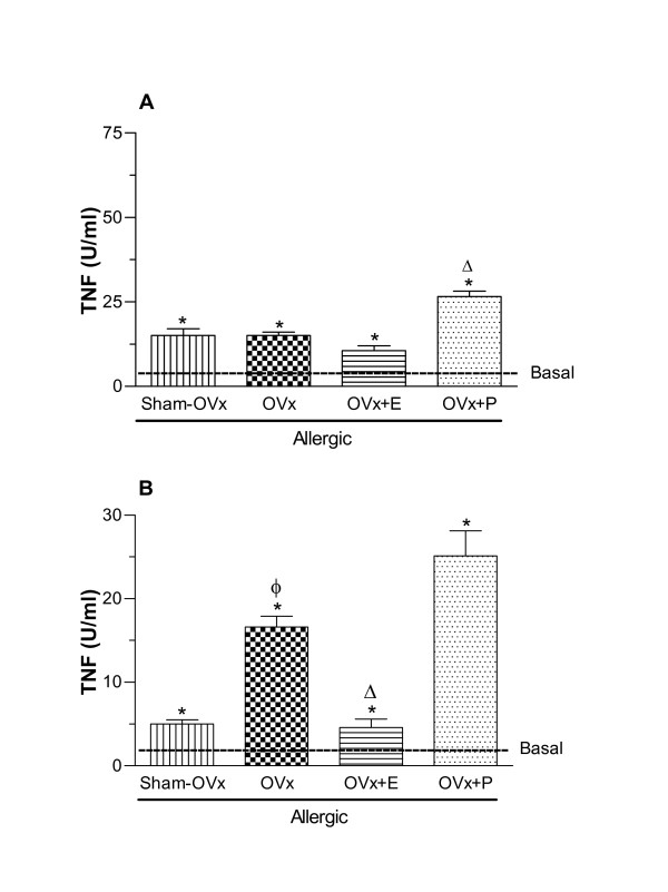
Results: We observed an exacerbated cell recruitment into the lungs of OVx rats, reduced blood leukocytes counting and increased the number of bone marrow cells. Estradiol-treated OVx allergic rats reduced, and those treated with progesterone increased, respectively, the number of cells in the BAL and bone marrow. Lungs of OVx allergic rats significantly increased the E-selectin expression, an effect prevented by estradiol but not by progesterone treatment. Systemically, estradiol treatment increased the number of peripheral blood leukocytes in OVx allergic rats when compared to non treated-OVx allergic rats. Cultured-BAL cells of OVx allergic rats released elevated amounts of LTB4 and nitrites while bone marrow cells increased the release of TNF-alpha and nitrites. Estradiol treatment of OVx allergic rats was associated with a decreased release of TNF-alpha, IL-10, LTB4 and nitrites by bone marrow cells incubates. In contrast, estradiol caused an increase in IL-10 and NO release by cultured-BAL cells. Progesterone significantly increased TNF- alpha by cultured BAL cells and bone marrow cells.

Conclusions: Data presented here suggest that upon hormonal oscillations the immune sensitization might trigger an allergic lung inflammation whose phenotype is under control of estradiol. Our data could contribute to the understanding of the protective role of estradiol in some cases of asthma symptoms in fertile ans post-menopausal women clinically observed.


Figures

Figure 1
Figure 1
Experimental design of ovariectomy (OVx) and ovalbumin-(OVA) sensitization. Anesthetized rats were subjected to ovariectomy (OVx) and at the indicated day 1 were ovalbumin (OVA) sensitized. Control group consisted of rats submitted to similar manipulations excepting the ovaries removal (Sham-OVx). Sensitized rats were boosted with OVA (Day 8), and the OVA-challenge performed at day 15. The rats were euthanized at day 16 (see material and methods for more details).
Figure 2
Figure 2
Total mononuclear cells and neutrophils (A) and eosinophil counts (B) in bronchoalveolar lavage (BAL) fluid of allergic rats (Sham-OVx and OVx). Basal values were obtained from nonmanipulated rats. Data are means ± SE from 5-8 experiments. *P < 0.05 compared with the basal group; ϕ P < 0.05 compared with the Sham-OVx allergic group.
Figure 3
Figure 3
Number of cells in peripheral blood (A) and in bone marrow (B) from allergic Sham-OVx and OVx rats. Basal values were obtained from nonmanipulated rats. Data are means ± SE from 5-8 experiments. *P < 0.05 compared with the basal group; ϕ P < 0.05 compared with the Sham-OVx allergic group.
Figure 4
Figure 4
Involvment of estradiol and progesterone in total cells, mononuclear cells and neutrophils (A) and eosinophils (B) recovered in BAL of rats subjected to allergic lung inflammation. Estradiol (280 μg s.c., single dose) and progesterone (200 μg s.c., single dose) replacement was performed in OVx rats 24 h before the antigen challenge. Data are means ± SE from 5-8 experiments. ϕ P < 0.05 compared with the Sham-OVx allergic group; Δ P < 0.05 compared to the OVx group.
Figure 5
Figure 5
Involvment of estradiol and progesterone in total cells in peripheral blood (A) and in bone marrow (B) from Sham-OVx and OVx allergic rats. Estradiol (280 μg s.c., single dose) and progesterone (200 μg s.c., single dose) replacement was performed in OVx rats 24 h before the antigen challenge. Data are means ± SE from 5-8 experiments. ϕ P < 0.05 compared with the Sham-OVx allergic group; Δ P < 0.05 compared to the OVx group.
Figure 6
Figure 6
TNF-alpha released by BAL (A) and bone marrow cells (B) 24 h after in vivo antigen challenge of allergic rats (Sham-OVx and OVx). Rats of OVx allergic groups were treated with estradiol or progesterone before the antigen challenge. Basal values were obtained from nonmanipulated rats. Data are means ± SE from 5-8 experiments. *P < 0.05 compared with the basal group; ϕ P < 0.05 compared with the Sham-OVx group; Δ P < 0.05 compared with the untreated OVx allergic group.
Figure 7
Figure 7
IL-10 released by BAL (A) and bone marrow cells (B) 24 h after in vivo antigen challenge of allergic rats (Sham-OVx and OVx). Rats of OVx allergic groups were treated with estradiol or progesterone before the antigen challenge. Basal values were obtained from nonmanipulated rats. Data are means ± SE from 5-8 experiments. *P < 0.05 compared with the basal group; ϕ P < 0.05 compared with the Sham-OVx group; Δ P < 0.05 compared with the untreated OVx allergic group.
Figure 8
Figure 8
LTB4 released by BAL (A) and bone marrow cells (B) 24 h after in vivo antigen challenge of allergic rats (Sham-OVx and OVx). Rats of OVx allergic groups were treated with estradiol or progesterone before the antigen challenge. Basal values were obtained from nonmanipulated rats. Data are means ± SE from 5-8 experiments. *P < 0.05 compared with the basal group; ϕ P < 0.05 compared with the Sham-OVx group; Δ P < 0.05 compared with the untreated OVx allergic group.
Figure 9
Figure 9
Nitrites released by BAL (A) and bone marrow cells (B) 24 h after in vivo antigen challenge of allergic rats (Sham-OVx and OVx). Rats of OVx allergic groups were treated with estradiol or progesterone before the antigen challenge. Basal values were obtained from nonmanipulated rats. Data are means ± SE from 5-8 experiments. *P < 0.05 compared with the basal group; ϕ P < 0.05 compared with the Sham-OVx group; Δ P < 0.05 compared with the untreated OVx allergic group.
Figure 10
Figure 10
Immunohystochemistry for lung E-Selectin expression of cells from BAL of allergic rats (Sham-OVx and OVx). Rats of OVx allergic groups were treated with estradiol or progesterone before the antigen challenge. Basal values were obtained from nonmanipulated rats. Data are means ± SE from 5-8 experiments. *P < 0.05 compared with the basal group; ϕ P < 0.05 compared with the Sham-OVx group; Δ P < 0.05 compared with the untreated OVx allergic group.

Similar articles

Cited by

References

    1. Haggerty CL, Ness RB, Kelsey S, Waterer GW. The impact of estrogen and progesterone on asthma. Ann Allergy Asthma Immunol. 2003;90(3):284–91. doi: 10.1016/S1081-1206(10)61794-2. - DOI - PubMed
    1. Stanford KI, Mickleborough TD, Ray S, Lindley MR, Koceja DM, Stager JM. Influence of menstrual cycle phase on pulmonary function in asthmatic athletes. Eur J Appl Physiol. 2006;96(6):703–10. doi: 10.1007/s00421-005-0067-7. - DOI - PubMed
    1. Frank RT. The hormonal causes of pre-menstrual tension. Arch Neurol Psychiatry. 1931;26:1053–1057.
    1. Gibbs CJ, Coutts II, Lock R, Finnegan OC, White RJ. Premenstrual exacerbation of asthma. Thorax. 1984;39(11):833–6. doi: 10.1136/thx.39.11.833. - DOI - PMC - PubMed
    1. Pauli BD, Reid RL, Munt PW, Wigle RD, Forkert L. Influence of the menstrual cycle on airway function in asthmatic and normal subjects. Am Rev Respir Dis. 1989;140(2):358–62. - PubMed

Publication types

LinkOut - more resources