

Umbilical endosalpingiosis: a case report
- PMID: 20735830
- PMCID: PMC2936926
- DOI: 10.1186/1752-1947-4-287
Umbilical endosalpingiosis: a case report
Abstract
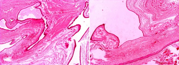
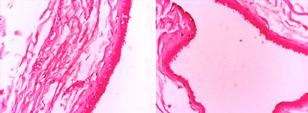
Introduction: Endosalpingiosis describes the ectopic growth of Fallopian tube epithelium. Pathology confirms the presence of a tube-like epithelium containing three types of cells: ciliated, columnar cells; non-ciliated, columnar secretory mucous cells; and intercalary cells.We report the case of a woman with umbilical endosalpingiosis and examine the nature and characteristics of cutaneous endosalpingiosis by reviewing and combining the other four cases existing in the international literature.
Case presentation: A 50-year-old Caucasian, Greek woman presented with a pale brown nodule in her umbilicus. The nodule was asymptomatic, with no cyclical discomfort or variation in size. Her personal medical, surgical and gynecologic history was uneventful. An excision within healthy margins was performed under local anesthesia. A cystic formation measuring 2.7×1.7×1 cm was removed. Histological examination confirmed umbilical endosalpingiosis.
Conclusions: Umbilical endosalpingiosis is a very rare manifestation of the non-neoplasmatic disorders of the Müllerian system. It appears with cyclic symptoms of pain and swelling of the umbilicus, but not always. The disease is diagnosed using pathologic findings and surgical excision is the definitive treatment.
Figures


Similar articles
-
Cutaneous endosalpingiosis arising from C-section scar: A case report with review of literature.J Cutan Pathol. 2023 Apr;50(4):310-315. doi: 10.1111/cup.14335. Epub 2022 Oct 14. J Cutan Pathol. 2023. PMID: 36169222 Review.
-
Florid cystic endosalpingiosis: Diagnostic challenges and management in the spectrum of müllerian anomalies - A case report.Int J Surg Case Rep. 2025 Feb;127:110840. doi: 10.1016/j.ijscr.2025.110840. Epub 2025 Jan 8. Int J Surg Case Rep. 2025. PMID: 39798339 Free PMC article.
-
Intramural florid cystic endosalpingiosis of the uterus after menopause.Pol J Pathol. 2018;69(3):321-324. doi: 10.5114/pjp.2018.79554. Pol J Pathol. 2018. PMID: 30509061
-
Vaginal Endosalpingiosis: A Case Report and Literature Review.Cureus. 2022 Mar 8;14(3):e22949. doi: 10.7759/cureus.22949. eCollection 2022 Mar. Cureus. 2022. PMID: 35411260 Free PMC article.
-
Refractory florid cystic endosalpingiosis: A case report with 5 years follow up and literature review.Int J Gynaecol Obstet. 2023 Oct;163(1):44-50. doi: 10.1002/ijgo.14764. Epub 2023 Apr 4. Int J Gynaecol Obstet. 2023. PMID: 37014527 Review.
Cited by
-
Vaginal Endosalpingiosis Case Report: A Rare Entity Presenting as Intermenstrual Bleeding.Case Rep Obstet Gynecol. 2017;2017:2424392. doi: 10.1155/2017/2424392. Epub 2017 Nov 9. Case Rep Obstet Gynecol. 2017. PMID: 29250450 Free PMC article.
-
Florid cystic endosalpingiosis, masquerading as malignancy in a young patient: a brief review.BMJ Case Rep. 2014 Jan 30;2014:bcr2013201645. doi: 10.1136/bcr-2013-201645. BMJ Case Rep. 2014. PMID: 24481015 Free PMC article.
-
Intraoperative Appearance of Endosalpingiosis: A Single-Center Experience of Laparoscopic Findings and Systematic Review of Literature.J Clin Med. 2022 Nov 27;11(23):7006. doi: 10.3390/jcm11237006. J Clin Med. 2022. PMID: 36498581 Free PMC article.
-
Endosalpingiosis of the Gallbladder: A Unique Complication of Ruptured Ectopic Pregnancy.Cureus. 2019 Aug 15;11(8):e5393. doi: 10.7759/cureus.5393. Cureus. 2019. PMID: 31620319 Free PMC article.
-
Imaging of Abdominal Wall Masses, Masslike Lesions, and Diffuse Processes.Radiographics. 2020 May-Jun;40(3):684-706. doi: 10.1148/rg.2020190170. Epub 2020 Apr 24. Radiographics. 2020. PMID: 32330085 Free PMC article. Review.
References
-
- Sampson JA. Postsalpingectomy endometriosis (endosalpingiosis) Am J Obstet Gynecol. 1930;20:443–480.
-
- Apostolidis S, Michalopoulos A, Papavramidis TS, Papadopoulos VN, Paramythiotis D, Harlaftis N. Inguinal endometriosis: three cases and literature review. South Med J. 2009;102:206–207. - PubMed
LinkOut - more resources
Full Text Sources
