. 2010 Aug 24:10:62.
doi: 10.1186/1472-6750-10-62.

Engineering of N. benthamiana L. plants for production of N-acetylgalactosamine-glycosylated proteins--towards development of a plant-based platform for production of protein therapeutics with mucin type O-glycosylation

Affiliations

Engineering of N. benthamiana L. plants for production of N-acetylgalactosamine-glycosylated proteins--towards development of a plant-based platform for production of protein therapeutics with mucin type O-glycosylation

Sasha M Daskalova et al. BMC Biotechnol. .

Abstract

Background: Mucin type O-glycosylation is one of the most common types of post-translational modifications that impacts stability and biological functions of many mammalian proteins. A large family of UDP-GalNAc polypeptide:N-acetyl-α-galactosaminyltransferases (GalNAc-Ts) catalyzes the first step of mucin type O-glycosylation by transferring GalNAc to serine and/or threonine residues of acceptor polypeptides. Plants do not have the enzyme machinery to perform this process, thus restricting their use as bioreactors for production of recombinant therapeutic proteins.

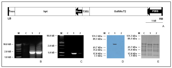
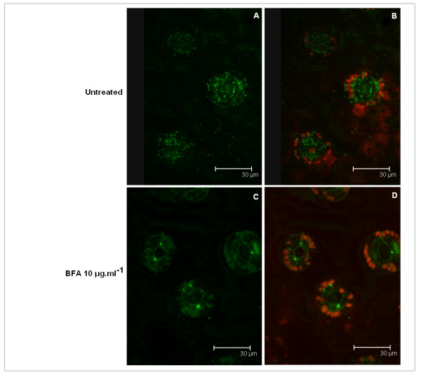
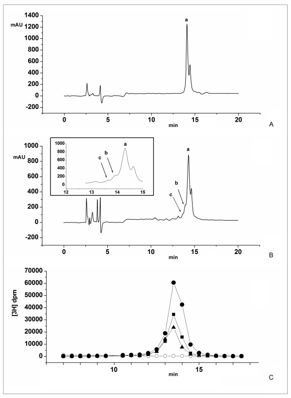
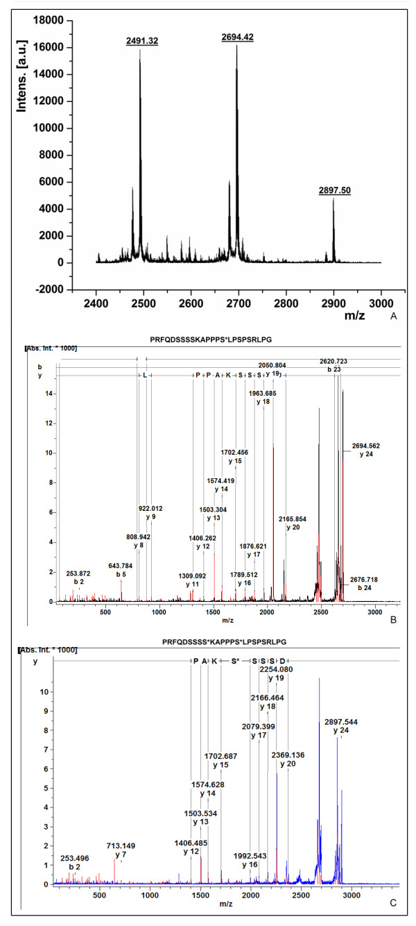
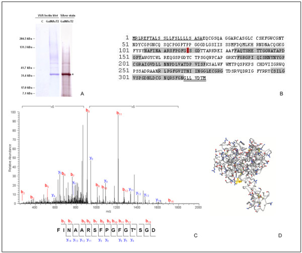
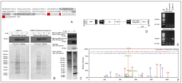
Results: The present study demonstrates that an isoform of the human GalNAc-Ts family, GalNAc-T2, retains its localization and functionality upon expression in N. benthamiana L. plants. The recombinant enzyme resides in the Golgi as evidenced by the fluorescence distribution pattern of the GalNAc-T2:GFP fusion and alteration of the fluorescence signature upon treatment with Brefeldin A. A GalNAc-T2-specific acceptor peptide, the 113-136 aa fragment of chorionic gonadotropin β-subunit, is glycosylated in vitro by the plant-produced enzyme at the "native" GalNAc attachment sites, Ser-121 and Ser-127. Ectopic expression of GalNAc-T2 is sufficient to "arm" tobacco cells with the ability to perform GalNAc-glycosylation, as evidenced by the attachment of GalNAc to Thr-119 of the endogenous enzyme endochitinase. However, glycosylation of highly expressed recombinant glycoproteins, like magnICON-expressed E. coli enterotoxin B subunit:H. sapiens mucin 1 tandem repeat-derived peptide fusion protein (LTBMUC1), is limited by the low endogenous UDP-GalNAc substrate pool and the insufficient translocation of UDP-GalNAc to the Golgi lumen. Further genetic engineering of the GalNAc-T2 plants by co-expressing Y. enterocolitica UDP-GlcNAc 4-epimerase gene and C. elegans UDP-GlcNAc/UDP-GalNAc transporter gene overcomes these limitations as indicated by the expression of the model LTBMUC1 protein exclusively as a glycoform.

Conclusion: Plant bioreactors can be engineered that are capable of producing Tn antigen-containing recombinant therapeutics.


Figures

Figure 1
Figure 1
Representative molecular analyses of N. benthamiana L. GalNAc-T2 transgenic plants. A - Schematic diagram of T-DNA of pH7WG2:GNT2 plant binary expression vector. Abbreviations: LB, left border; RB, right border; Pnos, nopaline synthase promoter; P35 S, CaMV 35 S promoter; hpt, hygromycin phosphotransferase gene; Tnos, 3' termination signal of nopaline synthase gene; T35 S, 3' termination signal of CaMV 35 S gene; B - PCR screen using 1 μg genomic DNA as a template and primers G1/G2 (see "Materials and Methods") amplifying human GalNAc-T2 gene (1.7 kb); C - RT-PCR using 0.5 μg total RNA as a template and primers G3/G4 (see "Materials and Methods) that amplify a 0.96 kb fragment of the GalNAc-T2 transcript; D - Western blot of total hydrophobic proteins probed with antibodies raised against human GalNAc-T2; E - The same protein samples separated by SDS-PAGE (4-20% gradient gel) after staining with Coomassie Brilliant Blue R-250; Abbreviations used: C - control plant; 1,2 - putative transgenic plants; M- markers: Promega 1 kb DNA ladder (B, C), BioRad Kaleidoscope prestained standards (D, E).
Figure 2
Figure 2
Localization of human GalNAc-T2 expressed in N. benthamiana L. by confocal microscopy. Image of leaf (guard cells) stably expressing GalNAc-T2:GFP fusion protein: A - image without chloroplasts, B - image with chloroplasts. Image of leaf (guard cells) stably expressing GalNAc-T2:GFP fusion protein after 1 h treatment with 10 μg/ml Brefeldin A: C - image without chloroplasts, D - image with chloroplasts. The green signal represents fluorescence of GalNAc-T2:GFP fusion protein, the red signal represents the autofluorescence of chloroplasts.
Figure 3
Figure 3
Functional analyses of human GalNAc-T2 expressed in N. benthamiana L. plants. GalNAc-T2 enzyme assay was set up in a final volume of 100 μl with UDP-GalNAc as the donor, and the hCG-β peptide as an acceptor (see "Materials and Methods"). A sample corresponding to 25 μl of the reaction mixture containing microsomal fraction of A - control plants, or B - GalNAc-T2 transgenic plants as an enzyme source, was subjected to RP-HPLC as described (see "Materials and Methods"): a - peak corresponding to the hCG-β peptide, b, c - peaks corresponding to the glycosylated forms of hCG-β peptide; C - monitoring the attachment of GalNAc to the hCG-β peptide by radioactive enzyme assay using [3H]UDP-GalNAc as a donor and 41 μg (●),, 20.5 μg (■) or 10.25 μg (▲) microsomal proteins from GalNAc-T2 plant, or 41 μg microsomal proteins from control plant (○) as an enzyme source. Aliquots of 0.5 ml from the collected fractions (2 ml fraction volume) were used to monitor the radioactivity.
Figure 4
Figure 4
Functional analyses of human GalNAc-T2 expressed in N. benthamiana L. plants. A - MALDI-TOF MS of glycosylated products (peaks b + c combined) of hCG-β peptide; B - MALDI-TOF/TOF MS spectrum of [M+H+] 2694.4 corresponding to the monoglycosylated hCG-β peptide - GalNAc is attached to Ser-15 (corresponding to Ser-127 of hCG-β); C - MALDI-TOF/TOF MS spectrum of [M+H+] 2897.5 corresponding to the diglycosylated hCG-β peptide - GalNAc is attached to Ser-9 and Ser-15 (corresponding to Ser-121 and Ser-127 of hCG-β, respectively).
Figure 5
Figure 5
Glycosylation of endogenous N. benthamiana L. proteins upon ectopic expression of human GalNAc-T2. A - VVA-lectin blot of the soluble proteins bound to VVA-agarose from control (C) and transgenic (GalNAc-T2) plants, and the corresponding silver staining for the GalNAc-T2 plant sample; B - sequence of tobacco endochitinase (GenBank Accession No P24091) identified with the 33 kDa band denoted in A by an asterisk: the signal peptide is underlined, the propeptide is double underlined, the covered regions after LC-MS/MS analysis of the chymotryptic fragments are gray-highlighted, the modified Thr-119 by 203 amu is red-highlighted; C - MS/MS analysis confirming the attachment of GalNAc (203 amu) to Thr-119; D - homology model of tobacco endochitinase built based on the 3 D structure of O. sativa L. japonica class I chitinase (pdb ID 2DKV); the position of the glycosylated Thr-119 is denoted by halos.
Figure 6
Figure 6
Characterization of LTBMUC1 produced in transgenic N. benthamiana L. plants expressing either human GalNAc-T2 or co-expressing human GalNAc-T2, Y. enterocolitica UDP-GlcNAc-4 epimerase, and C. elegans UDPGlcNAc/UDPGalNAc transporter. A - amino acid sequence of His-tagged LTBMUC1 fusion protein: MUC1 sequence is underlined, GPGP linker is italisized, experimentally confirmed sites for attachment of GalNAc by GalNAc-T2 are red-highlighted, covered regions after LC-MS/MS analysis of the chymotryptic fragments are gray-highlighted; B - purification of LTBMUC1 produced in GalNAc-T2 and GalNAc-T2//UDP-GlcNAc-4 epimerase//UDP-GlcNAc/UPD-GalNAc transporter plants by Ni2+ Sepharose Fast Flow chromatography: Fractions (1, 2, 3, 4 for LTBMUC1 produced in GalNAc-T2 plants; and 1', 2', 3', 4' for LTBMUC1 produced in the triple transgenic plants) eluted from the resin with 20 mM phosphate buffer pH 7.4 containing 0.5 M NaCl and 0.5 M imidazol, were screened by HisProbe-HRP (upper panels); the same blots were stained in parallel with Ponceau S (lower panels); C - VVA chromatography of LTBMUC1 produced in the tested plant systems after purification on Ni2+ Sepharose Fast Flow: identical aliquots of the volume-equalized samples 2 and 2' were screened by HisProbe-HRP before loading on VVA agarose gel (panel HisProbe-HRP detection before VVA agarose); bound proteins were eluted from the resin with 50 mM Tris HCl buffer pH 7.4 containing 0.1 M NaCl, 1 mM MgCl2 and 0.3 M GalNAc, and identical aliquots were screened by HisProbe-HRP (panel HisProbe-HRP detection after VVA agarose), VVA blotting (panel VVA blot after VVA agarose), and SDS-PAGE with subsequent silver-staining (panel silver staining after VVA agarose); asterisk denotes the position of LTBMUC1; D - schematic diagram of T-DNA of pBYR:GNE.GT plant binary expression vector. Abbreviations: LB, left border; RB, right border; Pnos, nopaline synthase promoter; P35 S, CaMV 35 S promoter; nptII, neomycin phosphotransferase gene; gne - Y. enterocolitica UDP-GlcNAc-4-epimerase gene; UDP-GalNAc - C. elegans UDP-GlcNAc/UDP-GalNAc transporter gene; Tnos, 3' termination signal of nopaline synthase gene; T35 S, 3' termination signal of CaMV 35 S gene; TvspB - 3' termination signal of soybean vegetative storage protein B gene; TrbcS - 3' termination signal of Rubisco small subunit gene; TEV HT - etch virus translational enhancer element; E - RT-PCR confirmation of the co-expression of human GalNAc-T2 (GalNAc-T2), C. elegans UDP-GlcNAc/UPD-GalNAc transporter (transporter), and Y. enetrocolitica UDP-GlcNAc-4 epimerase (gne) in N. benthamana L. plants using 0.5 μg total RNA as a template and primers G3/G4, GT3/GT4, and GE4/GE5, respectively (see "Materials and Methods). F - spectrum of a chymotryptic peptide of the 20/21 kDa band denoted in C by an asterisk, assisting in the identification of the band as LTBMUC1.

Similar articles

Cited by

References

    1. Sharma AK, Sharma MK. Plants as bioreactors: Recent developments and emerging opportunities. Biotechnol Adv. 2009;27:811–832. doi: 10.1016/j.biotechadv.2009.06.004. - DOI - PMC - PubMed
    1. Jin C, Altmann F, Strasser R, Mach L, Schaehs M, Kunert R, Rademancher T, Gloessl J, Steinkellner H. A plant-derived human monoclonal antibody induces an anti-carbohydrate immune response in rabbits. Glycobiology. 2008;18:235–241. doi: 10.1093/glycob/cwm137. - DOI - PubMed
    1. Bardor M, Faveeuw C, Fitchette AC, Gilbert D, Galas L, Trottein F, Faye L, Lerouge P. Immunoreactivity in mammals of two typical plant glycol-epitopes, core alpha(1,3)-fucose and core xylose. Glycobiology. 2003;13:427–434. doi: 10.1093/glycob/cwg024. - DOI - PubMed
    1. Fujiyama K, Misaki R, Sakai Y, Omasa T, Seki T. Change in glycosylation pattern with extension of endoplasmic reticulum retention signal sequence of mouse antibody produced by suspension-cultured tobacco BY2 cells. J Biosci Bioeng. 2009;107:165–172. doi: 10.1016/j.jbiosc.2008.09.016. - DOI - PubMed
    1. Strasser R, Altmann F, Mach L, Glössl J, Steinkellner H. Generation of Arabidopsis thaliana plants with complex N glycans lacking beta 1,2-linked xylose and core alpha 1,3-linked fucose. FEBS Lett. 2004;561:132–136. doi: 10.1016/S0014-5793(04)00150-4. - DOI - PubMed

Publication types

MeSH terms