

Cognitive and neural correlates of depression-like behaviour in socially defeated mice: an animal model of depression with cognitive dysfunction
- PMID: 20735879
- PMCID: PMC3432579
- DOI: 10.1017/S1461145710000945
Cognitive and neural correlates of depression-like behaviour in socially defeated mice: an animal model of depression with cognitive dysfunction
Abstract
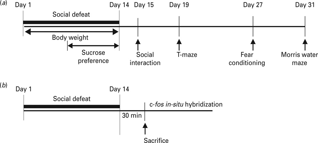
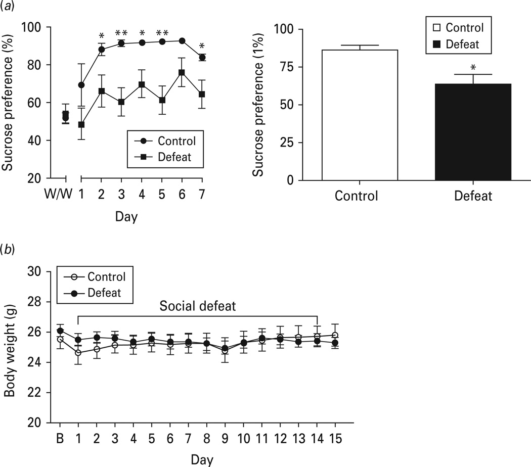
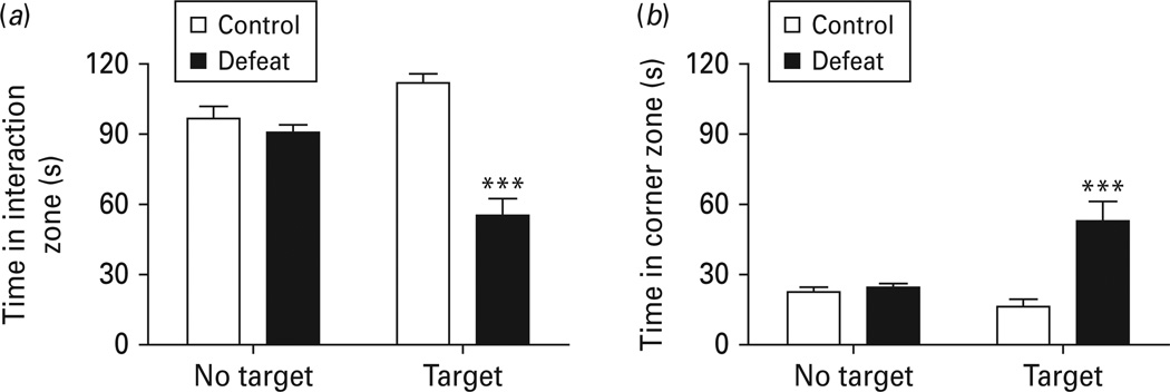
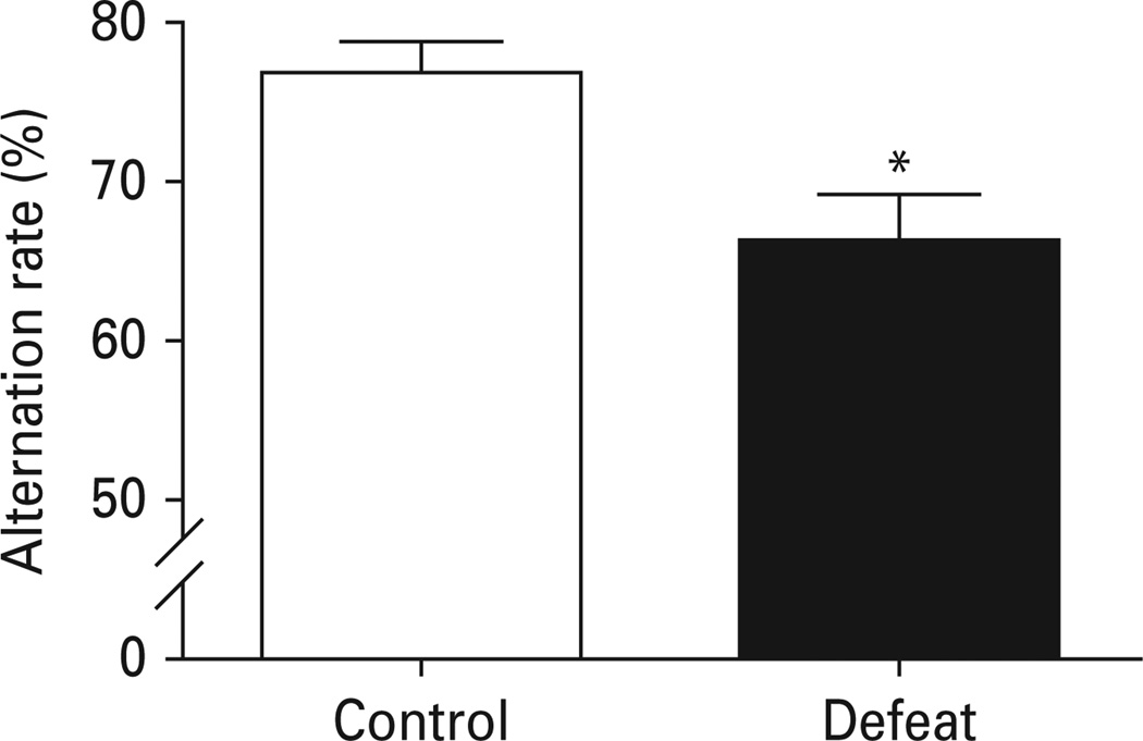
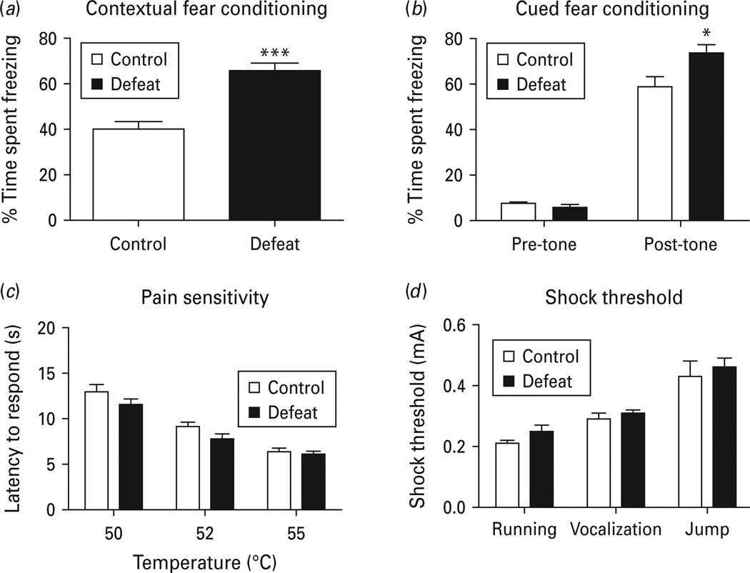
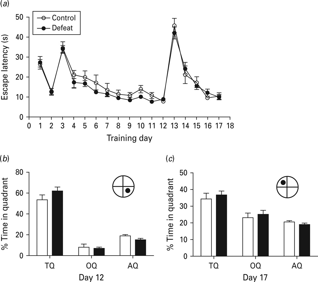
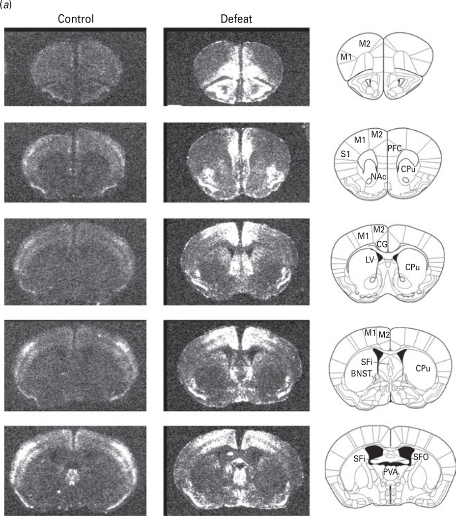
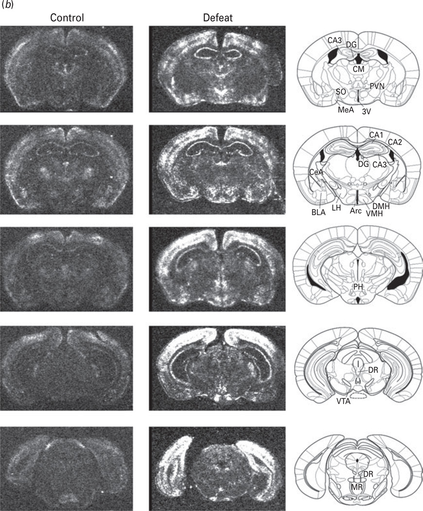
Human depression is associated with cognitive deficits. It is critical to have valid animal models in order to investigate mechanisms and treatment strategies for these associated conditions. The goal of this study was to determine the association of cognitive dysfunction with depression-like behaviour in an animal model of depression and investigate the neural circuits underlying the behaviour. Mice that were exposed to social defeat for 14 d developed depression-like behaviour, i.e. anhedonia and social avoidance as indicated by reduced sucrose preference and decreased social interaction. The assessment of cognitive performance of defeated mice demonstrated impaired working memory in the T-maze continuous alternation task and enhanced fear memory in the contextual and cued fear-conditioning tests. In contrast, reference learning and memory in the Morris water maze test were intact in defeated mice. Neuronal activation following chronic social defeat was investigated by c-fosin-situ hybridization. Defeated mice exhibited preferential neural activity in the prefrontal cortex, cingulate cortex, hippocampal formation, septum, amygdala, and hypothalamic nuclei. Taken together, our results suggest that the chronic social defeat mouse model could serve as a valid animal model to study depression with cognitive impairments. The patterns of neuronal activation provide a neural basis for social defeat-induced changes in behaviour.
Figures
Similar articles
-
Olfactory cues increase avoidance behavior and induce Fos expression in the amygdala, hippocampus and prefrontal cortex of socially defeated mice.Behav Brain Res. 2013 Nov 1;256:188-96. doi: 10.1016/j.bbr.2013.08.020. Epub 2013 Aug 19. Behav Brain Res. 2013. PMID: 23968590
-
Deletion of glutamate delta-1 receptor in mouse leads to enhanced working memory and deficit in fear conditioning.PLoS One. 2013;8(4):e60785. doi: 10.1371/journal.pone.0060785. Epub 2013 Apr 3. PLoS One. 2013. PMID: 23560106 Free PMC article.
-
The effects of social defeat on behavior and dopaminergic markers in mice.Neuroscience. 2015 Mar 12;288:167-77. doi: 10.1016/j.neuroscience.2014.12.043. Epub 2015 Jan 6. Neuroscience. 2015. PMID: 25575945
-
Cognitive dysfunction in the dystrophin-deficient mouse model of Duchenne muscular dystrophy: A reappraisal from sensory to executive processes.Neurobiol Learn Mem. 2015 Oct;124:111-22. doi: 10.1016/j.nlm.2015.07.006. Epub 2015 Jul 17. Neurobiol Learn Mem. 2015. PMID: 26190833
-
A robust animal model of state anxiety: fear-potentiated behaviour in the elevated plus-maze.Eur J Pharmacol. 2003 Feb 28;463(1-3):163-75. doi: 10.1016/s0014-2999(03)01279-2. Eur J Pharmacol. 2003. PMID: 12600708 Review.
Cited by
-
Social defeat drives hyperexcitation of the piriform cortex to induce learning and memory impairment but not mood-related disorders in mice.Transl Psychiatry. 2022 Sep 10;12(1):380. doi: 10.1038/s41398-022-02151-1. Transl Psychiatry. 2022. PMID: 36088395 Free PMC article.
-
Differential effects of chronic social stress and fluoxetine on meal patterns in mice.Appetite. 2013 May;64:81-8. doi: 10.1016/j.appet.2012.12.023. Epub 2013 Jan 11. Appetite. 2013. PMID: 23318656 Free PMC article.
-
Making Sense of Rodent Models of Anhedonia.Int J Neuropsychopharmacol. 2018 Nov 1;21(11):1049-1065. doi: 10.1093/ijnp/pyy083. Int J Neuropsychopharmacol. 2018. PMID: 30239762 Free PMC article. Review.
-
Molecular Signatures of Psychosocial Stress and Cognition Are Modulated by Chronic Lithium Treatment.Schizophr Bull. 2016 Jul;42 Suppl 1(Suppl 1):S22-33. doi: 10.1093/schbul/sbv194. Epub 2015 Dec 28. Schizophr Bull. 2016. PMID: 26714764 Free PMC article.
-
Social defeat stress induces depression-like behavior and alters spine morphology in the hippocampus of adolescent male C57BL/6 mice.Neurobiol Stress. 2016 Aug 21;5:54-64. doi: 10.1016/j.ynstr.2016.07.001. eCollection 2016 Dec. Neurobiol Stress. 2016. PMID: 27981196 Free PMC article.
References
-
- Airaksinen E, Larsson M, Lundberg I, Forsell Y. Cognitive functions in depressive disorders : evidence from a population-based study. Psychological Medicine. 2004;34:83–91. - PubMed
-
- Aisa B, Tordera R, Lasheras B, Del Rio J, et al. Cognitive impairment associated to HPA axis hyperactivity after maternal separation in rats. Psychoneuroendocrinology. 2007;32:256–266. - PubMed
-
- Alzoubi KH, Abdul-Razzak KK, Khabour OF, Al-Tuweiq GM, et al. Adverse effect of combination of chronic psychosocial stress and high fat diet on hippocampus-dependent memory in rats. Behavioural Brain Research. 2009;204:117–123. - PubMed
-
- Austin MP, Mitchell P, Goodwin GM. Cognitive deficits in depression: possible implications for functional neuropathology. British Journal of Psychiatry. 2001;178:200–206. - PubMed
Publication types
MeSH terms
Substances
Grants and funding
LinkOut - more resources
Full Text Sources
Other Literature Sources
Medical
