

Expression patterns of astrocyte elevated gene-1 (AEG-1) during development of the mouse embryo
- PMID: 20736086
- PMCID: PMC3165053
- DOI: 10.1016/j.gep.2010.08.004
Expression patterns of astrocyte elevated gene-1 (AEG-1) during development of the mouse embryo
Abstract
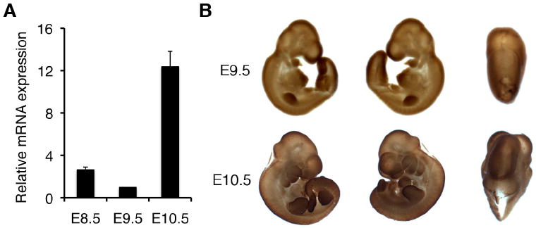
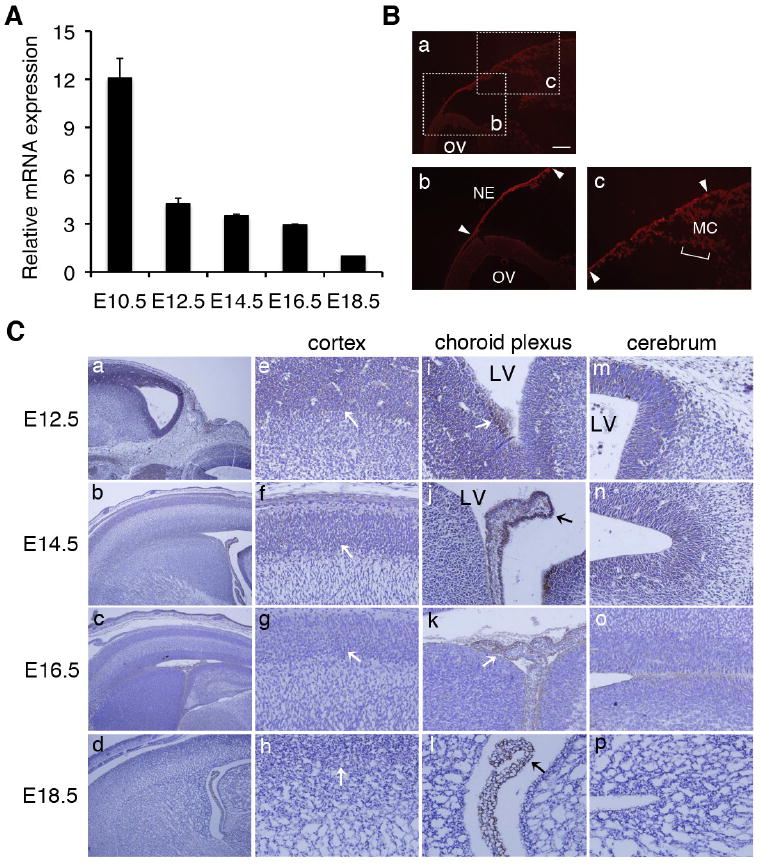
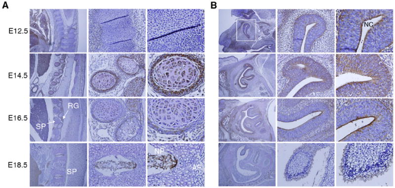
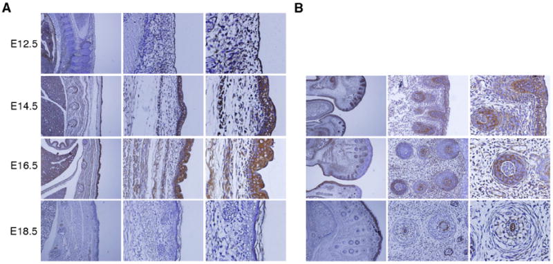
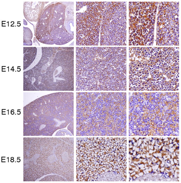
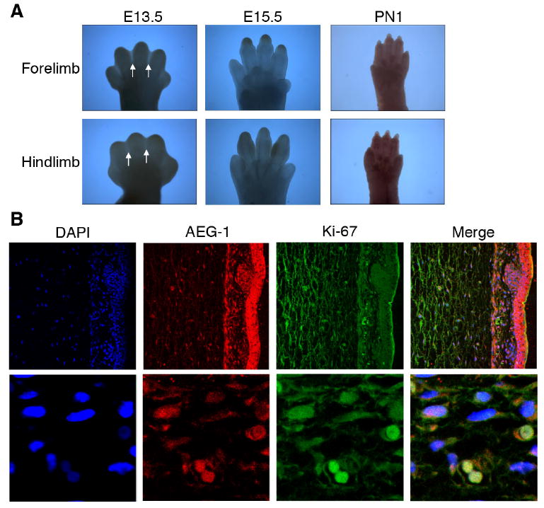
Expression of astrocyte elevated gene-1 (AEG-1) is elevated in multiple human cancers including brain tumors, neuroblastomas, melanomas, breast cancers, non-small cell lung cancers, liver cancers, prostate cancers, and esophageal cancers. This gene plays crucial roles in tumor cell growth, invasion, angiogenesis and progression to metastasis. In addition, over-expression of AEG-1 protects primary and transformed cells from apoptosis-inducing signals by activating PI3K-Akt signaling pathways. These results suggest that AEG-1 is intimately involved in tumorigenesis and may serve as a potential therapeutic target for various human cancers. However, the normal physiological functions of AEG-1 require clarification. We presently analyzed the expression pattern of AEG-1 during mouse development. AEG-1 was expressed in mid-to-hindbrain, fronto-nasal processes, limbs, and pharyngeal arches in the early developmental period from E8.5 to E9.5. In addition, at stages of E12.5-E18.5 AEG-1 was localized in the brain, and olfactory and skeletal systems suggesting a role in neurogenesis, as well as in skin, including hair follicles, and in the liver, which are organ sites in which AEG-1 has been implicated in tumor development and progression. AEG-1 co-localized with Ki-67, indicating a role in cell proliferation, as previously revealed in tumorigenesis. Taken together, these results suggest that AEG-1 may play a prominent role during normal mouse development in the context of cell proliferation as well as differentiation, and that temporal regulation of AEG-1 expression may be required during specific stages and in specific tissues during development.
Copyright © 2010 Elsevier B.V. All rights reserved.
Figures
Similar articles
-
Astrocyte elevated gene-1 regulates astrocyte responses to neural injury: implications for reactive astrogliosis and neurodegeneration.J Neuroinflammation. 2012 Aug 11;9:195. doi: 10.1186/1742-2094-9-195. J Neuroinflammation. 2012. PMID: 22884085 Free PMC article.
-
Expression patterns of MDA-9/syntenin during development of the mouse embryo.J Mol Histol. 2013 Apr;44(2):159-66. doi: 10.1007/s10735-012-9468-1. Epub 2012 Nov 20. J Mol Histol. 2013. PMID: 23180153 Free PMC article.
-
Emerging Role and Clinicopathological Significance of AEG-1 in Different Cancer Types: A Concise Review.Cells. 2021 Jun 15;10(6):1497. doi: 10.3390/cells10061497. Cells. 2021. PMID: 34203598 Free PMC article. Review.
-
Astrocyte elevated gene-1 (AEG-1) functions as an oncogene and regulates angiogenesis.Proc Natl Acad Sci U S A. 2009 Dec 15;106(50):21300-5. doi: 10.1073/pnas.0910936106. Epub 2009 Nov 25. Proc Natl Acad Sci U S A. 2009. PMID: 19940250 Free PMC article.
-
Astrocyte elevated gene-1: recent insights into a novel gene involved in tumor progression, metastasis and neurodegeneration.Pharmacol Ther. 2007 May;114(2):155-70. doi: 10.1016/j.pharmthera.2007.01.010. Epub 2007 Feb 24. Pharmacol Ther. 2007. PMID: 17397930 Free PMC article. Review.
Cited by
-
Astrocyte elevated gene-1 regulates astrocyte responses to neural injury: implications for reactive astrogliosis and neurodegeneration.J Neuroinflammation. 2012 Aug 11;9:195. doi: 10.1186/1742-2094-9-195. J Neuroinflammation. 2012. PMID: 22884085 Free PMC article.
-
Metadherin facilitates podocyte apoptosis in diabetic nephropathy.Cell Death Dis. 2016 Nov 24;7(11):e2477. doi: 10.1038/cddis.2016.335. Cell Death Dis. 2016. PMID: 27882943 Free PMC article.
-
AEG-1/MTDH/LYRIC: signaling pathways, downstream genes, interacting proteins, and regulation of tumor angiogenesis.Adv Cancer Res. 2013;120:75-111. doi: 10.1016/B978-0-12-401676-7.00003-6. Adv Cancer Res. 2013. PMID: 23889988 Free PMC article.
-
Astrocyte elevated gene-1 (AEG-1): A multifunctional regulator of normal and abnormal physiology.Pharmacol Ther. 2011 Apr;130(1):1-8. doi: 10.1016/j.pharmthera.2011.01.008. Epub 2011 Jan 20. Pharmacol Ther. 2011. PMID: 21256156 Free PMC article. Review.
-
The role of AEG-1/MTDH/LYRIC in the pathogenesis of central nervous system disease.Adv Cancer Res. 2013;120:159-92. doi: 10.1016/B978-0-12-401676-7.00006-1. Adv Cancer Res. 2013. PMID: 23889991 Free PMC article. Review.
References
-
- Bard JBL, Kaufman MH, Dubreuil C, Brune RM, Burger A, Baldock RA, Davidson DR. An internet-accessible database of mouse developmental anatomy based on a systematic nomenclature. Mech Dev. 1998;74:111–120. - PubMed
-
- Brown DM, Ruoslahti E. Metadherin, a cell surface protein in breast tumors that mediate lung metastasis. Cancer Cell. 2004;5:365–374. - PubMed
-
- DasGupta R, Fuchs E. Multiple roles for activated LEF/TCF transcription complexes during hair follicle development and differentiation. Development. 1999;126:4557–4568. - PubMed
Publication types
MeSH terms
Substances
Grants and funding
LinkOut - more resources
Full Text Sources
Molecular Biology Databases
