. 2010 Oct 29;285(44):34269-78.
doi: 10.1074/jbc.M110.143008. Epub 2010 Aug 24.

A novel cytoplasmic adaptor for retinoic acid receptor (RAR) and thyroid receptor functions as a Derepressor of RAR in the absence of retinoic acid

Affiliations

A novel cytoplasmic adaptor for retinoic acid receptor (RAR) and thyroid receptor functions as a Derepressor of RAR in the absence of retinoic acid

Ui-Hyun Park et al. J Biol Chem. .

Abstract

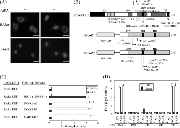
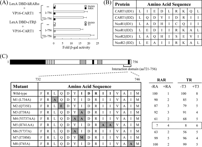
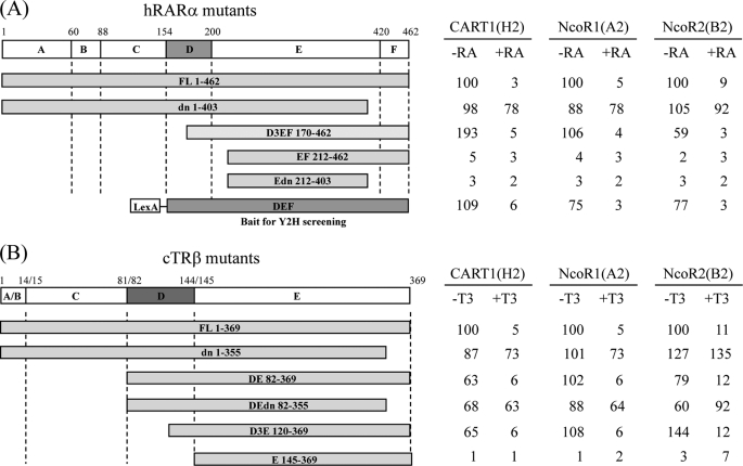
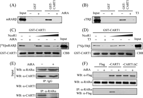
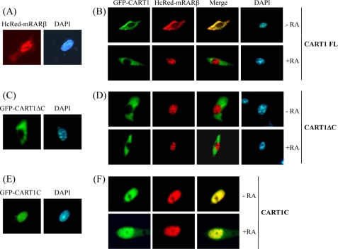
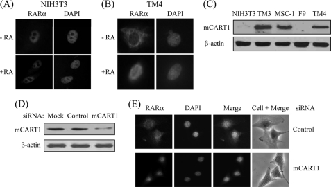
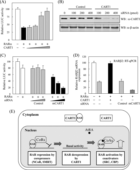
In most mammalian cells, the retinoic acid receptor (RAR) is nuclear rather than cytoplasmic, regardless of its cognate ligand, retinoic acid (RA). In testis Sertoli cells, however, RAR is retained in the cytoplasm and moves to the nucleus only when RA is supplied. This led us to identify a protein that regulates the translocation of RAR. From yeast two-hybrid screening, we identified a novel RAR-interacting protein called CART1 (cytoplasmic adaptor for RAR and TR). Systematic interaction assays using deletion mutants showed that the C-terminal CoRNR box of CART1 was responsible for the interaction with the NCoR binding region of RAR and TR. Such interaction was impaired in the presence of ligand RA, as further determined by GST pulldown assays in vitro and immunoprecipitation assays in vivo. Fluorescence microscopy showed that unliganded RAR was captured by CART1 in the cytoplasm, whereas liganded RAR was liberated and moved to the nucleus. Overexpression of CART1 blocked the transcriptional repressing activity of unliganded apoRAR, mediated by corepressor NCoR in the nucleus. CART1 siRNA treatment in a mouse Sertoli cell line, TM4, allowed RAR to move to the nucleus and blocked the derepressing function of CART1, suggesting that CART1 might be a cytoplasmic, testis-specific derepressor of RAR.


Figures

FIGURE 1.
FIGURE 1.
Isolation of CART1. A, shown is subcellular localization of RARα in TM4 cells. Mouse RARα in TM4 cells were visualized by staining with rabbit anti-RARα polyclonal antibody and Texas Red-conjugated anti-rabbit antibody in the absence and presence of ligand AtRA. Each bar represents 10 μm. B, shown are schematic representations of human CART1, NCoR1, and NCoR2 (SMRTe) isolated by yeast two-hybrid screening. Locations of identified clones and functional domains are indicated: EF, EF-hand motif; LZ, leucine zipper; ID, nuclear receptor interaction domain. C, interaction of CART1 with hRAR(DEF) is shown. Interactions were monitored by introducing LexA DBD-fused RAR(DEF) and Gal4 AD-fused test partners in yeast strain L40 and by β-galactosidase (β-gal) assays with transformed yeast extracts. -Fold β-galactosidase activity indicates relative value compared with the Gal4 AD empty control. In all experiments, 1 μm AtRA was added to the yeast culture. DMSO was used as a control for AtRA. Data are shown as the averages of three independent experiments (mean ± S.D.). D, shown is interaction of CART1 with other nuclear receptor family members. Yeast two-hybrid assays were performed with the other NRs indicated in the presence of their cognate ligands: RAR, 1 μm AtRA; RXR, 1 μm 9-cis RA; ER, 1 μm estradiol (E2); GR, 5 μm deoxycorticosterone; TR, 1 μm triiodo-l-thyronine (T3). Data are the average of three independent experiments (mean ± S.D.).
FIGURE 2.
FIGURE 2.
Mapping of RAR binding domain in CART1. To map the minimal region required for RAR and TR binding (A), CART1 deletions were fused to acidic VP16 AD using the pASV3 vector instead of Gal4 AD. Using these CART1 deletions and LexA DBD-RAR(DEF) or LexA DBD-TR(DE), yeast two-hybrid assays were performed in the absence (DMSO) and presence of AtRA (for RAR) or T3 (for TR). -Fold β-galactosidase activity indicates relative values compared with the VP16 AD empty control. B, shown is amino acid alignment of CART1, NCoR1, and NCoR2. Amino acid sequences of interacting domain (ID or CoRNR box) are listed. The consensus sequence corresponding to CoRNR box is (L/I)XX(I/V)IXXXL. C, shown is identification of critical amino acid residues of CART1 responsible for RAR or TR binding. The amino acid residues (aa) in the second putative CoRNR box (also designated as ID2) were mutated to alanine by PCR. Yeast two-hybrid assays were performed using CART1 mutants and LexA DBD-RAR(DEF) or LexA DBD-TR(DE). Numbers represent the relative percent of wild-type β-galactosidase activity shown by the average of three independent experiments (mean ± S.D.).
FIGURE 3.
FIGURE 3.
Comparison of the interaction modes of CART1, NCoR1, and NCoR2. The interactions were monitored by yeast two-hybrid assays using Gal4 AD-CART1 (clone H2), -NCoR1 (clone A2), or -NCoR2 (clone B2) and LexA DBD-RARα mutants (A) or LexA DBD-TRβ mutants (B). The range of amino acids in each mutant is indicated. Functional domains of RARα and TRβ were designated as A–F (or E for TR). Numbers represent the relative percent of wild-type β-galactosidase activity.
FIGURE 4.
FIGURE 4.
Confirmation of the interaction by GST pulldown and IP assays. A and B, shown is the interaction between CART1 and RAR or TR in vitro. In vitro synthesized 35S-hRARβ (A) or 35S-cTRβ (B) was incubated with 2 μg of GST-fused hCART1 (amino acids 699–756) in the presence of cognate ligand (2 μm). Bound proteins were visualized by SDS-PAGE and autoradiography. Input was 10% of the labeled sample used in assay. C and D, competition between CART1 and NCoR1 for RAR or TR binding is shown. For assays, 10 μl of in vitro translated 35S-hRARβ (C) or 35S-cTRβ (D) was mixed with purified GST-CART1 (amino acids 699–756) and further reacted with increasing volumes of NCoR1 (10, 20, and 40 μl). Then, GST pulldown assays were performed as described. Equal loading of beads was shown by Coomassie Brilliant Blue (CBB) staining of GST-CART1. E, endogenous interaction between CART1 and RARα is shown. TM4 cell lysates were prepared and immunoprecipitated with preimmune serum (IgG) or anti-RARα antibody. Precipitated proteins were revealed by WB using anti-CART1 antibody. F, requirement of the C-terminal region of CART1 for RAR binding in vivo is shown. NIH3T3 cells were transfected with FLAG-hCART1 or CART1 C-terminal truncation (CART1ΔC) expression vector and cultured in the absence and presence of 1 μm AtRA. The interaction was monitored by IP with anti-RARα antibody and WB using anti-FLAG antibody.
FIGURE 5.
FIGURE 5.
Cytoplasmic retention of RAR by CART1. NIH3T3 cells were transfected with HcRed-mRARβ and GFP-CART1 (or its variants) alone or together and treated with 1 μm AtRA. Cells were then fixed and observed under a fluorescence microscope. DAPI (1 μg/ml) was used to localize chromosomal DNA in the nucleus. A and B, localization of HcRed-RARβ alone (A) and together with GFP-CART1 (B) is shown. C and D, localization of CART1 C-terminal truncation (CART1 ΔC: amino acids 1–719) alone (C) and together with RAR (D) is shown. E and F, localization of the C-terminal region of CART1 (CART1C: amino acids 699–756) alone (E) and together with RAR (F) is shown.
FIGURE 6.
FIGURE 6.
Effect of CART1 knockdown on subcellular location of RAR in mouse Sertoli cells. A and B, subcellular location of endogenous RARα in NIH3T3 and TM4 Sertoli cells is shown. NIH3T3 (A) or TM4 (B) cells were fixed and permeabilized. Cells were then stained with rabbit anti-RARα polyclonal antibody and Texas Red-conjugated anti-rabbit antibody and observed by fluorescence microscopy. C, expression of CART1 is shown. Cellular extracts were prepared from mouse cell lines as indicated and subjected to WB using anti-CART1 antibody. β-Actin was used as an internal control. D and E, effect of CART1 knockdown on subcellular location of RAR in TM4 cells. TM4 cells were transfected with either control or CART1 siRNA (200 pmol) using Lipofectamine 2000 in the absence of AtRA. CART1 expression was monitored by WB (D). Endogenous RARα was visualized as described in B (E).
FIGURE 7.
FIGURE 7.
Effect of CART1 on transcriptional activity of RAR. A, shown is the effect of CART1 on transcriptional activity of RAR in the absence of AtRA. NIH3T3 cells were cotransfected with RARα (0.2 μg) and increasing amounts (0.1, 0.2, 0.4, 0.8 μg) of FLAG-CART1 together with the RARE-tk-luciferase reporter. Each bar represents the percent of mock treatment. The relative luciferase (Luc) activity was determined by luciferase assay after normalizing to the observed β-galactosidase activity. Data are the average of three independent experiments (mean ± S.D.). B, shown is the effect of siRNA on CART1 expression. After transfection into TM4 cells, using the amounts indicated, the expression of mouse CART1 was monitored by WB using an anti-CART1 polyclonal antibody. C, shown is the effect of CART1 knockdown on transcriptional activity of RAR. TM4 cells were cotransfected with RARα (0.2 μg) and increasing amounts of CART1 siRNA, as indicated in Fig. 7C, together with the RARE-tk-luciferase reporter. The relative luciferase activity is shown by the average of three independent experiments (mean ± S.D.). D, shown is the effect of CART1 overexpression or knockdown on the expression of endogenous RAR-regulated RARβ2 gene. Total RNA was extracted from TM4 cells transfected with control or CART1-specific siRNA (200 pmol) and subjected to real-time PCR coupled to reverse transcription. The expression levels were normalized using GAPDH as an internal standard. Relative expression (%) was defined as the variation relative to control. E, shown is a postulated model for the role of CART1 in RAR regulation. In testis cells, where CART1 is abundant, CART1 interacts with apoRAR in the cytoplasm, thus preventing its repressor function by cooperating with corepressors in the nucleus. In the presence of ligand AtRA, RAR dissociates from CART1 and moves to nucleus where it interacts with coactivators for transcriptional activation. qPCR, quantitative PCR.

Similar articles

Cited by

References

    1. Mangelsdorf D. J., Thummel C., Beato M., Herrlich P., Schütz G., Umesono K., Blumberg B., Kastner P., Mark M., Chambon P., Evans R. M. (1995) Cell 83, 835–839 - PMC - PubMed
    1. Chambon P. (1995) Recent Prog. Horm. Res. 50, 317–332 - PubMed
    1. Yamashita S. (1998) Histol. Histopathol. 13, 255–270 - PubMed
    1. DeFranco D. B. (1999) Cell. Biochem. Biophys. 30, 1–24 - PubMed
    1. Hager G. L., Lim C. S., Elbi C., Baumann C. T. (2000) J. Steroid Biochem. Mol. Biol. 74, 249–254 - PubMed

Publication types