. 2010 Nov 5;285(45):34909-21.
doi: 10.1074/jbc.M109.016287. Epub 2010 Aug 24.

Regulation of epidermal growth factor receptor ubiquitination and trafficking by the USP8·STAM complex

Affiliations

Regulation of epidermal growth factor receptor ubiquitination and trafficking by the USP8·STAM complex

Ilana Berlin et al. J Biol Chem. .

Abstract

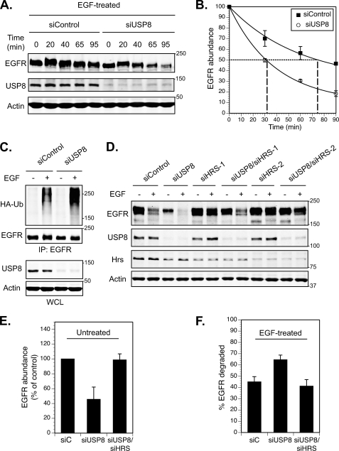
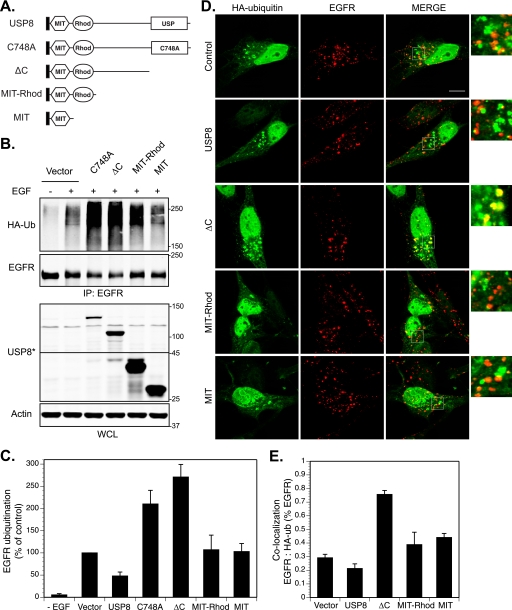
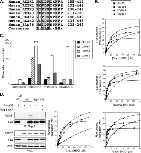
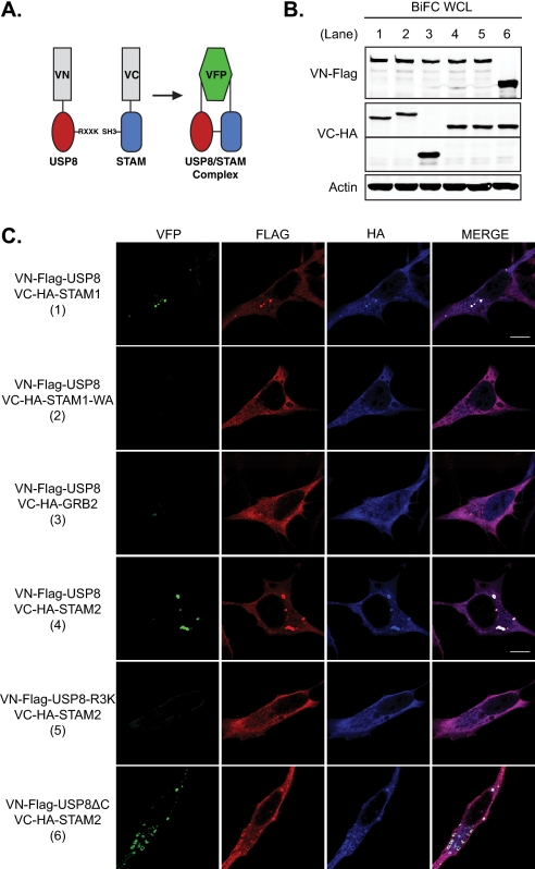
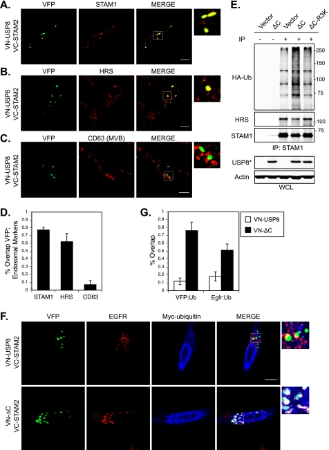
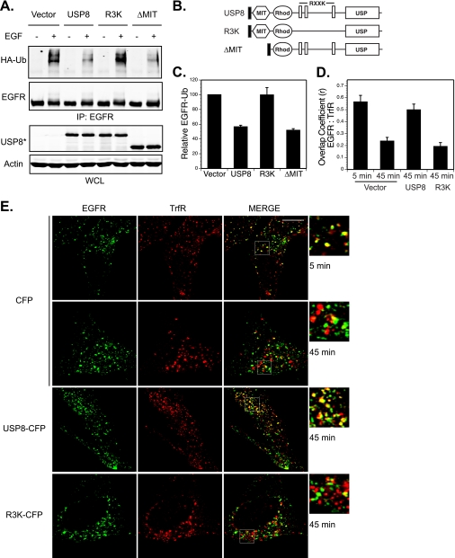
Reversible ubiquitination of activated receptor complexes signals their sorting between recycling and degradation and thereby dictates receptor fate. The deubiquitinating enzyme ubiquitin-specific protease 8 (USP8/UBPy) has been previously implicated in the regulation of the epidermal growth factor receptor (EGFR); however, the molecular mechanisms governing its recruitment and activity in this context remain unclear. Herein, we investigate the role of USP8 in countering ligand-induced ubiquitination and down-regulation of EGFR and characterize a subset of protein-protein interaction determinants critical for this function. USP8 depletion accelerates receptor turnover, whereas loss of hepatocyte growth factor-regulated substrate (Hrs) rescues this phenotype, indicating that USP8 protects EGFR from degradation via an Hrs-dependent pathway. Catalytic inactivation of USP8 incurs EGFR hyperubiquitination and promotes receptor localization to endosomes marked by high ubiquitin content. These phenotypes require the central region of USP8, containing three extended Arg-X-X-Lys (RXXK) motifs that specify direct low affinity interactions with the SH3 domain(s) of ESCRT-0 proteins, STAM1/2. The USP8·STAM complex critically impinges on receptor ubiquitination status and modulates ubiquitin dynamics on EGFR-positive endosomes. Consequently, USP8-mediated deubiquitination slows progression of EGFR past the early-to-recycling endosome circuit in a manner dependent upon the RXXK motifs. Collectively, these findings demonstrate a role for the USP8·STAM complex as a protective mechanism regulating early endosomal sorting of EGFR between pathways destined for lysosomal degradation and recycling.


Figures

FIGURE 1.
FIGURE 1.
USP8 depletion accelerates ligand-mediated EGFR degradation through an Hrs-dependent pathway. A and B, USP8 knockdown induces accelerated EGFR turnover in response to EGF. HeLa cells transfected with either control siRNA (siControl) or siRNA targeting human USP8 (siUSP8) were serum-starved and treated in the presence of 2.5 ng/ml EGF for the specified length of time. Western blot analysis of total cellular EGFR observed throughout the treatment time course and efficiency of USP8 knockdown are shown. B, shown is a graphic representation of EGFR degradation rates as a function of USP8 (see “Materials and Methods” for details). C, USP8 depletion results in elevated ligand-induced ubiquitination of EGFR. Cells co-transfected with HA-ubiquitin (HA-Ub) and either siControl or siUSP8 were serum-starved and treated in the absence (−) or presence (+) of 2.5 ng/ml EGF for 5 min. EGFR was immunoprecipitated, and ubiquitination was analyzed by Western blot against HA. D–F, USP8 modulates EGFR degradation through an Hrs-dependent pathway. Cells transfected with siControl alone, co-transfected with siUSP8, siHrs, or a combination of the two were serum-starved and treated in the absence (−) or presence (+) of 2.5 ng/ml EGF for 1 h. D, shown is a Western blot analysis of endogenous proteins. Quantification of changes in EGFR abundance (relative to siControl) (E) and down-regulation (expressed as % of total EGFR in untreated cells (−)) (F) are shown. All quantification was performed on the basis of three independent experiments (n = 3), with error bars corresponding to S.D.
FIGURE 2.
FIGURE 2.
The central region of USP8 is required to regulate EGFR ubiquitination. A, domain organization and mutational analysis of USP8 is shown. Boundaries of individual domains and residues critical for domain function are indicated using amino acid numbering of murine USP8 (GenBankTM accession no. BC050947). DUF, Domain of Unknown Function 1873; USP, ubiquitin-specific protease domain with catalytic Cys 748. Truncation mutants of USP8 with amino acid boundaries are designated in parentheses: ΔC, catalytic domain truncation (amino acid residues 1–735); MIT (amino acid residues 1–184); MIT-Rhod (amino acid residues 1–319). All USP8 mutants were constructed on the basis of available structural data (20, 33). B, catalytic inactivation of USP8 leads to EGFR hyperubiquitination in a manner dependent upon the central region of the enzyme. Cells co-transfected with HA-ubiquitin and vector control or USP8 mutants as indicated were serum-starved and treated in the absence (−) or presence (+) of 10 ng/ml EGF for 10 min. EGFR ubiquitination was assayed as in Fig. 1C and quantified relative to EGF-treated vector control samples; n = 3 (C). D, catalytically inactive USP8 results in enhanced localization of EGFR to intracellular ubiquitin and requires the central region of USP8. Cells were transfected as indicated, serum-starved, and treated with EGF as above. After treatment, cells were fixed and immunostained against HA-ubiquitin (green) and EGFR (red). Representative images are shown with 9× inset magnification; the scale bar corresponds to 10 μm. E, localization of EGFR to HA-ubiquitin was calculated as described under “Materials and Methods” and is represented as a fraction of EGFR overlapping HA-ubiquitin relative to total cellular EGFR; n = 2. All error bars correspond to S.D.
FIGURE 3.
FIGURE 3.
USP8 contains three RXXK motifs that constitute low affinity binding partners for non-canonical SH3 domains. A, ClustalW2 alignment of mouse and human USP8 protein sequences encompassing the three RXXK motifs of USP8 is shown. B, shown are SH3 domains of adaptor proteins exhibit low affinities for the RXXK peptides of USP8. Fluorescence polarization in millipolarization units (mP) was measured for fluorescein-tagged peptides corresponding to Slp76 (Fl-APSIDRSTKPA) and USP8-1 (Fl-KNVPQVDRTKKPA), USP8-2 (Fl-SGKVLSDRSTKPV), and USP8-3 (Fl-TVTPMVNRENKPT) as a function of GST-SH3 (measurements taken using Beakon 2000; data were plotted in DeltaGraph 5.7.5). C, shown is a graphic representation of equilibrium dissociation constants (KD) obtained using fluorescence polarization (see “Materials and Methods”). The corresponding numerical values can be found in supplemental Fig. S3C. D, The USP8·STAM complex co-immunoprecipitates in a manner dependent upon the interaction between the RXXK motifs of USP8 and the SH3 domain of STAM. Lysates from cells co-transfected with wild type USP8 (WT) or the triple RXXK mutant (R3K) and FLAG-STAM1 or its SH3 domain mutant, FLAG-STAM1-WA, were immunoprecipitated with anti-FLAG antibody, and samples were analyzed by Western blot against exogenous USP8 (USP8*). All error bars correspond to S.D.
FIGURE 4.
FIGURE 4.
Complex formation between USP8 and the SH3 domains of STAM proteins in their cellular context requires the RXXK/SH3 interaction. A, shown is a schematic representation of the BiFC assay. In this assay USP8 was fused to the N-terminal and SH3-containing adaptor proteins to the C-terminal fragments of VFP. VFP fluorescence was observed upon direct interaction between individually non-fluorescent VN and VC fusion proteins co-expressed in live cells. B, shown is BiFC fusion protein expression as analyzed by Western blot against VN-FLAG and VC-HA. C, complex formation between USP8 and STAM1/2 proteins requires the SH3 domain of STAM as well as the RXXK motifs of USP8 but not its catalytic domain (numbering of samples corresponds to the order of lanes in B). Cells were transfected with VN-FLAG-USP8 or its triple RXXK mutant, −R3K, in combination with either VC-VN-HA-STAM1, SH3 domain mutant -STAM1-WA (as shown in Fig. 3D, this mutant fails to interact with USP8), -GRB2, or -STAM2. After incubation adequate for visualization of BiFC complexes by VFP fluorescence (green), cells were fixed and immunostained against FLAG (red) and HA (blue). All images were taken under the same magnification, with the scale bar corresponding to 10 μm.
FIGURE 5.
FIGURE 5.
The USP8·STAM complex localizes to the ESCRT-0-complex and regulates ubiquitin dynamics on EGFR-positive endosomes. A–D, the USP8·STAM BiFC complex localizes to endosomes populated by the endogenous ESCRT-0 proteins, STAM1 (A) and Hrs (B), but not a late MVB marker, CD63 (C). Cells were co-transfected with VN-FLAG-USP8 and VC-HA-STAM2, fixed, and immunostained as indicated. D, quantification of colocalization between USP8·STAM BiFC and STAM1, Hrs, or CD63 is represented as a fraction of VFP fluorescence overlapping the indicated endosomal proteins; n = 3. E, overexpression of a catalytically inactive USP8 results in hyperubiquitination of the endogenous STAM·Hrs complex in a manner dependent upon the RXXK motifs. Cells were co-transfected with HA-ubiquitin (HA-Ub) and vector control or catalytically inactive USP8 containing either intact (ΔC) or mutated (ΔC-R3K) RXXK motifs. Endogenous ESCRT-0 complex was immunoprecipitated against the STAM1 protein as indicated, and its ubiquitination status was assessed by Western blot against HA. F, the USP8·STAM complex regulates ubiquitin dynamics on EGFR-positive endosomes. Cells expressing either VN-USP8 or VN-ΔC in combination with VC-STAM2 and Myc-ubiquitin were incubated under standard growth conditions to allow development of VFP fluorescence (green), briefly serum-starved, and treated in the presence of 10 ng/ml EGF for 30 min. After treatment, cells were fixed and immunostained against EGFR (red) and Myc (blue). Similar results were obtained with an EGF stimulation of 10 min (data not shown). All images are shown with 9× inset magnification and scale bars corresponding to 10 μm. G, co-localization of USP8·STAM BiFC and EGFR with Myc-ubiquitin is enhanced upon catalytic inactivation of USP8. Quantification of data is shown in F; n = 2. All error bars correspond to S.D.
FIGURE 6.
FIGURE 6.
The USP8·STAM complex regulates EGFR ubiquitination and trafficking. A, the RXXK motifs are essential for USP8-mediated deubiquitination of EGFR, whereas the MIT domain does not contribute to this function. Cells co-transfected with HA-ubiquitin (HA-Ub) and vector, USP8, R3K, or ΔMIT (constructs shown in B) were serum-starved and treated in the absence (−) or presence (+) of 10 ng/ml EGF for 10 min. EGFR ubiquitination was analyzed as in Fig. 2B. C, shown is quantification of the data in A; n = 3. D, USP8 requires the RXXK motifs to modulate transit of EGFR through the transferrin receptor (TrfR)-positive endosomes. Cells, transfected with CFP, USP8-CFP, or R3K-CFP were serum-starved, treated with 10 ng/ml EGF for 5 or 45 min, as indicated, fixed, and immunostained against TrfR (green) and EGFR (red). CFP-positive cells are shown (CFP fluorescence not provided) with 9× inset magnification and scale bars corresponding to 10 μm. E, colocalization between EGFR and TrfR is expressed in the form of an overlap coefficient, r (see “Materials and Methods” for details); n = 2. All error bars correspond to S.D.

Similar articles

Cited by

References

    1. Polo S., Di Fiore P. P. (2006) Cell 124, 897–900 - PubMed
    1. Pickart C. M. (2001) Annu. Rev. Biochem. 70, 503–533 - PubMed
    1. Haglund K., Sigismund S., Polo S., Szymkiewicz I., Di Fiore P. P., Dikic I. (2003) Nat. Cell Biol. 5, 461–466 - PubMed
    1. Hicke L., Riezman H. (1996) Cell 84, 277–287 - PubMed
    1. Duan L., Miura Y., Dimri M., Majumder B., Dodge I. L., Reddi A. L., Ghosh A., Fernandes N., Zhou P., Mullane-Robinson K., Rao N., Donoghue S., Rogers R. A., Bowtell D., Naramura M., Gu H., Band V., Band H. (2003) J. Biol. Chem. 278, 28950–28960 - PubMed

Publication types

MeSH terms

Substances