. 2010 Nov 12;285(46):35374-85.
doi: 10.1074/jbc.M110.148445. Epub 2010 Aug 24.

Direct association of Sprouty-related protein with an EVH1 domain (SPRED) 1 or SPRED2 with DYRK1A modifies substrate/kinase interactions

Affiliations

Direct association of Sprouty-related protein with an EVH1 domain (SPRED) 1 or SPRED2 with DYRK1A modifies substrate/kinase interactions

Dan Li et al. J Biol Chem. .

Abstract

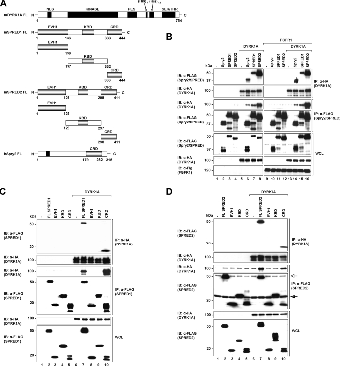
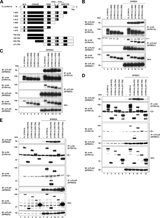
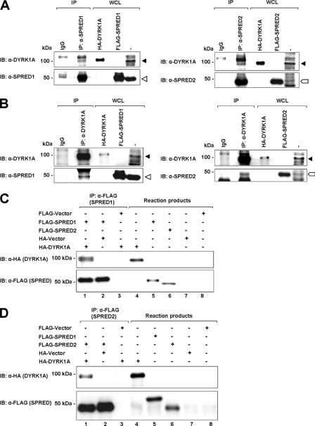
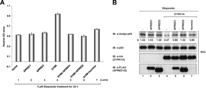
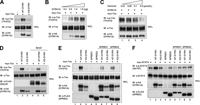
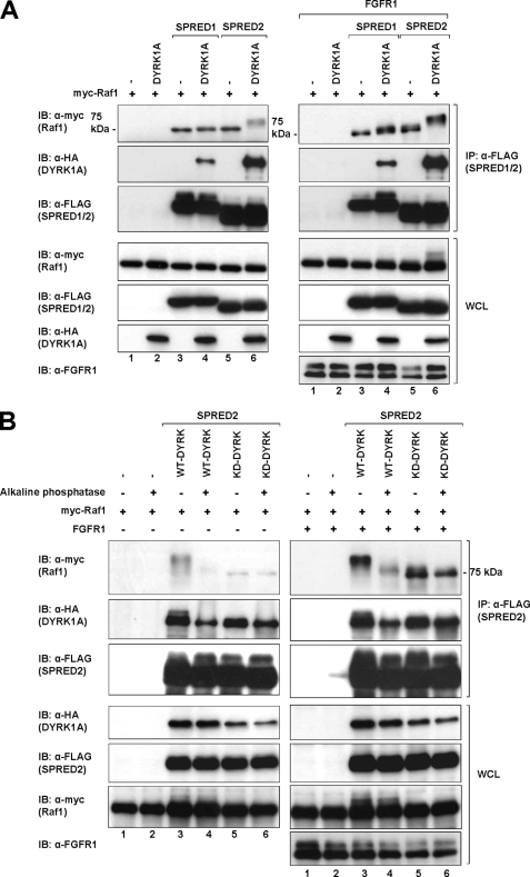
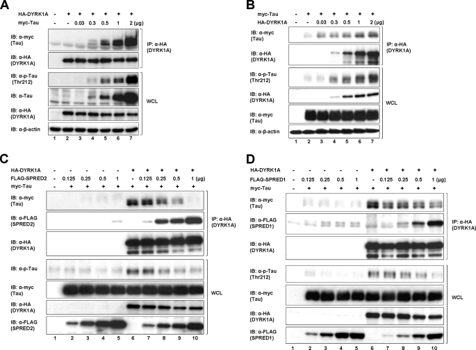
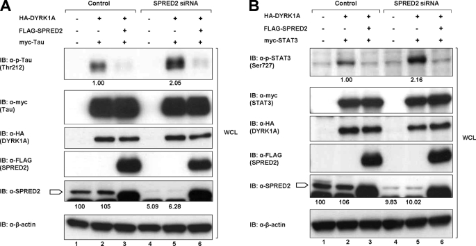
The mammalian SPRED (Sprouty-related protein with an EVH1 domain) proteins include a family of three members, SPRED1-3. Currently, little is known about their biochemistry. The best described, SPRED1, has been shown to inhibit the Ras/ERK pathway downstream of Ras. All three SPREDs have a cysteine-rich domain (CRD) that has high homology to the CRD of the Sprouty family of proteins, several of which are also Ras/ERK inhibitors. In the belief that binding partners would clarify SPRED function, we assayed for their associated proteins. Here, we describe the direct and endogenous interaction of SPRED1 and SPRED2 with the novel kinase, DYRK1A. DYRK1A has become the subject of recent research focus as it plays a central role in Caenorhabditis elegans oocyte maturation and egg activation, and there is strong evidence that it could be involved in Down syndrome in humans. Both SPRED1 and SPRED2 inhibit the ability of DYRK1A to phosphorylate its substrates, Tau and STAT3. This inhibition occurs via an interaction of the CRD of the SPREDs with the kinase domain of DYRK1A. DYRK1A substrates must bind to the kinase to enable phosphorylation, and SPRED proteins compete for the same binding site to modify this process. Our accumulated evidence indicates that the SPRED proteins are likely physiological modifiers of DYRK1A.


Figures

FIGURE 1.
FIGURE 1.
SPRED1 and -2 interact with DYRK1A through the Cys-rich domain. A, schematic diagram showing the domain structures of human Spry2, mouse SPRED1, mouse SPRED2, and mouse DYRK1A. CRD, NLS, KINASE (kinase catalytic domain), PEST (domain rich in proline, glutamate, serine, and threonine), His (histidine repeat sequence), SER/THR (serine/threonine-rich region), EVH1 (Ena-VASP homology-1 domain), and KBD (c-Kit binding domain). B, HEK293 cells were transfected with the indicated plasmids (FLAG-Spry2, FLAG-SPRED1, FLAG-SPRED2, and HA-DYRK1A) or a control pXJ40 vector. 24 h post-transfection cell lysates were subjected to immunoprecipitation (IP) using anti-FLAG or rat anti-HA. The immunoprecipitates were separated on SDS-PAGE and immunoblotted with the antibodies indicated on the left. Whole cell lysates (WCL) were immunoblotted (IB) to verify equal protein expression levels in all the samples tested. C and D, different fragments of SPRED1 (C) and SPRED2 (D) as indicated in A were co-expressed with DYRK1A in 293 cells. Lysates were subjected to immunoprecipitation followed by immunoblotting of the immunoprecipitates and WCL samples with the antibodies indicated to the left. FL, full-length protein; open arrow indicates the immunoglobulin heavy chain (SPRED2 band is the darker band that runs marginally lower than the immunoglobulin heavy chain). Half-open arrow indicates the immunoglobulin light chain (the c-Kit-binding domain band is above the immunoglobulin light chain). There is a faint nonspecific band that runs across the immunoprecipitated DYRK1A in the anti-FLAG immunoprecipitated complex (3rd panel, lanes 7 and 10; the DYRK1A band in these samples show as higher intensity bands).
FIGURE 2.
FIGURE 2.
SPRED1 and -2 interact with the kinase domain of DYRK1A. A, schematic diagram showing full-length and different truncation constructs of DYRK1A that were used in the subsequent studies. B and C, full-length, C-terminal truncated mutants (1–663, 1–649, 1–610, and 1–589) of DYRK1A and full-length SPRED1 (B) and SPRED2 (C) were transfected in 293 cells. Cell lysates were immunoprecipitated (IP) with FLAG or rat HA antibodies. Immunoprecipitates and WCL were resolved by SDS-PAGE and immunoblotted (IB) with the antibodies indicated on the left. Arrow indicates a nonspecific band. The band of the FL DYRK1A protein in the anti-FLAG complex seen in lane 8 (B, 3rd panel) is higher in intensity compared with the nonspecific band. D and E, 293 cells were co-transfected with full-length DYRK1A, the kinase domain of DYRK1A(150–470), and three N-terminal truncates of DYRK1A (150–754, 323–754, and 471–754) as indicated. Cell lysates were immunoprecipitated with anti-FLAG and rat anti-HA, and the precipitated proteins were separated on SDS-PAGE and analyzed by Western blotting techniques with the indicated antibodies. Open arrow indicates the immunoglobulin heavy chain.
FIGURE 3.
FIGURE 3.
SPRED1 and -2 interact endogenously and directly with DYRK1A. A, PC-3 cell lysates were immunoprecipitated with anti-SPRED1 and -2 antibodies, and the immunoprecipitated (IP) proteins and the WCL were subjected to SDS-PAGE and immunoblotted (IB) with anti-DYRK1A antibody. B, PC-3 cell lysates were immunoprecipitated with anti-DYRK1A antibody. The immunoprecipitates and the WCL were separated on SDS-PAGE and analyzed by immunoblotting with anti-SPRED1 and -SPRED2, respectively. Closed triangle indicates DYRK1A band; open triangle indicates SPRED1; block arrow indicates SPRED2. The HA-tagged DYRK1A and FLAG-tagged SPRED1 and -2 in the WCL are used as positive controls to indicate that the correct protein bands are present in the immunocomplex. C and D, direct association of DYRK1A and SPRED1 and -2 in vitro. Binding of in vitro translated HA-tagged DYRK1A to FLAG-tagged SPRED1 (C) and SPRED2 (D) proteins in the transcription and translation (TnT) assay. TnT reaction products and anti-FLAG immunoprecipitates were subjected to SDS-PAGE and immunoblotted with anti-HA and anti-FLAG.
FIGURE 4.
FIGURE 4.
SPRED1 and -2 inhibit DYRK1A-mediated cell proliferation by suppressing p53 deacetylation. A, 293 cells were transfected with combination of FLAG-tagged SPRED1, SPRED2 and HA-tagged DYRK1A. 24 h post-transfection, the cells were treated with 5 μm etoposide, or both 5 μm etoposide and 100 nm harmine for 24 h. Cell viability of the treated cells was analyzed as described under “Experimental Procedures” (n = 3, p < 0.05). B, levels of acetylated p53, total p53, HA-DYRK1A, and FLAG-SPRED1 and -2 in the WCL mentioned in A were determined by Western analysis with the indicated antibodies. Asterisk indicates the quantification results of acetyl-p53. IB, immunoblot.
FIGURE 5.
FIGURE 5.
SPRED1, SPRED2, and Spry2 disrupt the DYRK1A-directed substrate phosphorylation. A, cell lysates from 293 cells transfected with the indicated plasmids (WT-DYRK1A, KD-DYRK1A, and vector control) were subjected to SDS-PAGE and immunoblotted with anti-phospho-Tau Thr-212 (p-Tau Thr-212), anti-Tau, and anti-HA. B, cell lysates from 293 cells transfected with increasing amounts of DYRK1A and a constant amount of Tau were subjected to SDS-PAGE and immunoblotted (IB) with anti-phospho-Tau (p-Tau Thr-212), anti-Tau, and anti-HA. C, 293 cells were transfected with constant amount of WT-DYRK1A and Tau. The cells were treated with increasing concentrations of harmine for 30 min after 24 h post-transfection. The cell lysates were subjected to SDS-PAGE and immunoblotted with the antibodies indicated on the left. D, cell lysates from 293 cells co-transfected with WT-, KD-DYRK1A, Spry2, and Tau were subjected to SDS-PAGE and immunoblotted with the indicated antibodies. E, cell lysates from 293 cells transfected with combinations of WT-, KD-DYRK1A, SPRED1, SPRED2, and Tau were subjected to SDS-PAGE and immunoblotted with the antibodies shown on the left. The relative quantities of phospho-Tau for each lane in B–E (top panel) were indicated in the bar charts (supplemental Fig. 5, A–D, respectively). F, cell lysates from 293 cells co-transfected with WT-, KD-DYRK1A, SPRED1, SPRED2, and STAT3 were subjected to SDS-PAGE and immunoblotted with the indicated antibodies. The relative levels of phospho-STAT3 are indicated in the bar chart (supplemental Fig. 5E).
FIGURE 6.
FIGURE 6.
SPRED proteins do not inhibit the kinase activity of DYRK1A. A, HEK293 cells were transfected with the indicated plasmids (FLAG-SPRED1/2, HA-DYRK1A, and myc-Raf1) or a control pXJ40 vector. 24 h post-transfection cell lysates were subjected to immunoprecipitation (IP) using anti-FLAG. The immunoprecipitates were separated on SDS-PAGE and immunoblotted with the antibodies indicated on the left. Whole cell lysates (WCL) were immunoblotted (IB) to verify equal protein expression levels in all the samples tested. B, HEK293 cells were transfected with the indicated plasmids (FLAG-SPRED2, HA-WT-DYRK1A or HA-KD-DYRK1A, and myc-Raf1). Lysates obtained using the above setup were treated with alkaline phosphatase or BSA as a control and resolved by SDS-PAGE as mentioned in A.
FIGURE 7.
FIGURE 7.
SPRED1 and -2 compete with Tau for binding to DYRK1A. A, HEK293 cells were transfected with constant amount of the HA-DYRK1A plasmids and an increasing amount of myc-Tau. Lysates were immunoprecipitated (IP) using anti-HA and subjected to SDS-PAGE immunoblot (IB) analysis as indicated in Fig. 1. B, HEK293 cells were transfected with constant amounts of myc-Tau and an increasing amount of HA-DYRK1A. Lysates were immunoprecipitated using anti-HA, separated on SDS-PAGE, and immunoblotted using the antibodies indicated on the left. C, HEK293 cells were transfected with constant amounts of HA-DYRK1A and myc-Tau together with an increasing amount of FLAG-SPRED2 plasmids. Lysates were immunoprecipitated using anti-HA and subjected to SDS-PAGE and immunoblotted analysis as indicated. D, HEK293 cells were treated as described in C using FLAG-SPRED1 instead of FLAG-SPRED2. Cell lysates were likewise treated and analyzed as in C.
FIGURE 8.
FIGURE 8.
Knockdown of SPRED2 increases the DYRK1A-dependent phosphorylation of Tau and STAT3. A, 293 cells were transfected with vector control, nonspecific siRNA, or a pool of four SPRED2 siRNAs using Lipofectamine 2000 as described under “Experimental Procedures.” 48 h post-transfection of the siRNA, the cells were transfected with the other cDNA plasmids as indicated. Cells were harvested 24 h after the second transfection. Cell lysates were separated on SDS-PAGE and immunoblotted (IB) with antibodies indicated on the left. B, 293 cells were treated in the same way as described in A, but with the replacement of myc-Tau with myc-STAT3. Cell lysates were subsequently treated similarly as in A. Block arrow indicates SPRED2.

Similar articles

Cited by

References

    1. Schlessinger J. (2000) Cell 103, 211–225 - PubMed
    1. Schlessinger J. (2002) Cell 110, 669–672 - PubMed
    1. Zwick E., Bange J., Ullrich A. (2001) Endocr.-Relat. Cancer 8, 161–173 - PubMed
    1. Shaw R. J., Cantley L. C. (2006) Nature 441, 424–430 - PubMed
    1. Midgley R. S., Kerr D. J. (2002) Crit. Rev. Oncol. Hematol. 44, 109–120 - PubMed

Publication types

MeSH terms