. 2010 Nov 5;285(45):34319-29.
doi: 10.1074/jbc.M110.162925. Epub 2010 Aug 23.

Double strand break unwinding and resection by the mycobacterial helicase-nuclease AdnAB in the presence of single strand DNA-binding protein (SSB)

Affiliations

Double strand break unwinding and resection by the mycobacterial helicase-nuclease AdnAB in the presence of single strand DNA-binding protein (SSB)

Mihaela-Carmen Unciuleac et al. J Biol Chem. .

Abstract

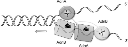
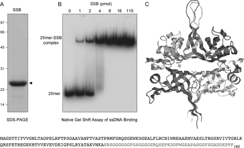
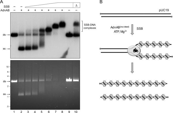
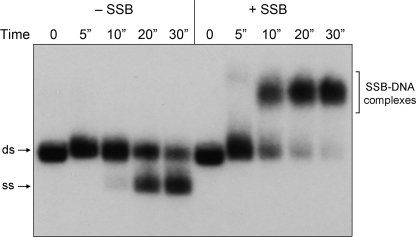
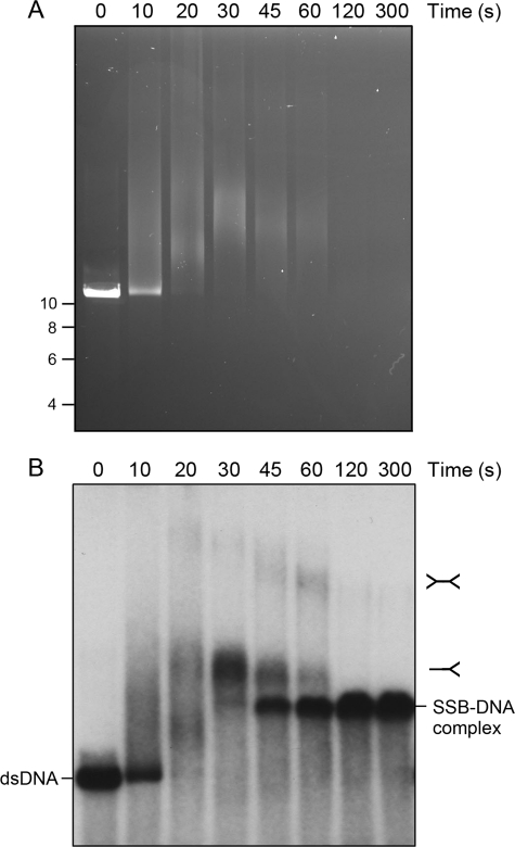
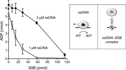
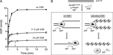
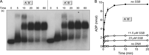
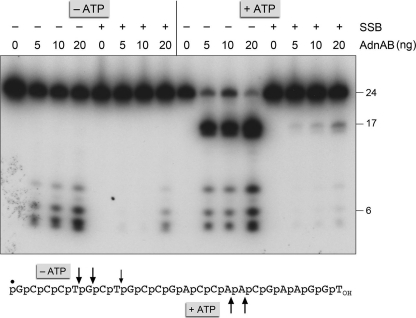
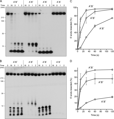
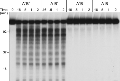
Mycobacterial AdnAB is a heterodimeric DNA helicase-nuclease and 3' to 5' DNA translocase implicated in the repair of double strand breaks (DSBs). The AdnA and AdnB subunits are each composed of an N-terminal motor domain and a C-terminal nuclease domain. Inclusion of mycobacterial single strand DNA-binding protein (SSB) in reactions containing linear plasmid dsDNA allowed us to study the AdnAB helicase under conditions in which the unwound single strands are coated by SSB and thereby prevented from reannealing or promoting ongoing ATP hydrolysis. We found that the AdnAB motor catalyzed processive unwinding of 2.7-11.2-kbp linear duplex DNAs at a rate of ∼250 bp s(-1), while hydrolyzing ∼5 ATPs per bp unwound. Crippling the AdnA phosphohydrolase active site did not affect the rate of unwinding but lowered energy consumption slightly, to ∼4.2 ATPs bp(-1). Mutation of the AdnB phosphohydrolase abolished duplex unwinding, consistent with a model in which the "leading" AdnB motor propagates a Y-fork by translocation along the 3' DNA strand, ahead of the "lagging" AdnA motor domain. By tracking the resection of the 5' and 3' strands at the DSB ends, we illuminated a division of labor among the AdnA and AdnB nuclease modules during dsDNA unwinding, whereby the AdnA nuclease processes the unwound 5' strand to liberate a short oligonucleotide product, and the AdnB nuclease incises the 3' strand on which the motor translocates. These results extend our understanding of presynaptic DSB processing by AdnAB and engender instructive comparisons with the RecBCD and AddAB clades of bacterial helicase-nuclease machines.


Figures

FIGURE 1.
FIGURE 1.
Model for division of labor during AdnAB unwinding and processing DSB ends. Binding of AdnAB to the DSB end initiates ATP-dependent translocation of the tandem motor in the 3′ to 5′ direction along one of the DNA strands, with the dominant AdnB motor domain in the vanguard. Unwinding of the DNA duplex by the advancing motor results in pumping of the 3′ ssDNA strand into the AdnB nuclease domain and threading of the 5′ ssDNA strand through the AdnA nuclease domain. The model is based on experiments in Ref. and is interrogated further by this study.
FIGURE 2.
FIGURE 2.
Recombinant M. smegmatis SSB. A, purification. An aliquot (10 μg) of the Superdex-200 fraction of recombinant SSB was analyzed by SDS-PAGE. The Coomassie Blue-stained gel is shown, with the positions and sizes (kDa) of marker polypeptide indicated on the left. B, DNA binding. Reaction mixtures (10 μl) containing 20 mm Tris-HCl, pH 8.0, 0.1 μm (1 pmol) 5′ 32P-labeled 25-mer ssDNA (5′-CCAGAACAAACTCATCGTCGTCTAC), 5% glycerol, and increasing amounts of SSB as specified (expressed as pmol of SSB monomers) were incubated for 30 min at 4 °C. The mixtures were adjusted to 15% glycerol and then analyzed by electrophoresis through a 15-cm native 6% polyacrylamide gel containing 22.5 mm Tris borate, 0.625 mm EDTA. The gel was run at 110 V in the cold room for 3 h and then dried under vacuum on DE81 paper. The free 32P-labeled DNA and slower migrating protein·[32P]DNA complexes were visualized by autoradiography of the dried gel. C, crystal structure of the M. smegmatis SSB-(1–120) tetramer is shown (Protein Data Bank code 1X3E); the terminal residues of the SSB-(1–120) protomer at top right are indicated by N and C*, respectively. The primary structure of the M. smegmatis SSB polypeptide is shown at the bottom of the figure.
FIGURE 3.
FIGURE 3.
SSB captures the strands unwound by the AdnAB motor. A, reaction mixtures (10 μl) containing 20 mm Tris-HCl, pH 8.0, 1 mm DTT, 2 mm MgCl2, 1 mm ATP, 200 ng of 5′ 32P-labeled pUC19 DNA (BamHI-digested; 230 fmol of DSB ends), 1.06 pmol of nuclease-dead AdnAB (where indicated by +), and increasing amounts of SSB, either 3.6 (lane 3), 7.2 (lane 4), 14.4 (lane 5), 28.8 (lane 6), 57.5 (lane 7), or 115 (lanes 8 and 9) pmol of SSB monomer, were incubated for 10 min at 37 °C. The reactions were quenched by adjusting the mixtures to 50 mm EDTA, 15% glycerol, 0.125% Orange-G dye. The mixture in lane 10, lacking AdnAB, was heated for 5 min at 95 °C (Δ) and then chilled on ice prior to adding SSB (115 pmol). The reaction products were analyzed by electrophoresis through a 0.8% native agarose gel in 50 mm Tris acetate, 2.5 mm EDTA. After visualizing the DNA by staining with ethidium bromide (bottom panel), the gel was dried under vacuum on DE81 paper, and radiolabeled DNA was visualized by autoradiography of the dried gel (top panel). B, reaction scheme is illustrated, whereby SSB tetramers bind to the single-stranded DNA formed in the wake of the advancing AdnAB motor to yield SSB·ssDNA complexes as end products. ds, double strand; ss, single strand.
FIGURE 4.
FIGURE 4.
Estimation of the rate of pUC19 unwinding by the AdnAB motor. Reaction mixtures (50 μl) contained 20 mm Tris-HCl, pH 8.0, 1 mm DTT, 2 mm MgCl2, 1 mm ATP, 1 μg of 5′ 32P-labeled pUC19 DNA (BamHI-digested; 1.14 pmol of DSB ends), either no SSB (−SSB) or 11.5 μm SSB monomers (+SSB), and 3.8 pmol (76 nm) of nuclease-dead AdnAB. The reactions were initiated by adding AdnAB to reaction mixtures prewarmed to 37 °C. Aliquots (10 μl) were then withdrawn after incubation at 37 °C for the times specified, and the reactions were quenched immediately with EDTA. The time 0 samples were taken prior to adding AdnAB. The products were analyzed by native agarose gel electrophoresis. Radiolabeled DNA was visualized by autoradiography of the dried gel. ds, double strand; ss, single strand.
FIGURE 5.
FIGURE 5.
Unwinding of an 11.2-kb dsDNA by the AdnAB motor. Reaction mixtures (80 μl) contained 20 mm Tris-HCl, pH 8.0, 1 mm DTT, 2 mm MgCl2, 1 mm ATP, 0.8 μg of 5′ 32P-labeled pUC-H DNA (SmaI-digested; 910 fmol of DSB ends), either no SSB (−SSB) or 23 μm SSB monomers (+SSB), and 9.7 pmol (120 nm) of nuclease-dead AdnAB. Aliquots (10 μl) were withdrawn after incubation at 37 °C for the times specified and then quenched immediately with EDTA. The reaction products were analyzed by electrophoresis through a 0.6% native agarose gel. DNA was visualized by staining with ethidium bromide (A) before the gel was dried, and radiolabeled DNA was visualized by autoradiography (B). The positions and sizes (kbp) of linear dsDNA markers are indicated on the left in A.
FIGURE 6.
FIGURE 6.
SSB inhibits ssDNA-triggered ATP hydrolysis by the AdnAB motor. Reaction mixtures (10 μl) containing 20 mm Tris-HCl, pH 8.0, 0.5 mm DTT, 1 mm MgCl2, 1 mm [α-32P]ATP, 1 or 3 μm 24-mer ssDNA (5′-GCCCTGCTGCCGACCAACGAAGGT) as specified, 8.5 nm nuclease-dead AdnAB, and increasing amounts of SSB polypeptide as specified (pmol of SSB monomer) were incubated for 10 min at 37 °C. The reactions were quenched with 2 μl of 5 m formic acid. Aliquots (2 μl) were analyzed by ascending PEI-cellulose TLC with 0.45 m ammonium sulfate as the mobile phase. [α-32P]ATP and [α-32P]ADP were quantified by scanning the TLC plate with a Fujix BAS2500 imager. The extents of [α-32P]ADP formation are plotted as a function of input SSB. Each datum is the average of three experiments (±S.E.). The schematics at right illustrate how ssDNA-triggered ATP hydrolysis is coupled to translocation of the AdnAB motor and how sequestration of the ssDNA cofactor by SSB progressively masks its ability to trigger the motor ATPase.
FIGURE 7.
FIGURE 7.
Estimating the coupling of ATP hydrolysis and duplex unwinding. A, reaction mixtures (80 μl) containing 20 mm Tris-HCl, pH 8.0, 1 mm DTT, 2 mm MgCl2, 1 mm [α-32P]ATP, 1.6 μg of pUC19 DNA (BamHI-digested; 1.82 pmol of DSB ends), 9.7 pmol (120 nm) of nuclease-dead AdnAB, and either no SSB, 11.5 μm SSB, or 23 μm SSB as specified were incubated at 37 °C. Aliquots (10 μl, containing 10 nmol input ATP) were withdrawn at the times specified and quenched with formic acid. The products were analyzed by PEI-cellulose TLC. The extents of conversion of [α-32P]ATP to [α-32P]ADP are plotted as a function of time. Each datum is the average of three experiments ±S.E. B, illustration of the ATP reaction outcomes in the absence of SSB, where unwound ssDNA is available to trigger ongoing ATP hydrolysis, versus in the presence of saturating SSB, where the unwound SSB-coated DNA strands are largely unavailable to serve as platforms for further ATP hydrolysis.
FIGURE 8.
FIGURE 8.
Duplex unwinding by AdnAB with a crippled AdnA phosphohydrolase module. A, reaction mixtures (60 μl) containing 20 mm Tris-HCl, pH 8.0, 1 mm DTT, 2 mm MgCl2, 1 mm ATP, 1.2 μg of 5′ 32P-labeled pUC19 DNA (BamHI-digested; 1.37 pmol DSB ends), 11.5 μm SSB, and 7.3 pmol (120 nm) of nuclease-dead AdnAB heterodimers with either two wild-type motor domains (A+B+) or a wild-type AdnB motor plus a AdnAD255A mutant motor (AB+) were incubated at 37 °C. Aliquots (10 μl) were withdrawn at the times specified and quenched with EDTA. The mixtures were analyzed by native agarose gel electrophoresis, and the radiolabeled DNA was visualized by autoradiography of the dried gel. B, reaction mixtures (80 μl) containing 20 mm Tris-HCl, pH 8.0, 1 mm DTT, 2 mm MgCl2, 1 mm [α-32P]ATP, 1.6 μg of pUC19 DNA (BamHI-digested; 1.82 pmol of DSB ends), 9.7 pmol (120 nm) of nuclease-dead AdnAB+ motor, and either no SSB, 11.5 μm SSB, or 23 μm SSB as specified were incubated at 37 °C. The “no DNA” control reaction mixture contained AdnAB+ motor, 23 μm SSB, and other components but no pUC19 DNA. Aliquots (10 μl, containing 10 nmol of input ATP) were withdrawn at the times specified and quenched with formic acid. The products were analyzed by PEI-TLC. The extents of conversion of [α-32P]ATP to [α-32P]ADP are plotted as a function of time. Each datum is the average of three experiments ±S.E.
FIGURE 9.
FIGURE 9.
SSB inhibits the ssDNA nuclease activities of AdnAB. Reaction mixtures (10 μl) containing 20 mm Tris-HCl, pH 8.0, 0.5 mm DTT, 2 mm MgCl2, 1 mm ATP, 100 nm (1 pmol) 5′ 32P-labeled 24-mer ssDNA (shown at bottom with the 5′-label denoted by ●), 4 pmol of SSB (where indicated by +), and AdnAB as specified (5, 10 or 20 ng of AdnB subunit, corresponding to 50, 100, or 200 fmol of AdnAB heterodimer) were incubated for 5 min at 37 °C. The reactions were quenched with formamide/EDTA, heated for 5 min at 95 °C, and then analyzed by electrophoresis through a 15-cm 18% polyacrylamide gel containing 7 m urea, 45 mm Tris borate, 1.25 mm EDTA. An autoradiograph of the gel is shown; oligonucleotide sizes are noted on the right. The principal sites of AdnAB incision of the 24-mer DNA in the absence of ATP are indicated by arrows above the oligonucleotide sequence; the cleavage sites induced by ATP are indicated below.
FIGURE 10.
FIGURE 10.
AdnAB nuclease action at 5′-labeled DSB ends. Reaction mixtures (50 μl) containing 20 mm Tris-HCl, pH 8.0, 1 mm DTT, 2 mm MgCl2, 1 mm ATP, 1 μg of 5′ 32P-labeled pUC19 DNA (BamHI-digested; 1.14 pmol DSB ends), and 6 pmol (120 nm) of AdnAB heterodimers with either two wild-type nuclease domains (A+B+), a wild-type AdnB nuclease plus an inactive AdnAD934A mutant nuclease (AB+), a wild-type AdnA nuclease plus an inactive AdnBD1014A mutant nuclease (A+B), or two inactive mutant nucleases (AB), and either no SSB (A) or 23 μm SSB (B) were incubated at 37 °C. The reactions were initiated by adding AdnAB to reaction mixtures prewarmed to 37 °C. Aliquots (10 μl) were withdrawn at the times specified and quenched with formamide/EDTA. The mixtures were heated for 5 min at 95 °C and then analyzed by electrophoresis through 15-cm 15% polyacrylamide gels containing 7 m urea, 45 mm Tris borate, 1.25 mm EDTA. Autoradiographs of the gels are shown. The positions and sizes (in nucleotides) of heat-denatured 5′-labeled DNA markers (generated by restriction endonuclease digestion of the 5′ 32P-labeled pUC19 DNA substrate) are indicated on the left. C and D, extents of 5′ strand cleavage to generate short oligonucleotides were quantified by scanning the gels with a phosphorimager and are plotted as a function of time for reactions containing no SSB (C) or 23 μm SSB (D). Each datum is the average of two experiments; error bars indicate the range.
FIGURE 11.
FIGURE 11.
AdnAB nuclease action at 3′-labeled DSB ends. Reaction mixtures (50 μl) containing 20 mm Tris-HCl, pH 8.0, 1 mm DTT, 2 mm MgCl2, 1 mm ATP, 1 μg of 3′ 32P-labeled pUC19 DNA (EcoRI-digested and 3′-labeled with [32P]dAMP; 1.14 pmol of DSB ends), 23 μm SSB, and 6 pmol (120 nm) of AdnAB heterodimers as indicated were incubated at 37 °C. Aliquots (10 μl) were withdrawn at the times specified and quenched with formamide/EDTA. The mixtures were heated for 5 min at 95 °C and then analyzed by electrophoresis through a 15-cm 15% polyacrylamide gel containing 7 m urea, 45 mm Tris borate, 1.25 mm EDTA. An autoradiograph of the gel is shown. The positions and sizes (in nucleotides) of heat-denatured 3′-labeled DNA markers (generated by restriction endonuclease digestion of the 3′ 32P-labeled pUC19 DNA substrate) are indicated on the left.
FIGURE 12.
FIGURE 12.
AdnAB product analysis by alkaline-agarose gel electrophoresis. Reaction mixtures (25 μl) containing 20 mm Tris-HCl, pH 8.0, 1 mm DTT, 2 mm MgCl2, 1 mm ATP, either 0.5 μg of 5′ 32P-labeled pUC19 (BamHI-digested; 570 fmol of DSB ends; A) or 0.5 μg of 3′ 32P-labeled pUC19 (EcoRI-digested; 570 fmol of DSB ends; B), 3 pmol (120 nm) of AdnAB heterodimers as indicated, and either no SSB (−) or 23 μm SSB (+) were incubated at 37 °C. After 10 or 30 s, 10-μl aliquots were withdrawn and quenched immediately by adjustment to 72 mm EDTA and 0.85% SDS. The mixtures were supplemented with proteinase K (0.8 units; Sigma) and incubated at 37 °C for 15 min. The samples were then adjusted to 100 mm NaOH, 4% glycerol, and 1% bromphenol blue and analyzed by electrophoresis (10 h at 20 V at room temperature) through a 1.5% alkaline-agarose gel in 50 mm NaOH, 2 mm EDTA. The gel was rinsed twice for 10 min in 5% trichloroacetic acid and then dried under vacuum on DE81 paper. Labeled DNA was visualized by autoradiography of the dried gels. The positions and sizes (in nucleotides) of heat-denatured 5′- or 3′-labeled DNA markers (generated by restriction endonuclease digestion of the 32P-labeled pUC19 DNA substrates) are indicated on the right.

Similar articles

Cited by

References

    1. Cromie G. A. (2009) J. Bacteriol. 191, 5076–5084 - PMC - PubMed
    1. Singleton M. R., Dillingham M. S., Gaudier M., Kowalczykowski S. C., Wigley D. B. (2004) Nature 432, 187–193 - PubMed
    1. Dillingham M. S., Kowalczykowski S. C. (2008) Microbiol. Mol. Biol. Rev. 72, 642–671 - PMC - PubMed
    1. Haijema B. J., Venema G., Kooistra J. (1996) J. Bacteriol. 178, 5086–5091 - PMC - PubMed
    1. Haijema B. J., Meima R., Kooistra J., Venema G. (1996) J. Bacteriol. 178, 5130–5137 - PMC - PubMed

Publication types

MeSH terms

LinkOut - more resources