

Axolotl Nanog activity in mouse embryonic stem cells demonstrates that ground state pluripotency is conserved from urodele amphibians to mammals
- PMID: 20736286
- PMCID: PMC2926951
- DOI: 10.1242/dev.049262
Axolotl Nanog activity in mouse embryonic stem cells demonstrates that ground state pluripotency is conserved from urodele amphibians to mammals
Abstract
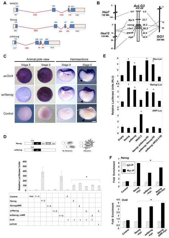
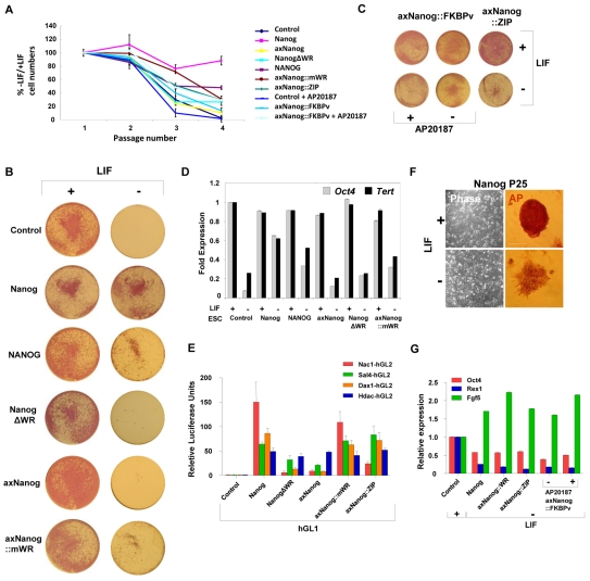
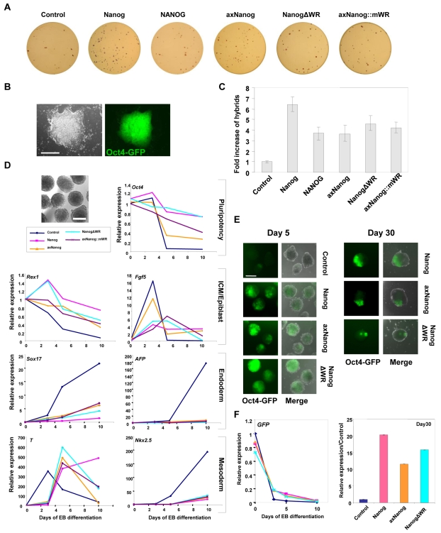
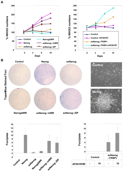
Cells in the pluripotent ground state can give rise to somatic cells and germ cells, and the acquisition of pluripotency is dependent on the expression of Nanog. Pluripotency is conserved in the primitive ectoderm of embryos from mammals and urodele amphibians, and here we report the isolation of a Nanog ortholog from axolotls (axNanog). axNanog does not contain a tryptophan repeat domain and is expressed as a monomer in the axolotl animal cap. The monomeric form is sufficient to regulate pluripotency in mouse embryonic stem cells, but axNanog dimers are required to rescue LIF-independent self-renewal. Our results show that protein interactions mediated by Nanog dimerization promote proliferation. More importantly, they demonstrate that the mechanisms governing pluripotency are conserved from urodele amphibians to mammals.
Figures
Similar articles
-
Conserved long noncoding RNAs transcriptionally regulated by Oct4 and Nanog modulate pluripotency in mouse embryonic stem cells.RNA. 2010 Feb;16(2):324-37. doi: 10.1261/rna.1441510. Epub 2009 Dec 21. RNA. 2010. PMID: 20026622 Free PMC article.
-
[OCT4 and NANOG are the key genes in the system of pluripotency maintenance in mammalian cells].Genetika. 2008 Dec;44(12):1589-608. Genetika. 2008. PMID: 19178078 Review. Russian.
-
The Oct4 homologue PouV and Nanog regulate pluripotency in chicken embryonic stem cells.Development. 2007 Oct;134(19):3549-63. doi: 10.1242/dev.006569. Development. 2007. PMID: 17827181
-
Control of ground-state pluripotency by allelic regulation of Nanog.Nature. 2012 Feb 12;483(7390):470-3. doi: 10.1038/nature10807. Nature. 2012. PMID: 22327294
-
Concise review: pursuing self-renewal and pluripotency with the stem cell factor Nanog.Stem Cells. 2013 Jul;31(7):1227-36. doi: 10.1002/stem.1384. Stem Cells. 2013. PMID: 23653415 Free PMC article. Review.
Cited by
-
Targeted expression profiling reveals distinct stages of early canine fibroblast reprogramming are regulated by 2-oxoglutarate hydroxylases.Stem Cell Res Ther. 2020 Dec 9;11(1):528. doi: 10.1186/s13287-020-02047-1. Stem Cell Res Ther. 2020. PMID: 33298190 Free PMC article.
-
Primordial germ cells: the first cell lineage or the last cells standing?Development. 2015 Aug 15;142(16):2730-9. doi: 10.1242/dev.113993. Development. 2015. PMID: 26286941 Free PMC article. Review.
-
Reprogramming capacity of Nanog is functionally conserved in vertebrates and resides in a unique homeodomain.Development. 2011 Nov;138(22):4853-65. doi: 10.1242/dev.068775. Development. 2011. PMID: 22028025 Free PMC article.
-
Characterization of Danio rerio Nanog and functional comparison to Xenopus Vents.Stem Cells Dev. 2012 May 20;21(8):1225-38. doi: 10.1089/scd.2011.0285. Epub 2011 Oct 3. Stem Cells Dev. 2012. PMID: 21967637 Free PMC article.
-
On the origin and evolutionary history of NANOG.PLoS One. 2014 Jan 17;9(1):e85104. doi: 10.1371/journal.pone.0085104. eCollection 2014. PLoS One. 2014. PMID: 24465486 Free PMC article.
References
-
- Anderson J. S., Reisz R. R., Scott D., Frobisch N. B., Sumida S. S. (2008). A stem batrachian from the early Permian of Texas and the origin of frogs and salamanders. Nature 453, 515-518 - PubMed
-
- Bachvarova R. F., Masi T., Drum M., Parker N., Mason K., Patient R., Johnson A. D. (2004). Gene expression in the axolotl germ line: Axdazl, Axvh, Axoct-4, and Axkit. Dev. Dyn. 231, 871-880 - PubMed
-
- Bachvarova R. F., Crother B. I., Johnson A. D. (2009a). Evolution of germ cell development in tetrapods: comparison of urodeles and amniotes. Evol. Dev. 11, 603-609 - PubMed
-
- Bachvarova R. F., Crother B. I., Manova K., Chatfield J., Shoemaker C. M., Crews D. P., Johnson A. D. (2009b). Expression of Dazl and Vasa in turtle embryos and ovaries: evidence for inductive specification of germ cells. Evol. Dev. 11, 525-534 - PubMed
Publication types
MeSH terms
Substances
Grants and funding
LinkOut - more resources
Full Text Sources
Other Literature Sources
Research Materials
Miscellaneous
