. 2010 Sep;137(18):3107-17.
doi: 10.1242/dev.046573.

HLH54F is required for the specification and migration of longitudinal gut muscle founders from the caudal mesoderm of Drosophila

Affiliations

HLH54F is required for the specification and migration of longitudinal gut muscle founders from the caudal mesoderm of Drosophila

Afshan Ismat et al. Development. 2010 Sep.

Abstract

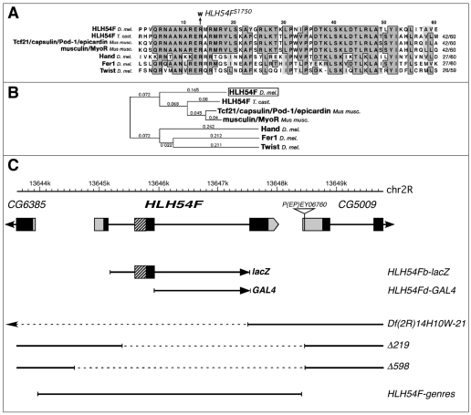
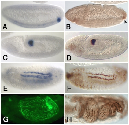
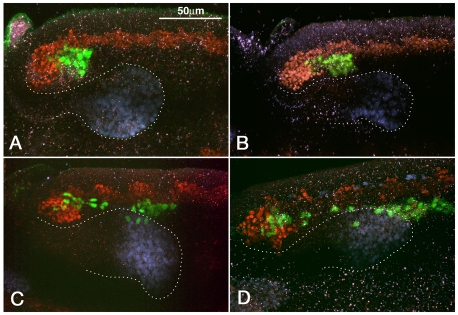
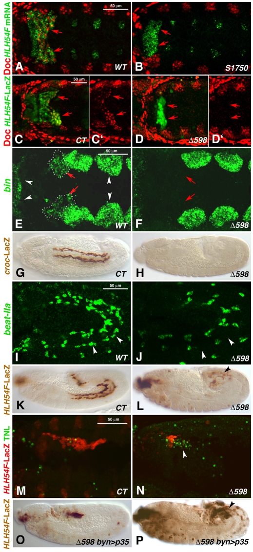
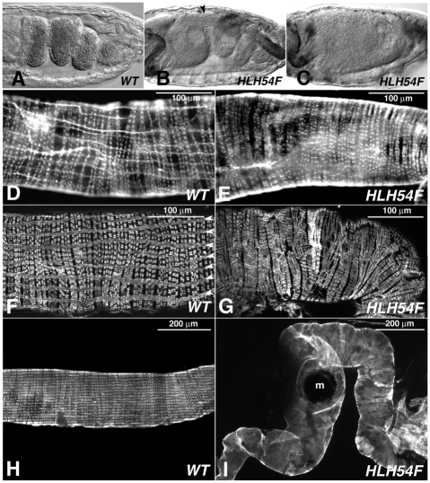
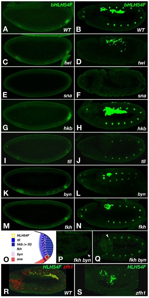
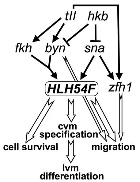
HLH54F, the Drosophila ortholog of the vertebrate basic helix-loop-helix domain-encoding genes capsulin and musculin, is expressed in the founder cells and developing muscle fibers of the longitudinal midgut muscles. These cells descend from the posterior-most portion of the mesoderm, termed the caudal visceral mesoderm (CVM), and migrate onto the trunk visceral mesoderm prior to undergoing myoblast fusion and muscle fiber formation. We show that HLH54F expression in the CVM is regulated by a combination of terminal patterning genes and snail. We generated HLH54F mutations and show that this gene is crucial for the specification, migration and survival of the CVM cells and the longitudinal midgut muscle founders. HLH54F mutant embryos, larvae, and adults lack all longitudinal midgut muscles, which causes defects in gut morphology and integrity. The function of HLH54F as a direct activator of gene expression is exemplified by our analysis of a CVM-specific enhancer from the Dorsocross locus, which requires combined inputs from HLH54F and Biniou in a feed-forward fashion. We conclude that HLH54F is the earliest specific regulator of CVM development and that it plays a pivotal role in all major aspects of development and differentiation of this largely twist-independent population of mesodermal cells.


Figures

Fig. 1.
Fig. 1.
HLH54F protein sequence comparison, gene locus and mutations. (A) Alignment of the bHLH domain of HLH54F with the most closely related bHLH domains from mouse, Tribolium and Drosophila. The R-to-W exchange in the predicted protein from EMS allele HLH54FS1750 is indicated. (B) Phylogenetic analysis of data from A. (C) HLH54F gene map showing translated regions (black), the bHLH domain (hatched) and UTRs (gray). Beneath are shown the genomic regions used for the lacZ construct, GAL4 construct and the genomic rescue construct HLH54F-genres, as well as the molecularly defined breakpoints (dashed lines) of Df(2R)14H10W-21 and the P-excision alleles HLH54FΔ219 and HLH54FΔ598.
Fig. 2.
Fig. 2.
An HLH54F-lacZ reporter recapitulates the expression of HLH54F. Wild-type Drosophila embryos hybridized with HLH54F digoxygenin RNA (A,C,E,G) and comparable HLH54Fb-lacZ embryos stained with β-galactosidase (β-gal) antibodies (B,D,F,H). (A,B) Blastoderm stage embryo (stage 5, A) and embryo at early gastrulation (stage 6, B). (C,D) Stage 9 embryos during germ band elongation. (E,F) In stage 13 embryos, the cells expressing HLH54F mRNA (E) and β-gal (F) have migrated anteriorly and spread along either side of trunk visceral mesoderm. (G) Stage 15 embryo with fluorescent detection of HLH54F mRNA, showing newly formed and elongating syncytia distributed over the midgut. (H) Stage 16 embryo showing HLH54F-lacZ-stained longitudinal muscle fibers.
Fig. 3.
Fig. 3.
Early phase of migration of caudal visceral mesoderm cells. Sagittal views of posterior portion of elongated germ bands of HLH54Fb-lacZ Drosophila embryos stained with antibodies against β-gal (green; CVM), Twist (red; trunk mesoderm) and Hb9 (blue; midgut endoderm). (A) In stage 9 embryos, the posterior portion of the mesoderm bends around internally such that the CVM becomes positioned internally and slightly more anteriorly. (B) At stage 10, the CVM separates from the remaining mesoderm. (C) At late stage 10, individual cells leave the CVM clusters and start migrating anteriorly between posterior midgut (dotted outline) and trunk mesoderm. (D)By the end of stage 11, most CVM cells have surpassed the migrating endoderm anteriorly and are associated with trunk mesoderm.
Fig. 4.
Fig. 4.
Embryonic HLH54F mutant phenotypes. (A,C,C′,E,G,I,K,M) Wild-type (WT) or control (CT) Drosophila embryos and (B,D,D′,F,H,J,L,N-P) HLH54F mutant embryos (alleles as indicated). (A-F) Dorsal views; (G-P) lateral views. (A) Stage 10 WT embryo stained for Doc protein in bilateral CVM clusters (arrows, red) and HLH54F mRNA (green). (B) HLH54FS1750 embryo stained as in A, which lacks Doc signals specifically in the CVM areas marked by mutant HLH54F mRNA (arrows). (C,C′) Stage 10 HLH54Fb-lacZ embryo stained for Doc protein (red, shown singly in C′) and β-gal (green) in CVM (arrows). (D,D′) HLH54FΔ598 embryo with HLH54Fb-lacZ stained as in C is missing Doc expression in β-gal-stained caudal mesoderm. (E) Late stage 10 WT embryo stained for bin mRNA in the CVM (red arrows, dotted outlines) and in TVM and HVM primordia (white arrowheads). (F) HLH54FΔ598 mutant, in which bin mRNA is missing in corresponding areas. (G) Stage 14 WT embryo expressing croc-lacZ in migrating CVM cells. (H) HLH54F; croc-lacZ mutant without croc-lacZ expression. (I) Stage 12 embryo showing beat-IIa mRNA in migrating CVM cells adjacent to the TVM (arrowheads) and in unidentified cells. (J) HLH54FΔ598 mutant embryo without beat-IIa mRNA signals adjacent to the TVM. (K) Stage 12 control embryo expressing HLH54Fb-lacZ in the migrating CVM. (L) Stage 12 HLH54FΔ598 mutant embryo expressing HLH54Fb-lacZ, showing a large decrease in the number of positive cells, which are not migrating (arrowhead). (M) Early stage 12 HLH54Fb-lacZ control embryo stained for β-gal (CVM, red) and by TUNEL assay (green), which shows few apoptotic cells in CVM areas. (N) Stage 12 HLH54FΔ598, HLH54Fb-lacZ mutant embryo with many TUNEL-positive cells (arrowhead). (O) Stage 11 HLH54FΔ598 mutant embryo expressing HLH54Fb-lacZ and byn-GAL4-driven anti-apoptotic p35 with normal numbers of internalized lacZ-positive cells (see Fig. 2D). (P) Stage 12 HLH54FΔ598 mutant embryo with apoptosis inhibition as in O and an increased number of HLH54Fb-lacZ-containing cells as compared with mutants without forced p35 expression (see L), which fail to migrate and differentiate.
Fig. 5.
Fig. 5.
HLH54F mutant phenotypes of embryonic midgut and midgut musculature in larvae and adults. (A) Stage 15 wild-type (WT) Drosophila embryo with constricted and looping midgut. (B) Stage 15 HLH54FS1750/HLH54FΔ598 embryo with incomplete midgut constriction (arrowhead); also stained for GFP reporter for foregut and hindgut muscles, which are unaffected. (C) HLH54F mutant embryo as in B without any midgut constrictions. (D) Section of WT third-instar larval midgut stained with Rhodamine-conjugated phalloidin to visualize both the circular and the longitudinal visceral muscles. (E) Homozygous HLH54FΔ598 third-instar larval gut with absent longitudinal visceral muscles. (F) Section of WT adult midgut stained for Tropomyosin, showing regularly spaced longitudinal and circular muscle fibers. (G) Adult midgut section from a HLH54FS1750/HLH54FΔ589 fly stained for Tropomyosin, showing the lack of longitudinal midgut muscles and circular muscles with irregular morphologies and interruptions. (H,I) WT (H, from F) and mutant (I, from G) adult midguts at lower magnification. Unlike the smooth and linear surface of WT midguts, midguts from mutants have an undulating surface and are curled. m, melanotic mass.
Fig. 6.
Fig. 6.
HLH54F and Bin as co-regulators of a caudal visceral mesoderm-specific enhancer from the Doc locus. (A-H) lacZ (A-D) or nuclear GFP (E-H) reporter gene expression from DocF4s1 and derivatives is shown in early stage 12 Drosophila embryos as detected with anti-β-gal (diaminobenzidine) and anti-GFP (fluorescence) antibodies (dorsal views). (A) DocF4s1-lacZ in a wild-type background showing expression in bilateral CVM clusters at the beginning of cell migration (right). Additional activity is seen in bilateral clusters of the procephalic neuroectoderm (left). (B-D) DocF4s1-lacZ in embryos with UAS-HLH54F (B), UAS-bin (C) and UAS-HLH54F + UAS-bin (D), driven pan-mesodermally by twi-GAL4. (E) Control DocF4s1-GFP expression. (F) DocF4s1 Hmut-GFP expression (all E-boxes mutated). (G) DocF4s1 Bmut-GFP expression (all Bin binding motifs mutated). (H) DocF4s1 H+Bmut-GFP (all HLH54F and Bin binding motifs mutated). (I) Map of the DocF4s1 enhancer within the Doc locus and of HLH54F (H) and Bin (B) binding motifs within the enhancer.
Fig. 7.
Fig. 7.
Regulation of early HLH54F expression. HLH54F mRNA staining of late blastoderm stage Drosophila embryos (A,C,E,G,I,K,M,P) and in stage 11 embryos (B,D,F,H,J,L,N,Q,S,R) of wild type and mutants as indicated. (A,B) Wild-type HLH54F expression. (C,D) Slightly reduced HLH54F expression and mis-migrating CVM cells at stage 11 in twi mutants. (E,F) Complete absence of HLH54F expression in sna mutants. (G,H) Posterior expansion of HLH54F expression at blastoderm stage and increased CVM at stage 11 in hkb mutants. (I,J) Almost complete absence of HLH54F expression at blastoderm and absence of HLH54F-expressing CVM at stage 11 in tll mutants. (K-N) Reduced HLH54F expression domains in byn and fkh mutant blastoderm embryos and reduced HLH54F-labeled CVM at stage 11. (O) Schematic of the expression domains of the tested genes at late blastoderm stage. (P) Early gastrulation stage fkh byn double mutant showing only low, residual levels of HLH54F expression (arrowhead). (Q) Stage 11 fkh byn double mutant with very few HLH54F-expressing cells (arrowhead). (R) Early stage 12 wild-type embryo co-stained for HLH54F mRNA (green) and Zfh1 protein (red) in the migrating CVM. (S) Stage 12 zfh1-deficient embryo stained as in R. HLH54F is expressed in CVM cells, but cell migration is disrupted.
Fig. 8.
Fig. 8.
Regulatory interactions during caudal visceral mesoderm development. There is high-level expression of zfh1 in presumptive caudal visceral mesoderm. Although shown separately, the developmental outputs downstream of HLH54F are intimately connected. Black arrows, gene regulation; white arrows, developmental regulation. cvm, caudal visceral mesoderm; lvm, longitudinal visceral muscle.

Similar articles

Cited by

References

    1. Azpiazu N., Frasch M. (1993). tinman and bagpipe: two homeo box genes that determine cell fates in the dorsal mesoderm of Drosophila. Genes Dev. 7, 1325-1340 - PubMed
    1. Barolo S., Carver L. A., Posakony J. W. (2000). GFP and β-galactosidase transformation vectors for promoter/enhancer analysis in Drosophila. Biotechniques 29, 726-732 - PubMed
    1. Bischof J., Maeda R. K., Hediger M., Karch F., Basler K. (2007). An optimized transgenesis system for Drosophila using germ-line-specific phiC31 integrases. Proc. Natl. Acad. Sci. USA 104, 3312-3317 - PMC - PubMed
    1. Bonini N. M., Fortini M. E. (1999). Surviving Drosophila eye development: integrating cell death with differentiation during formation of a neural structure. BioEssays 21, 991-1003 - PubMed
    1. Brand A. H., Perrimon N. (1993). Targeted gene expression as a means of altering cell fates and generating dominant phenotypes. Development 118, 401-415 - PubMed

Publication types

MeSH terms

Substances