. 2010 Sep;137(18):3119-28.
doi: 10.1242/dev.048785.

Moesin1 and Ve-cadherin are required in endothelial cells during in vivo tubulogenesis

Affiliations

Moesin1 and Ve-cadherin are required in endothelial cells during in vivo tubulogenesis

Ying Wang et al. Development. 2010 Sep.

Abstract

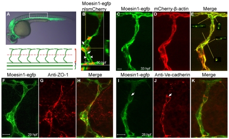
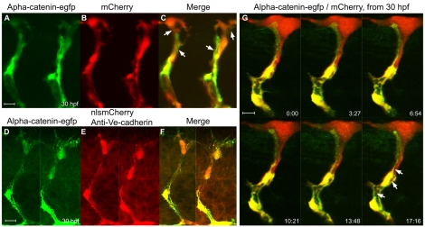
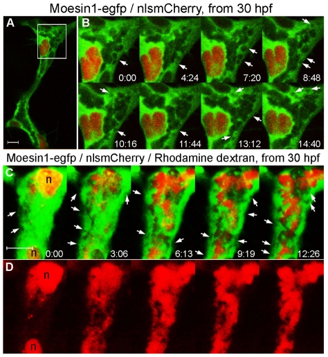
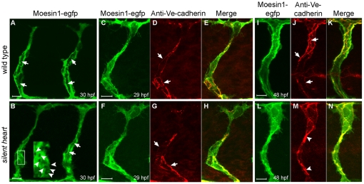
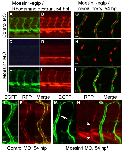
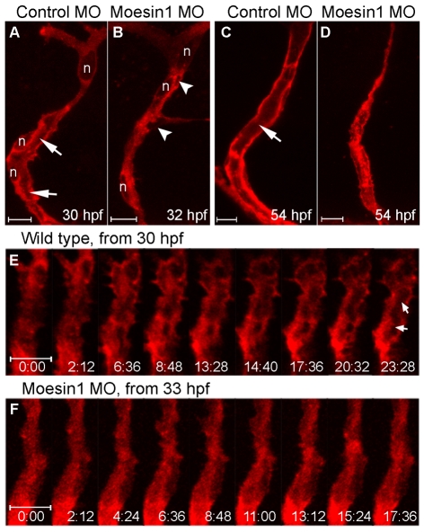
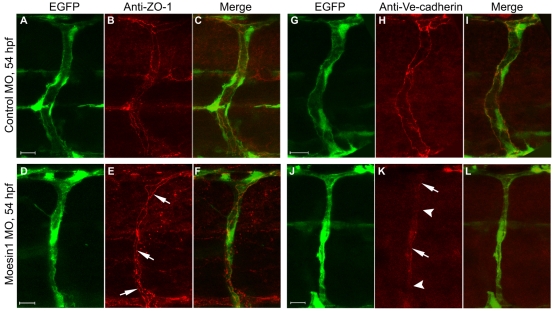
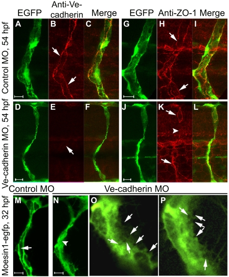
Endothelial tubulogenesis is a crucial step in the formation of functional blood vessels during angiogenesis and vasculogenesis. Here, we use in vivo imaging of living zebrafish embryos expressing fluorescent fusion proteins of beta-Actin, alpha-Catenin, and the ERM family member Moesin1 (Moesin a), to define a novel cord hollowing process that occurs during the initial stages of tubulogenesis in intersegmental vessels (ISVs) in the embryo. We show that the primary lumen elongates along cell junctions between at least two endothelial cells during embryonic angiogenesis. Moesin1-EGFP is enriched around structures that resemble intracellular vacuoles, which fuse with the luminal membrane during expansion of the primary lumen. Analysis of silent heart mutant embryos shows that initial lumen formation in the ISVs is not dependent on blood flow; however, stabilization of a newly formed lumen is dependent upon blood flow. Zebrafish moesin1 knockdown and cell transplantation experiments demonstrate that Moesin1 is required in the endothelial cells of the ISVs for in vivo lumen formation. Our analyses suggest that Moesin1 contributes to the maintenance of apical/basal cell polarity of the ISVs as defined by adherens junctions. Knockdown of the adherens junction protein Ve-cadherin disrupts formation of the apical membrane and lumen in a cell-autonomous manner. We suggest that Ve-cadherin and Moesin1 function to establish and maintain apical/basal polarity during multicellular lumen formation in the ISVs.


Figures

Fig. 1.
Fig. 1.
Moesin1-EGFP and mCherry-β-Actin fusion proteins are enriched at apical membranes and cellular junctions. (A) A 30 hpf zebrafish embryo from Tg(flk1:moesin1-egfp) with the box indicating the area shown beneath. Red, black and green brackets indicate the intersegmental vessels (ISVs), dorsal aorta (DA) and posterior caudal vein (PCV), respectively. The red dashed line designates the boundary between the dorsal (top) or ventral (bottom) ISVs. (B) An ISV in a Tg(flk1:moesin1-egfp)/Tg(flk1:nlsmCherry) embryo showing the primary lumen (arrows) forming at 30 hpf. Moesin1-EGFP is enriched at the apical membrane (arrowheads). The nuclei are in red. Virtual cross-sections are at the bottom and to the right of the main panel. Yellow lines designate the planes of the cross-sections. (C-E)A Tg(flk1:moesin1-egfp)/Tg(flk1:mCherry-β-actin) embryo at 33 hpf displayed considerable overlap of fluorescence in the ISV. Virtual cross-sections in E show the lumen. (F-H) Moesin1-EGFP and the tight junction protein ZO-1 co-localized at 29 hpf. (I-K) At 28 hpf, Moesin1-EGFP is enriched at adherens junctions, as labeled with anti-Ve-cadherin antibody, although not always (arrows). (A-K) Lateral images, dorsal is up, anterior is to the left. Scale bars: 10 μm in all figures.
Fig. 2.
Fig. 2.
α-Catenin-EGFP localizes to adherens junctions associated with the primary lumen in the ISVs. (A-C) Confocal images of the ISVs in a living Tg(flk1:α-catenin-egfp)/Tg(flk1:mCherry) zebrafish embryo at 30 hpf. Arrows in C point to long putative adherens junctions between cells. (D-F) Labeling of adherens junctions in the ISVs with anti-Ve-cadherin antibody in a Tg(flk1:α-catenin-egfp)/Tg(flk1:nlsmCherry) double-transgenic embryo. (G) Time-lapse confocal images of an ISV in a Tg(flk1:α-catenin-egfp)/Tg(flk1:mCherry) embryo showing the putative primary lumen (arrows) forming between the cellular junctions. The time format is minutes:seconds.
Fig. 3.
Fig. 3.
Vacuoles fuse with luminal membranes during initial lumen expansion. (A) An ISV from a living Tg(flk1:moesin1-egfp)/Tg(flk1:nlsmCherry) zebrafish embryo with the box indicating the area shown in B. (B) Time-lapse confocal images showing formation of primary lumen in an ISV with the integration of vacuoles (arrows) from 30 hpf. (C,D) A dorsal ISV from a living Tg(flk1:moesin1-egfp)/Tg(flk1:nlsmCherry) embryo injected intravenously with labeled dextran (D) showing the intracellular vacuoles without labeled dextran (C, arrows). The time is in minutes:seconds. n, nucleus.
Fig. 4.
Fig. 4.
Primary endothelial lumen formation in the ISVs does not require blood flow. (A,B) Formation of the primary lumen (arrows) at 30 hpf is observed with the Tg(flk1:moesin1-egfp) line in either wild-type (A) or silent heart mutants (B). (B) silent heart embryos also displayed vacuoles during tubulogenesis (arrowheads in inset). (C-H) Ve-cadherin-labeled junctions (arrows) appeared normal at 29 hpf in silent heart mutants. (I-N) At 48 hpf, Ve-cadherin-labeled junctions (arrows) are often clustered in the silent heart embryos (arrowheads), which is likely to reflect the collapse of the primary lumen.
Fig. 5.
Fig. 5.
Moesin1 is required in the endothelial cells for tubulogenesis. (A-F) The Tg(flk1:moesin1-egfp) line partially rescues the Moesin1 knockdown phenotype. (A,B) A Tg(flk1:moesin1-egfp) zebrafish embryo injected with 4 ng control MO. All the ISVs were perfused with tetramethylrhodamine-dextran (TMRD, red). (C,D) Wild-type sibling embryo injected with 4 ng moesin1 MO. Most ISVs are not perfused with TMRD, although circulation of TMRD is observed in the axial vessels. (E,F) A Tg(flk1:moesin1-egfp) embryo injected with 4 ng moesin1 MO. Most ISVs are perfused with TMRD. (G-I) Same experiment as in A-F in Tg(flk1:moesin1-egfp)/Tg(flk1:nlsmCherry) embryos shows that the endothelial cells are present in the ISVs despite a lack of circulation at 54 hpf. (J-O) Transplantation of endothelial cells was used to determine autonomous verses non-autonomous effects. (J-L) Normal endothelial tubulogenesis occurs when the donor embryo was injected with control MO. (M-O) Transplanted endothelial cells from Moesin1 knockdown embryos fail to undergo normal tubulogenesis (arrowheads), whereas the ISV in the recipient embryo (arrow) completes lumen formation.
Fig. 6.
Fig. 6.
Moesin1 is required during ISVs tubulogenesis. (A-D) Confocal images of ISVs from Tg(flk1:mCherry-β-actin) living zebrafish embryos. (A,C) Embryos injected with 4 ng control MO. The lumen (arrows) is seen at 30 hpf (A) and is well formed at 54 hpf (C). (B,D) Embryos injected with 4 ng moesin1 MO. The primary lumen is not observed at 32 hpf (B), nor at 54 hpf (D), although a few putative vacuoles or intercellular spaces are observed (B, arrowheads). (E,F) Confocal time-lapse images of wild-type and Moesin1 knockdown Tg(flk1:mCherry-β-actin) embryos. The time format is minutes:seconds. (E) In wild-type embryos, formation of the primary lumen is observed (arrows) at 30 hpf. (F) In Moesin1 knockdown embryos, mCherry-β-Actin remains throughout the cytoplasm and the primary lumen is not observed at 33 hpf. n, nucleus.
Fig. 7.
Fig. 7.
Knockdown of Moesin1 results in extensive loss of adherens junctions without affecting ZO-1-associated junctions. (A-F) Confocal images of ISVs in the Tg(fli1:egfp)y1 zebrafish embryos that were probed for ZO-1 (red) at 54 hpf. (A-C) ZO-1-associated junctions are seen in a control embryo. (D-F) The ZO-1-associated junctions (arrow) are observed in Moesin1 knockdown embryos but are not well separated from one another. (G-L) Confocal images of ISVs in Tg(fli1:egfp)y1 embryos labeled with Ve-cadherin (red) at 54 hpf. (G-I) Ve-cadherin labeling in a control embryo. (J-L) Moesin1 knockdown causes an extensive loss of Ve-cadherin labeling (arrowheads); some weak Ve-cadherin signals are observed (arrows).
Fig. 8.
Fig. 8.
Knockdown of Ve-cadherin impairs lumen formation. (A-F) Confocal images of ISVs in Tg(fli1:egfp)y1 zebrafish embryos probed for Ve-cadherin (red) at 54 hpf. Ve-cadherin labeling (arrows) is observed in control embryos (A-C), but not in Ve-cadherin knockdown embryos (D-F). (G-L) Confocal images of ISVs in Tg(fli1:egfp)y1 embryos labeled with ZO-1 antibody (red) at 54 hpf. ZO-1-associated junctions (arrows) are observed in control embryos (G-I), but are not detected in some regions of the ISV (arrowhead) in a Ve-cadherin knockdown embryo (J-L). (M-P) Confocal images of ISVs in Tg(flk1:moesin1-egfp) embryos at 32 hpf. The primary lumen (arrow) is forming in a control embryo (M). Putative vacuoles or intercellular spaces (arrows in the higher magnification images O and P) are observed throughout the cytoplasm in endothelial cells in Ve-cadherin knockdown embryos (N-P), without formation of primary lumen (N, arrowhead).

Similar articles

Cited by

References

    1. Andrew D. J., Ewald A. J. (2010). Morphogenesis of epithelial tubes: insights into tube formation, elongation, and elaboration. Dev. Biol. 341, 34-55 - PMC - PubMed
    1. Bagnat M., Cheung I. D., Mostov K. E., Stainier D. Y. (2007). Genetic control of single lumen formation in the zebrafish gut. Nat. Cell Biol. 9, 954-960 - PubMed
    1. Bayless K. J., Davis G. E. (2002). The Cdc42 and Rac1 GTPases are required for capillary lumen formation in three-dimensional extracellular matrices. J. Cell Sci. 115, 1123-1136 - PubMed
    1. Bayless K. J., Davis G. E. (2004). Microtubule depolymerization rapidly collapses capillary tube networks in vitro and angiogenic vessels in vivo through the small GTPase Rho. J. Biol. Chem. 279, 11686-11695 - PubMed
    1. Berryman M., Franck Z., Bretscher A. (1993). Ezrin is concentrated in the apical microvilli of a wide variety of epithelial cells whereas moesin is found primarily in endothelial cells. J. Cell Sci. 105, 1025-1043 - PubMed

Publication types

MeSH terms