

Regional control of tumor growth
- PMID: 20736295
- PMCID: PMC3044487
- DOI: 10.1158/1541-7786.MCR-10-0047
Regional control of tumor growth
Abstract
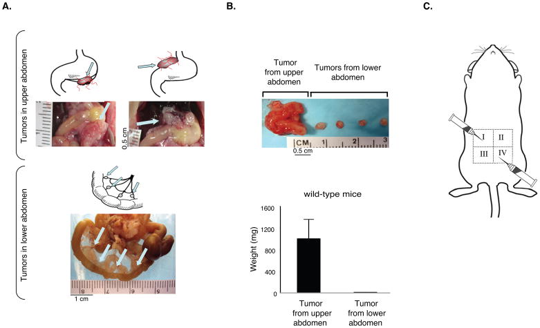
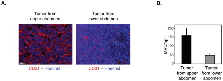
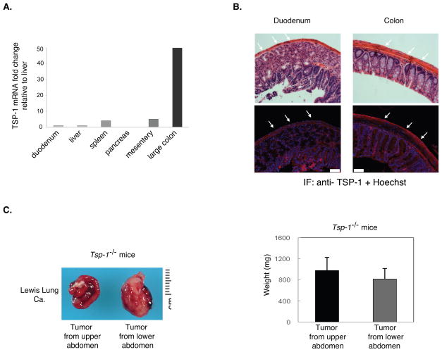
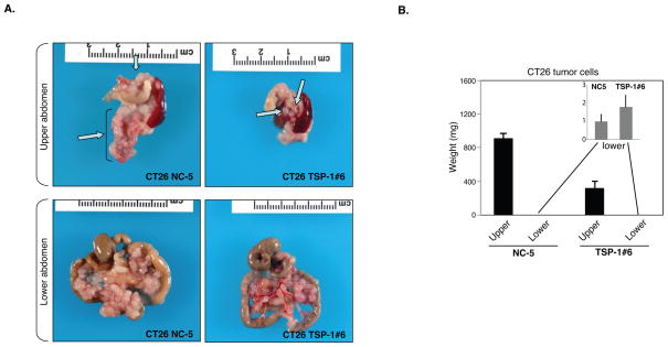
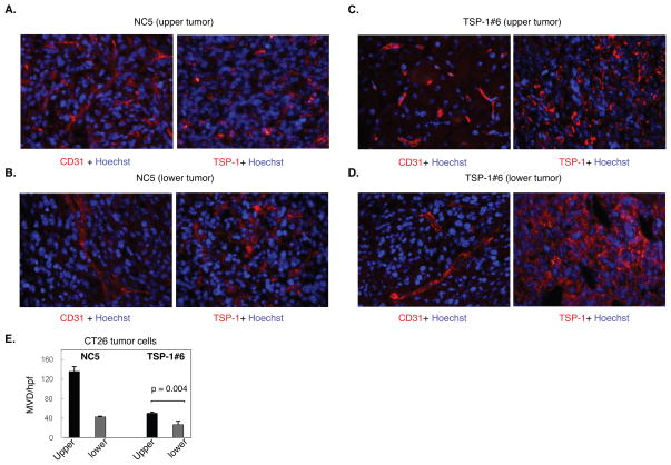
Tumors implanted near the scapulae have been shown to grow four times faster than the same tumors implanted at the iliac crest. Although there were marked differences in the vascularization of tumors from these two different sites, the mechanism controlling regional angiogenesis was not identified. Here, we show site-specific growth of intraperitoneal tumor implants in the mouse abdomen. Our data indicate that the angiogenic response of the host differs significantly between the upper and lower sites in the mouse abdomen and reveal that the expansion of tumor mass is restricted to sites with low angiogenic responses, such as the bowel mesentery in the lower abdomen. We show that, in this model, this suppression of angiogenesis is due to an expression gradient of thrombospondin-1 (TSP-1), a potent endogenous angiogenesis inhibitor. Mice with a targeted deletion of TSP-1 no longer show regional restriction of tumor growth. The physiologic relevance of these findings may be seen in patients with peritoneal carcinomatosis, whereby tumors spread within the peritoneal cavity and show differential growth in the upper and lower abdomen. We hypothesize that the difference in tumor growth in these patients may be due to a gradient of TSP-1 expression in stroma. Finally, our studies suggest that upregulation of TSP-1 in tumor cells is one method to suppress the growth of tumors in the upper abdomen.
© 2010 AACR.
Figures
Similar articles
-
Context dependent role of the CD36--thrombospondin--histidine-rich glycoprotein axis in tumor angiogenesis and growth.PLoS One. 2012;7(7):e40033. doi: 10.1371/journal.pone.0040033. Epub 2012 Jul 10. PLoS One. 2012. PMID: 22808089 Free PMC article.
-
Thrombospondin-1 as an endogenous inhibitor of angiogenesis and tumor growth.J Cell Mol Med. 2002 Jan-Mar;6(1):1-12. doi: 10.1111/j.1582-4934.2002.tb00307.x. J Cell Mol Med. 2002. PMID: 12003665 Free PMC article. Review.
-
The N-terminal domain of thrombospondin-1: a key for the dual effect of TSP-1 in angiogenesis and cancer progression?ScientificWorldJournal. 2009 Feb 15;9:133-6. doi: 10.1100/tsw.2009.11. ScientificWorldJournal. 2009. PMID: 19219378 Free PMC article.
-
Expression of thrombospondin-1 by tumor cells in patient-derived ovarian carcinoma xenografts.Connect Tissue Res. 2015;56(5):355-63. doi: 10.3109/03008207.2015.1045065. Epub 2015 Jun 15. Connect Tissue Res. 2015. PMID: 25943461
-
Regulation of tumor angiogenesis by thrombospondin-1.Biochim Biophys Acta. 2006 Apr;1765(2):178-88. doi: 10.1016/j.bbcan.2005.11.002. Epub 2005 Dec 21. Biochim Biophys Acta. 2006. PMID: 16406676 Review.
Cited by
-
Thrombospondins: old players, new games.Curr Opin Lipidol. 2013 Oct;24(5):401-9. doi: 10.1097/MOL.0b013e3283642912. Curr Opin Lipidol. 2013. PMID: 23892609 Free PMC article. Review.
-
From Oncogenic Signaling Pathways to Single-Cell Sequencing of Immune Cells: Changing the Landscape of Cancer Immunotherapy.Molecules. 2021 Apr 14;26(8):2278. doi: 10.3390/molecules26082278. Molecules. 2021. PMID: 33920054 Free PMC article. Review.
-
Advances in Anti-metabolic Disease Treatments Targeting CD47.Curr Pharm Des. 2022;28(46):3720-3728. doi: 10.2174/1381612828666221006123144. Curr Pharm Des. 2022. PMID: 36201266 Review.
-
Invoking the power of thrombospondins: regulation of thrombospondins expression.Matrix Biol. 2014 Jul;37:69-82. doi: 10.1016/j.matbio.2014.02.001. Epub 2014 Feb 25. Matrix Biol. 2014. PMID: 24582666 Free PMC article. Review.
-
Accelerated Endosomal Escape of Splice-Switching Oligonucleotides Enables Efficient Hepatic Splice Correction.ACS Appl Mater Interfaces. 2025 Feb 12;17(6):9000-9018. doi: 10.1021/acsami.4c19340. Epub 2025 Jan 28. ACS Appl Mater Interfaces. 2025. PMID: 39873730 Free PMC article.
References
-
- Twort J, Twort CC. The variable sensitivity of different sites of the skin of mice to carcinogenic agents. J Pathol Bacteriol. 1936;42:303–16.
-
- Kobayashi K. Regional differences in mitotic activity due to injury in mouse skin. Cell and tissue research. 1976;175(3):319–24. - PubMed
-
- Sugarbaker PH. Peritoneum as the first-line of defense in carcinomatosis. Journal of surgical oncology. 2007;95(2):93–6. - PubMed
-
- Sano T. Is peritoneal carcinomatosis an incurable disease or controllable locoregional condition?--Challenge of surgeons with intraperitoneal hyperthermic chemotherapy. Japanese journal of clinical oncology. 2001;31(12):571–2. - PubMed
-
- Jacquet P, Vidal-Jove J, Zhu B, Sugarbaker P. Peritoneal carcinomatosis from gastrointestinal malignancy: natural history and new prospects for management. Acta chirurgica Belgica. 1994;94(4):191–7. - PubMed
Publication types
MeSH terms
Substances
Grants and funding
LinkOut - more resources
Full Text Sources
Molecular Biology Databases
Miscellaneous
