. 2010 Oct;8(10):1375-87.
doi: 10.1158/1541-7786.MCR-09-0537. Epub 2010 Aug 24.

Cyclin-dependent kinase 4-mediated phosphorylation inhibits Smad3 activity in cyclin D-overexpressing breast cancer cells

Affiliations

Cyclin-dependent kinase 4-mediated phosphorylation inhibits Smad3 activity in cyclin D-overexpressing breast cancer cells

Stanislav Zelivianski et al. Mol Cancer Res. 2010 Oct.

Abstract

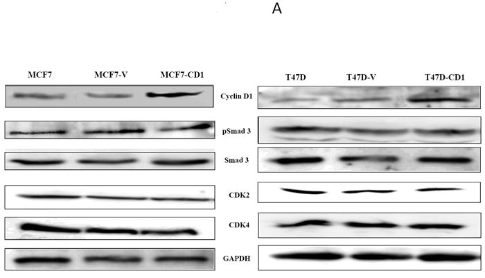
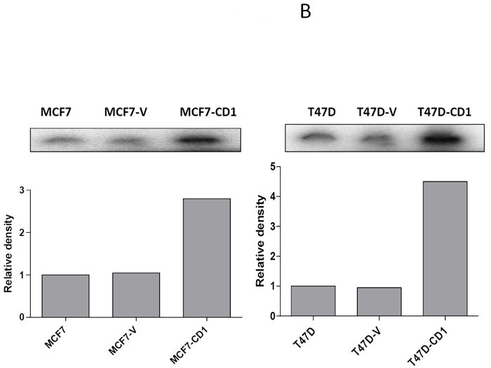
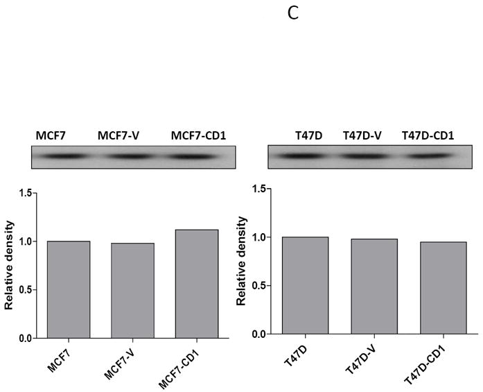
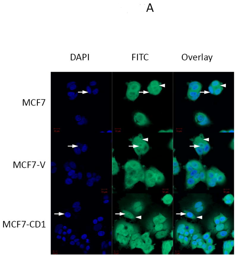
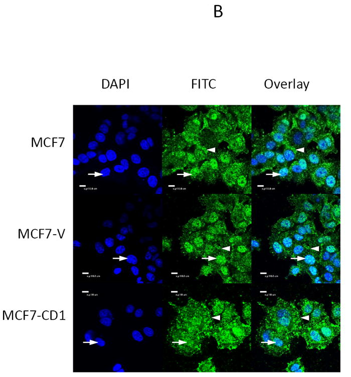
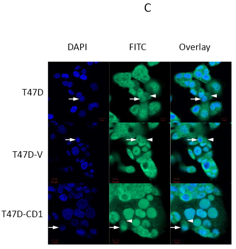
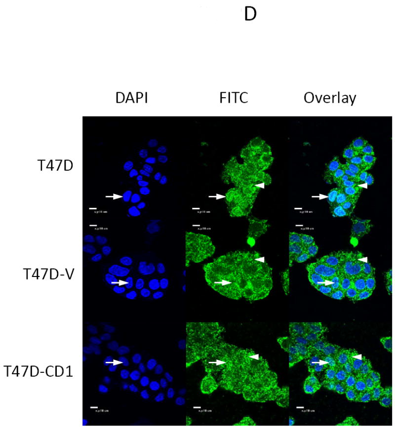
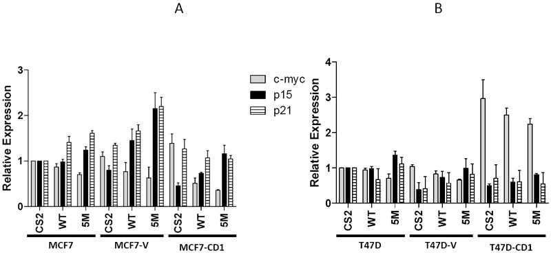
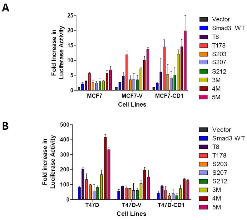
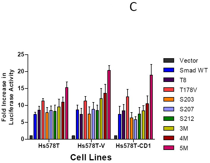
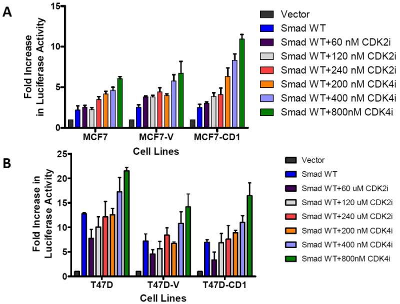
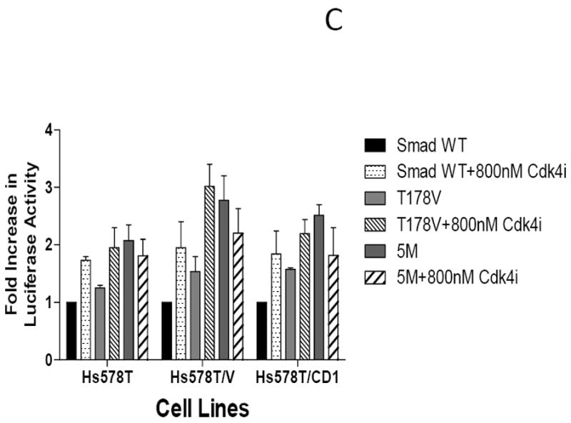
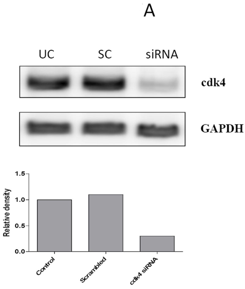
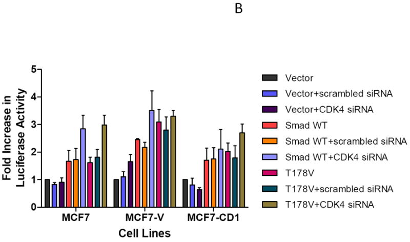
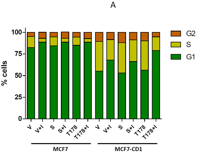
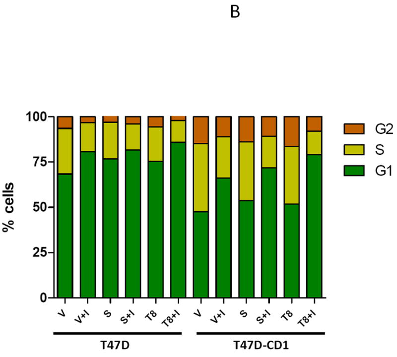
Smad3, a component of the transforming growth factor β signaling cascade, contributes to G(1) arrest in breast cancer cells. Cyclin D1/cyclin-dependent kinase 4 (CDK4) promotes G(1)-S-phase transition, and CDK phosphorylation of Smad3 has been associated with inhibition of Smad3 activity. We hypothesized that overexpression of cyclin D1 exerts tumorigenic effects in breast cancer cells through CDK4-mediated phosphorylation and inhibition of Smad3 and release of G(1) arrest. Real-time quantitative reverse transcription-PCR and immunoblotting were used to evaluate expression of study proteins in cyclin D1-overexpressing breast cancer cells. Smad3 transcriptional activity and cell cycle control were examined in cells transfected with wild-type (WT) Smad3 or Smad3 with single or multiple CDK phosphorylation site mutations (M) in the presence or absence of the CDK4 inhibitor or cotransfection with cdk4 small interfering RNA (siRNA). Transfection of the Smad3 5M construct resulted in decreased c-myc and higher p15(INK4B) expression. Compared with WT Smad3, overexpression of the Smad3 T8, T178, 4M, or 5M mutant constructs resulted in higher Smad3 transcriptional activity. Compared with cells transfected with WT Smad3, Smad3 transcriptional activity was higher in cells overexpressing Smad3 mutant constructs and treated with the CDK4 inhibitor or transfected with cdk4 siRNA. Cells transfected with Smad3 T8 or T178 and treated with the CDK4 inhibitor showed an increase in the G(1) cell population. Inhibition of CDK-mediated Smad3 phosphorylation released cyclin D1-regulated blockade of Smad3 transcriptional activity and recovered cell cycle arrest in breast cancer cells. Targeted inhibition of CDK4 activity may have a role in the treatment of cyclin D-overexpressing breast cancers.


Figures

FIGURE 1
FIGURE 1
Cyclin D expression and activity in MCF7 and T47D stably transfected cells. (A) Protein extracts from stably transfected MCF7 or T47D clones were separated (30 μg/lane) on SDS/PAGE and subjected to immunoblot analysis with the indicated antibodies. (B) CDK4 kinase assay radiographs and densitometric quantification in MCF7 and T47D study cell lines.
FIGURE 1
FIGURE 1
Cyclin D expression and activity in MCF7 and T47D stably transfected cells. (A) Protein extracts from stably transfected MCF7 or T47D clones were separated (30 μg/lane) on SDS/PAGE and subjected to immunoblot analysis with the indicated antibodies. (B) CDK4 kinase assay radiographs and densitometric quantification in MCF7 and T47D study cell lines.
FIGURE 2
FIGURE 2
Smad3 and phospho-Smad3 localization in cyclin D-overexpressing MCF7 and T47D cells. Smad3 localized to the nucleus in MCF7 (A, B) and T47D (B, C) parental, vector control (V), and cyclin D-overexpressing (CD1) cell lines. Endogenous Smad3 and phospho-Smad3 were visualized by immunofluorescence microscopy with an anti-Smad3 polyclonal antibody and FITC-conjugated secondary antibody. Cell nuclei were counterstained with DAPI. Scale bar is 10 μm. Arrows denote nuclei. Arrowheads denote cytoplasmic Smad3.
FIGURE 2
FIGURE 2
Smad3 and phospho-Smad3 localization in cyclin D-overexpressing MCF7 and T47D cells. Smad3 localized to the nucleus in MCF7 (A, B) and T47D (B, C) parental, vector control (V), and cyclin D-overexpressing (CD1) cell lines. Endogenous Smad3 and phospho-Smad3 were visualized by immunofluorescence microscopy with an anti-Smad3 polyclonal antibody and FITC-conjugated secondary antibody. Cell nuclei were counterstained with DAPI. Scale bar is 10 μm. Arrows denote nuclei. Arrowheads denote cytoplasmic Smad3.
FIGURE 3
FIGURE 3
Expression of Smad3-regulated genes in study cell lines. MCF7 (A) and T47D (B) cell lines were transfected with wild type Smad3 (WT) or the 5M Smad3 CDK phosphorylation site mutant, and transcript levels of c-myc, p15, and p21 were measured by real-time quantitative RT-PCR.
FIGURE 4
FIGURE 4
Relative transcriptional activity of various Smad3 constructs in cyclin D1 overexpressing MCF7 (A), T47D (B), and Hs578T (C) cell lines. Cells were co-transfected with the Smad-responsive CAGA-luc reporter construct and Renilla luciferase reporter, in addition to the indicated Smad3 expression constructs. Firefly and Renilla luciferase activities were determined. Data are shown as fold increase in normalized luciferase activity (firefly/Renilla) compared with empty vector-transfected MCF7, T47D, or Hs578T cells. Error bars indicate standard deviation from the mean of normalized luciferase activity for each study condition.
FIGURE 4
FIGURE 4
Relative transcriptional activity of various Smad3 constructs in cyclin D1 overexpressing MCF7 (A), T47D (B), and Hs578T (C) cell lines. Cells were co-transfected with the Smad-responsive CAGA-luc reporter construct and Renilla luciferase reporter, in addition to the indicated Smad3 expression constructs. Firefly and Renilla luciferase activities were determined. Data are shown as fold increase in normalized luciferase activity (firefly/Renilla) compared with empty vector-transfected MCF7, T47D, or Hs578T cells. Error bars indicate standard deviation from the mean of normalized luciferase activity for each study condition.
FIGURE 5
FIGURE 5
Dose-dependent increase in Smad3 transcriptional activity in MCF7 and T47D cells treated with increasing concentrations of CDK2 or CDK4 inhibitors. MCF7 (A) or T47D (B) study cell lines were transfected with the Smad-responsive CAGA-luc reporter construct, Renilla luciferase reporter, and wild type (WT) Smad3 expression vector, then treated with the indicated concentrations of CDK2 inhibitor (CDK2i) or CDK4 inhibitor (CDK4i). Firefly and Renilla luciferase activities were determined. Data are shown as fold increase in normalized luciferase activity (firefly/Renilla) compared with empty vector-transfected MCF7 or T47D cells. Error bars indicate standard deviation from the mean of normalized luciferase activity for each study condition.
FIGURE 6
FIGURE 6
Restoration of Smad3 transcriptional activity with inhibition of CDK4 activity. MCF7 (A), T47D (B), and Hs578T (C) study cell lines were transfected with the Smad-responsive CAGA-luc reporter construct, Renilla luciferase reporter, and either wild type (WT) Smad3, 5M Smad3, T178 Smad3 (MCF7 and Hs578T), or T8 Smad3 (T47D only) expression vectors. The cells were treated with vehicle or 800 nM CDK4 inhibitor (CDK4i). Firefly and Renilla luciferase activities were determined. Data are shown as fold increase in normalized luciferase activity (firefly/Renilla) compared with empty vector-transfected MCF7, T47D, or Hs578T cells. Error bars indicate standard deviation from the mean of normalized luciferase activity for each study condition. For Figure 6B * denotes value too low to graphically represent.
FIGURE 6
FIGURE 6
Restoration of Smad3 transcriptional activity with inhibition of CDK4 activity. MCF7 (A), T47D (B), and Hs578T (C) study cell lines were transfected with the Smad-responsive CAGA-luc reporter construct, Renilla luciferase reporter, and either wild type (WT) Smad3, 5M Smad3, T178 Smad3 (MCF7 and Hs578T), or T8 Smad3 (T47D only) expression vectors. The cells were treated with vehicle or 800 nM CDK4 inhibitor (CDK4i). Firefly and Renilla luciferase activities were determined. Data are shown as fold increase in normalized luciferase activity (firefly/Renilla) compared with empty vector-transfected MCF7, T47D, or Hs578T cells. Error bars indicate standard deviation from the mean of normalized luciferase activity for each study condition. For Figure 6B * denotes value too low to graphically represent.
FIGURE 7
FIGURE 7
Restoration of Smad3 transcriptional activity with siRNA knockdown of CDK4. (A) MCF7 cells were transfected with scrambled (SC) or cdk4 siRNA (siRNA) for 48 hours, then the cells were lysed and the level of CDK4 protein was determined in untransfected cells (UC) and transfected cells by immunoblot analysis using anti-CDK4 antibody. GAPDH was used as loading control. (B) MCF7 study cell lines were co-transfected with the Smad-responsive CAGA-luc reporter construct and Renilla luciferase reporter; siRNA or cdk4 siRNA; and either empty vector, wild type (WT) Smad3, or T178 Smad3 mutant expression vectors. Firefly and Renilla luciferase activities were determined. Data are shown as fold increase in normalized luciferase activity (firefly/Renilla) compared with empty vector-transfected MCF7 or T47D cells. Error bars indicate standard deviation from the mean of normalized luciferase activity for each study condition.
FIGURE 7
FIGURE 7
Restoration of Smad3 transcriptional activity with siRNA knockdown of CDK4. (A) MCF7 cells were transfected with scrambled (SC) or cdk4 siRNA (siRNA) for 48 hours, then the cells were lysed and the level of CDK4 protein was determined in untransfected cells (UC) and transfected cells by immunoblot analysis using anti-CDK4 antibody. GAPDH was used as loading control. (B) MCF7 study cell lines were co-transfected with the Smad-responsive CAGA-luc reporter construct and Renilla luciferase reporter; siRNA or cdk4 siRNA; and either empty vector, wild type (WT) Smad3, or T178 Smad3 mutant expression vectors. Firefly and Renilla luciferase activities were determined. Data are shown as fold increase in normalized luciferase activity (firefly/Renilla) compared with empty vector-transfected MCF7 or T47D cells. Error bars indicate standard deviation from the mean of normalized luciferase activity for each study condition.
FIGURE 8
FIGURE 8
Inhibition of CDK4-mediated phosphorylation of Smad3 leads to G1 arrest in MCF7 parental and cyclin D-overexpressing cell lines. (A) MCF7 and MCF7-CD1 cells were transfected with empty vector (V), wild type Smad3 (S), or the T178 Smad3 mutant expression vectors. (B) T47D and T47D-CD1 cells were transfected with empty vector (V), wild type Smad3 (S), or the T8 Smad3 mutant expression vectors. The cells were then treated with vehicle or CDK4 inhibitor (I). Cells were harvested and the cell cycle profile was assessed by FACS analysis. Figures show one representative experiment from 3 separate data sets.
FIGURE 8
FIGURE 8
Inhibition of CDK4-mediated phosphorylation of Smad3 leads to G1 arrest in MCF7 parental and cyclin D-overexpressing cell lines. (A) MCF7 and MCF7-CD1 cells were transfected with empty vector (V), wild type Smad3 (S), or the T178 Smad3 mutant expression vectors. (B) T47D and T47D-CD1 cells were transfected with empty vector (V), wild type Smad3 (S), or the T8 Smad3 mutant expression vectors. The cells were then treated with vehicle or CDK4 inhibitor (I). Cells were harvested and the cell cycle profile was assessed by FACS analysis. Figures show one representative experiment from 3 separate data sets.

Similar articles

Cited by

References

    1. Brown KA, Pietenpol JA, Moses HL. A tale of two proteins: differential roles and regulation of Smad2 and Smad3 in TGF-beta signaling. J Cell Biochem. 2007;101(1):9–33. - PubMed
    1. Yue J, Mulder KM. Transforming growth factor-beta signal transduction in epithelial cells. Pharmacol Ther. 2001;91(1):1–34. - PubMed
    1. Massague J. TGF-beta signal transduction. Annu Rev Biochem. 1998;67:753–91. - PubMed
    1. Nakao A, Imamura T, Souchelnytskyi S, et al. TGF-beta receptor-mediated signalling through Smad2, Smad3 and Smad4. Embo J. 1997;16(17):5353–62. - PMC - PubMed
    1. Matsuura I, Chiang KN, Lai CY, et al. Pin1 promotes TGF-beta-induced migration and invasion. J Biol Chem. 2009 - PMC - PubMed

Publication types

MeSH terms