. 2010 Sep 15;123(Pt 18):3102-11.
doi: 10.1242/jcs.064774. Epub 2010 Aug 24.

The fibroblast-derived paracrine factor neuregulin-1 has a novel role in regulating the constitutive color and melanocyte function in human skin

Affiliations

The fibroblast-derived paracrine factor neuregulin-1 has a novel role in regulating the constitutive color and melanocyte function in human skin

Wonseon Choi et al. J Cell Sci. .

Abstract

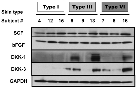
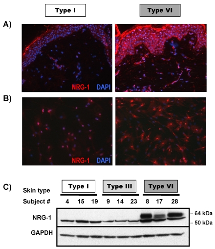
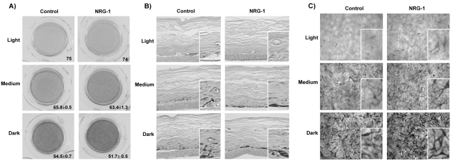
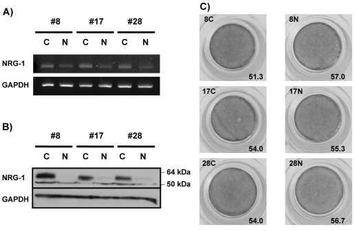
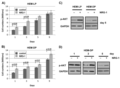
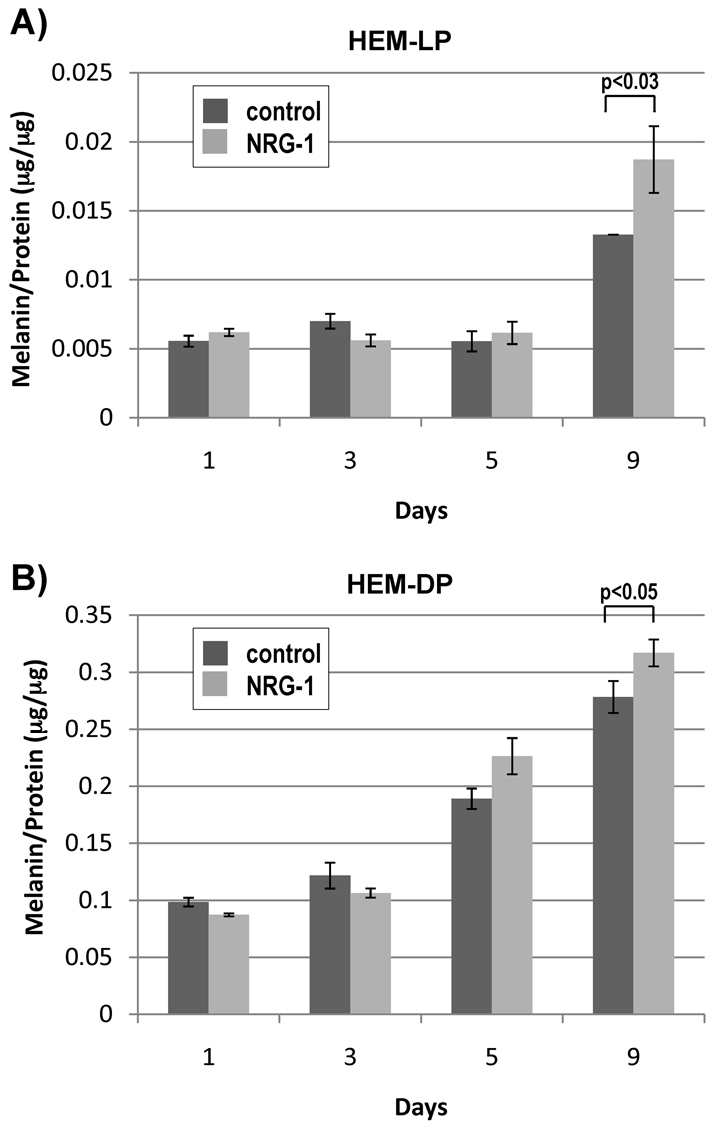
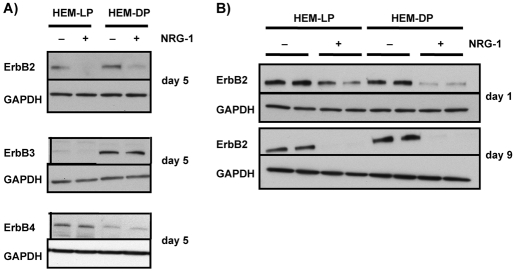
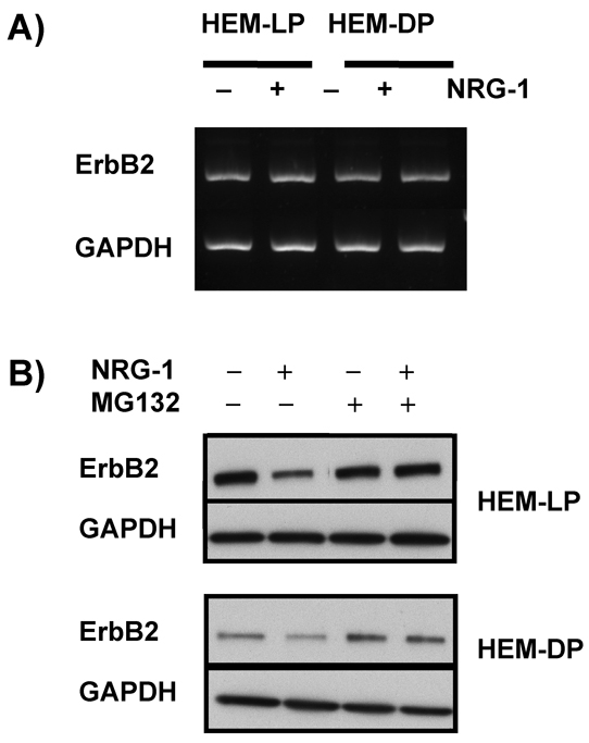
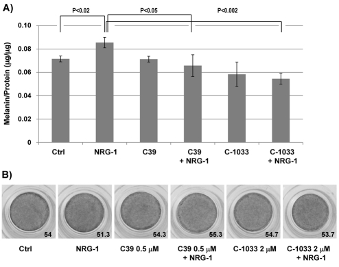
Interactions between melanocytes and neighboring cells in the skin are important in regulating skin color in humans. We recently demonstrated that the less pigmented and thicker skin on the palms and soles is regulated by underlying fibroblasts in those areas, specifically via a secreted factor (DKK1) that modulates Wnt signaling. In this study, we tested the hypothesis that dermal fibroblasts regulate the constitutive skin color of individuals ranging from very light to very dark. We used microarray analysis to compare gene expression patterns in fibroblasts derived from lighter skin types compared to darker skin types, with a focus on secreted proteins. We identified a number of genes that differ dramatically in expression and, among the expressed proteins, neuregulin-1, which is secreted by fibroblasts derived from dark skin, effectively increases the pigmentation of melanocytes in tissue culture and in an artificial skin model and regulates their growth, suggesting that it is one of the major factors determining human skin color.


Figures

Fig. 1.
Fig. 1.
Protein expression levels of known fibroblast-derived melanogenic paracrine factors. Proteins were extracted from each of the 15 fibroblast cell lines, and western blotting was used to detect levels of SCF, bFGF, DKK-1, DKK-3, and GAPDH (used as a loading control). Three representative fibroblast lines for each of three skin types are shown here.
Fig. 2.
Fig. 2.
Expression of NRG-1 in normal human skin and in cultured normal human fibroblasts. (A) Frozen sections of normal human skin from type I and type VI phototypes were used to examine expression levels of NRG-1. Images are representative of three different subjects of each phototype, which all showed similar results. (B) Fibroblasts were cultured in chamber-slides and stained with NRG-1 antibody. Images are representative of three different fibroblast cell lines of each phototype, which all showed similar results. (C) Proteins were extracted from each of 15 fibroblast cell lines and western blotting was used to detect the level of NRG-1 and GAPDH (used as a loading control). Three representative fibroblast lines for each of three skin types are shown.
Fig. 3.
Fig. 3.
Effect of NRG-1 on the pigmentation of 3D reconstructed skins. (A) Light (from Caucasian skin, n=1), medium (from Asian skin, n=4) and dark (from African-American skin, n=2) MelanoDerms were treated with or without NRG-1 (50 ng/ml) for 9 days. Images were taken of the inverted MelanoDerms at the end of the protocol. The L value shown for each MelanoDerm was calculated as the average of three spots measured in the center of each MelanoDerm; results are the averages of L values in the number of experiments indicated above ± s.d. (B) Melanin content in human skin equivalents analyzed by Fontana–Masson staining; insets show areas at threefold magnification. (C) Overhead bright field microscopic view of MelanoDerms; insets show areas at threefold magnification.
Fig. 4.
Fig. 4.
Effect of NRG-1 knockdown by shRNA transduction. (A) mRNA levels and (B) protein levels of NRG-1 in three different fibroblasts (#8, 17, and 28) from type VI skin after control shRNA (C) or NRG-1 shRNA (N) transduction. (C) Change of MelanoDerm pigmentation when fibroblasts were transduced with control shRNA or NRG-1 shRNA. L values shown are the average of three spots measured in the center of each MelanoDerm.
Fig. 5.
Fig. 5.
Effect of NRG-1 on the proliferation of human melanocytes in culture. (A) Lightly pigmented (HEM-LP) and (B) darkly pigmented (HEM-DP) melanocytes were treated with or without NRG-1 (50 ng/ml) for 1, 3, 5, or 9 days in 10-cm culture dishes, and the total cell numbers counted. Results are the averages of three experiments ± s.d. (C) Phosphorylated Akt (p-AKT) levels were detected by western blotting for HEM-LP and HEM-DP treated with NRG-1 (50 ng/ml) for 5 days compared to vehicle-treated controls; GAPDH was used as a loading control. (D) HEM-DP were treated with NRG-1 (50 ng/ml) for 1, 3, or 5 days and phosphorylated Akt levels were detected by western blotting compared to vehicle-treated controls; GAPDH was used as a loading control. Results shown are representative of three experiments; statistically significant differences are indicated.
Fig. 6.
Fig. 6.
Effect of NRG-1 on the pigmentation of cultured human melanocytes. (A) Lightly pigmented (HEM-LP) and (B) Darkly pigmented (HEM-DP) melanocytes were treated with or without NRG-1 (50 ng/ml) for 1, 3, 5, or 9 days and proteins were extracted from each sample. After centrifugation at 10,000 g for 15 minutes, precipitates were analyzed for melanin content, and the supernatants were analyzed for protein content. Results are the average of three experiments ± s.d.; statistically significant differences are indicated.
Fig. 7.
Fig. 7.
Expression of ErbBs after NRG-1 treatment. (A) HEM-LP and HEM-DP were treated with or without NRG-1 (50 ng/ml) for 5 days, and levels of ErbB2, ErbB3, or ErbB4 were detected by western blotting. Results shown are representative of three experiments. (B) Expression of ErbB2 was examined in HEM-LP and in HEM-DP after 1 or 9 days or treatment with NRG-1 compared to vehicle-treated controls; the results are representative of two independent experiments.
Fig. 8.
Fig. 8.
Mechanism of ErbB2 abrogation after NRG-1 treatment. (A) ErbB2 mRNA levels in HEM-LP and in HEM-DP were detected by RT-PCR after treatment with or without NRG-1 (50 ng/ml) for 1 day. (B) MG132 (120 nM) was added to the melanocyte culture medium to inhibit proteasomal degradation during treatment of HEM-LP and HEM-DP with or without NRG-1 (50 ng/ml). Proteins were extracted for each sample, and ErbB2 levels were detected by western blotting. Results are representative of three experiments.
Fig. 9.
Fig. 9.
Effect of pan-ErbB inhibitors on NRG-1 action on human melanocytes and MelanoDerms. (A) HEM-DP were treated for 9 days with 50 ng/ml NRG-1 in the presence of C39 (0.5 μM) or CI-1033 (2 μM) or vehicle (DMSO) alone. The results are the average of three independent experiments ± s.d. (B) Dark (from African-American skin) MelanoDerms were treated for 9 days with 50 ng/ml NRG-1 in the presence of C39 (0.5 μM) or CI-1033 (2 μM) or vehicle (DMSO) alone. L values shown are the average of three spots measured in the center of each MelanoDerm.

References

    1. Ako E., Yamashita Y., Ohira M., Yamazaki M., Hori T., Kubo N., Sawada T., Hirakawa K. (2007). The pan-erbB tyrosine kinase inhibitor CI-1033 inhibits human esophageal cancer cells in vitro and in vivo. Oncol. Rep. 17, 887-893 - PubMed
    1. Ando H., Watabe H., Valencia J. C., Yasumoto K., Furumura M., Funasaka Y., Oka M., Ichihashi M., Hearing V. J. (2004). Fatty acids regulate pigmentation via proteasomal degradation of tyrosinase-a new aspect of ubiquitin-proteasome function. J. Biol. Chem. 279, 15427-15433 - PubMed
    1. Ando H., Wen Z.-M., Kim H.-Y., Salem N., Valencia J. C., Costin G. E., Watabe H., Yasumoto K., Niki Y., Kondoh H., et al. (2006). Intracellular composition of fatty acid affects the processing and function of tyrosinase through the ubiquitin-proteasome pathway. Biochem. J. 394, 43-50 - PMC - PubMed
    1. Beer J. Z., Hearing V. J. (2007). Skin color, melanin, race/ethnicity and UV-induced DNA damage. In Biophysical and Physiological Effects of Solar Radiation on Human Skin (ed. Giacomoni P. U.), pp. 99-126 Cambridge: Royal Society of Chemistry;
    1. Bothwell M. (1997). Neurotrophin function in skin. J. Investig. Dermatol. Symp. Proc. 2, 27-30 - PubMed

Publication types

Substances