. 2010 Sep 15;123(Pt 18):3136-45.
doi: 10.1242/jcs.071837. Epub 2010 Aug 24.

Leiomodin-2 is an antagonist of tropomodulin-1 at the pointed end of the thin filaments in cardiac muscle

Affiliations

Leiomodin-2 is an antagonist of tropomodulin-1 at the pointed end of the thin filaments in cardiac muscle

Takehiro Tsukada et al. J Cell Sci. .

Abstract

Regulation of actin filament assembly is essential for efficient contractile activity in striated muscle. Leiomodin is an actin-binding protein and homolog of the pointed-end capping protein, tropomodulin. These proteins are structurally similar, sharing a common domain organization that includes two actin-binding sites. Leiomodin also contains a unique C-terminal extension that has a third actin-binding WH2 domain. Recently, the striated-muscle-specific isoform of leiomodin (Lmod2) was reported to be an actin nucleator in cardiomyocytes. Here, we have identified a function of Lmod2 in the regulation of thin filament lengths. We show that Lmod2 localizes to the pointed ends of thin filaments, where it competes for binding with tropomodulin-1 (Tmod1). Overexpression of Lmod2 results in loss of Tmod1 assembly and elongation of the thin filaments from their pointed ends. The Lmod2 WH2 domain is required for lengthening because its removal results in a molecule that caps the pointed ends similarly to Tmod1. Furthermore, Lmod2 transcripts are first detected in the heart after it has begun to beat, suggesting that the primary function of Lmod2 is to maintain thin filament lengths in the mature heart. Thus, Lmod2 antagonizes the function of Tmod1, and together, these molecules might fine-tune thin filament lengths.


Figures
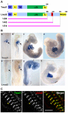
Fig. 1.
Fig. 1.
Structure, expression and localization of chicken Lmod2. (A) Domain structure of Tmod1 and Lmod2. Tmod1 domain structure was determined based on biochemical, morphological and structural analysis (Kostyukova, 2008). Lmod2 domain structure was determined based on amino acid sequence homology to Tmod1 (Chereau et al., 2008). Lmod2 and Tmod1 share actin-capping (A1,A2), tropomyosin-binding (TM1) and leucine-rich repeat (LRR) domains. The second tropomyosin-binding domain (TM2) is unique to Tmod1. Lmod2 contains a C-terminal extension, which includes proline-rich (P) and actin-binding Wiskott–Aldrich syndrome protein homology 2 (WH2) domains. Truncated fragments used in this study are indicated by pink lines (residues 1–394, 1–443 and 1–514). (B) Whole-mount in situ hybridization localization of TMOD1 (a–d) and LMOD2 (e–h) transcripts in developing chicken embryos. At HH stage 11, TMOD1 expression was detected in the looping heart (a, arrow), whereas expression of LMOD2 was undetectable (e). By HH stage 14, TMOD1 and LMOD2 were both detected in the myocardium (b,f, arrows), although TMOD1 staining was consistently more intense than that of LMOD2. By HH Stage 17 (c,g), TMOD1 and LMOD2 transcripts were localized to the myocardium and to the somites. Similar patterns were observed at HH stage 19 (d,h, asterisks). (C) Tmod1 (green) and Lmod2 (red) both localized to thin filament pointed ends in cardiomyocytes. Merged image revealed some non-overlap of Tmod1 and Lmod2 staining. Scale bar: 5 μm.
Fig. 2.
Fig. 2.
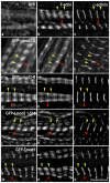
Expression of full-length Lmod2 results in uniform elongation of actin-thin filaments from their pointed ends; its WH2 domain appears to be required for this effect. Cardiomyocytes expressing GFP alone (a), GFP-Lmod2 (d,g), GFP-Lmod2(1–514) (j), or GFP-Tmod1 (m) were stained with Phalloidin (b,e,h,k,n) and for α-actinin to label the Z-discs (c,f,i,l,o). Expression of full-length GFP-Lmod2 resulted in elongation of thin filament pointed ends as discerned by a narrow gap, or sometimes the absence of a gap, in phalloidin staining in the middle of the sarcomere (e,h). GFP-Lmod2-A is an example of myofibrils with no detectable gap; GFP-Lmod2-B is an example of myofibrils with a discernible narrow gap. Conversely, expression of GFP-Lmod2(1–514), which lacks the actin-binding WH2 domain, resulted in a clear, wide gap of Phalloidin staining in the H-zone, comparable with that seen following the expression of GFP-Tmod1. Doublets of GFP-Lmod2(1–514) were clearly observed at the pointed ends (note: no doublets were observed in the cells expressing full-length GFP-Lmod2). Pointed ends, yellow arrowheads; Z-discs, red arrowheads. Scale bar: 5 μm.
Fig. 3.
Fig. 3.
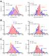
Expression of full-length Lmod2 results in longer actin filaments, whereas expression of Lmod2(1–514) or Tmod1 results in shorter actin filaments. Cardiomyocytes expressing GFP alone, GFP-Lmod2, GFP-Lmod2(1–514) or GFP-Tmod1 were stained with phalloidin to measure thin filament lengths (from pointed end to pointed end: n=150, 60, 75, 89, respectively) and for α-actinin to measure sarcomere lengths (from Z-disc to Z-disc: n=54, 55, 70, 45, respectively). In the cells expressing full-length GFP-Lmod2, only myofibrils displaying distinguishable F-actin gaps were used for measurements. Data are from 2–5 cultures and the values are presented as a percentage (mean ± s.d.). (A) Thin filament lengths were significantly longer in cells expressing GFP-Lmod2 compared with those in cells expressing GFP alone (*P<0.01, Student's t-test). (B,C) By contrast, thin filament lengths were significantly reduced in cells expressing GFP-Lmod2(1–514) or GFP-Tmod1 (*P<0.01). No significant differences in sarcomere lengths were observed (right panels).
Fig. 4.
Fig. 4.
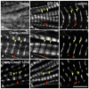
Expression of Lmod2 also results in longer thin filaments marked by GFP-tropomyosin. Cardiomyocytes expressing GFP-tropomyosin (b,e,h) and mCherry alone (a), mCherry-Lmod2 (d), or mCherry-Lmod2(1–514) (g) were stained for α-actinin (c,f,i). In cells expressing mCherry alone, GFP-tropomyosin assembled in its characteristic pattern along the thin filaments with clear gaps visible at the pointed and barbed ends (b, red and yellow arrowheads, respectively). However, in cells expressing mCherry-Lmod2, the gap in GFP-tropomyosin assembly at the H-zone disappeared indicating the thin filaments had elongated (note, the increase in intensity at the center of the sarcomere indicates overlap of GFP-tropomyosin from opposite sarcomeres; yellow arrowheads). Interestingly, expression of mCherry-Lmod2(1–514) resulted in a clear, wide gap of GFP-tropomyosin assembly at both the H-zone and Z-disc, analogous to expression of mCherry alone. Yellow arrowheads, pointed ends; red arrowheads, Z-discs. Scale bar: 5 μm.
Fig. 5.
Fig. 5.
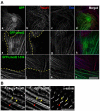
Lmod2 and Lmod2(1–514) can displace Tmod1 from the pointed ends. (A) Cardiomyocytes expressing GFP alone (a) GFP-Lmod2 (e), or GFP-Lmod2(1–514) (i) were stained for Tmod1 (red, b,f,j) and Z-disc titin (blue, c,g,k). Little or no detectable Tmod1 localization was observed in cells expressing full-length GFP-Lmod2 or GFP-Lmod2(1–514) (f,j). This suggests that both proteins can compete with Tmod1 for pointed-end association. Yellow dashed lines indicate boundaries between cells. Scale bar: 15 μm. (B) Pointed-end assembly of GFP-Lmod2 was not observed in cardiomyocytes co-expressing mCherry-Tmod1 (left) and GFP-Lmod2 (center). These data suggest that Tmod1 can outcompete Lmod2 for pointed-end binding. Yellow arrowheads, pointed ends; red arrowheads, Z-discs. Scale bar: 5 μm.
Fig. 6.
Fig. 6.
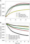
Lmod2 binds to, but does not cap, the pointed ends of preformed actin filaments; tropomyosin enhances this binding. Pyrene fluorescence, which measures actin polymerization, is plotted vs. time. Initial protein concentrations were 6 nM gelsolin-capped filaments and 1.1 μM G-actin. (A) Lmod2 (20 nM) was able to nucleate G-actin (red triangles); however, its addition to gelsolin-capped actin seeds reduced this ability, indicating that it bound the seeds (i.e. was not available to nucleate) (green squares). (B) The addition of stTM (1 μM) enhanced the binding of Lmod2 (red triangles). Lmod2 association with the gelsolin-capped seeds could be partially removed by adding 20 nM Tmod1 (green squares), whereas the addition of Tmod1 (20 nM) to the seeds without Lmod2 resulted in a significant decrease in polymerization (yellow diamonds). (C) At a molar concentration that was lower than the concentration of the seeds (6 nM), Lmod2 (5 nM) mostly bound to the seeds (green squares). Note that 5 nM Lmod2 could still nucleate G-actin in the absence of seeds (red triangles). (D) At 5 nM, Lmod2 completely bound to the seeds in the presence of tropomyosin (1 μM) (red triangles). As before, Lmod2 association with the seeds could be partially removed by adding 20 nM Tmod1 (green squares). Control, A/G, is actin plus gelsolin-capped actin filaments (red circles).
Fig. 7.
Fig. 7.
Tropomyosin increases the affinity of Lmod2 for actin seeds and enhances the capping activity of Lmod2(1–514). The polymerization data were fitted to a single exponential equation and initial polymerization rates (Rexp) were calculated. Rexp values were then normalized by the initial rate obtained for polymerization of actin on gelsolin-capped actin seeds (Rcontrol) (open hexagon). Initial protein concentrations were 6 nM gelsolin-capped filaments and 1.1 μM G-actin. In the presence of tropomyosin (1 μM), Lmod2 decreased the rate of actin polymerization (red triangles). This suggests that the affinity of Lmod2 for the seeds increased in the presence of tropomyosin. At 5, 20 and 50 nM, tropomyosin-dependent capping activity of Lmod2(1–514) was observed (yellow diamonds), although no actin capping activity was seen in the absence of tropomyosin (green squares). Although not dramatically different, the capping activity of Lmod2(1–514) is lower than that of Tmod1 (blue triangles).
Fig. 8.
Fig. 8.
Lmod2(1–514), similarly to Tmod1, can inhibit actin polymerization and depolymerization in the presence of tropomyosin. (A) Lmod2(1–514) (20 nM) had weak nucleation activity when added to actin alone (red triangles). When added to stTM-decorated filaments, Lmod2(1–514) (20 nM) remarkably prevented actin polymerization (yellow diamonds), whereas addition to filaments without stTM only resulted in a slight reduction of polymerization (green squares). Initial protein concentrations were 6 nM gelsolin-capped filaments, 1.1 μM G-actin, and 1 μM stTM. Control, A/G, is actin plus gelsolin-capped actin filaments. (B) Lmod2(1–514) prevented depolymerization from the pointed ends in a dose-dependent manner (downward red triangles: 1 nM, green squares: 5 nM, yellow diamonds: 20 nM). At 20 nM Lmod(1–514) inhibited polymerization similarly to 20 nM Tmod1 (upward blue triangles). The graph shows the depolymerization of actin at the pointed ends of gelsolin-capped actin filaments (0.6 μM F-actin, 6 nM gelsolin) in the presence of 0.75 μM stTM, 20 nM Tmod1 or different concentrations of Lmod2(1–514).
Fig. 9.
Fig. 9.
Model of the function of Lmod2 at thin filament pointed ends. (A) During early stages of heart development when the myofibrils have formed, only Tmod1 is expressed, resulting in short thin filaments. (B) At a later stage of development, Lmod2 is expressed. Lmod2 assembles at the pointed ends of the thin filaments and competes for binding with Tmod1. Since Lmod2 is unable to cap, the thin filaments elongate. (C) Interactions between Tmod1, Lmod2, tropomyosin, and actin at the pointed ends probably contribute to maintaining the final lengths of the thin filaments.

Similar articles

Cited by

References

    1. Almenar-Queralt A., Lee A., Conley C. A., Ribas de Pouplana L., Fowler V. M. (1999). Identification of a novel tropomodulin isoform, skeletal tropomodulin, that caps actin filament pointed ends in fast skeletal muscle. J. Biol. Chem. 274, 28466-28475 - PubMed
    1. Babcock G. G., Fowler V. M. (1994). Isoform-specific interaction of tropomodulin with skeletal muscle and erythrocyte tropomyosins. J. Biol. Chem. 269, 27510-27518 - PubMed
    1. Bai J., Hartwig J. H., Perrimon N. (2007). SALS, a WH2-domain-containing protein, promotes sarcomeric actin filament elongation from pointed ends during Drosophila muscle growth. Dev. Cell 13, 828-842 - PubMed
    1. Baker R. K., Antin P. B. (2003). Ephs and ephrins during early stages of chick embryogenesis. Dev. Dyn. 228, 128-142 - PubMed
    1. Bernstein B. W., Bamburg J. R. (1982). Tropomyosin binding to F-actin protects the F-actin from disassembly by brain actin-depolymerizing factor (ADF). Cell Motil. 2, 1-8 - PubMed

Publication types