

Activating stress-activated protein kinase-mediated cell death and inhibiting epidermal growth factor receptor signaling: a promising therapeutic strategy for prostate cancer
- PMID: 20736346
- PMCID: PMC2948854
- DOI: 10.1158/1535-7163.MCT-10-0180
Activating stress-activated protein kinase-mediated cell death and inhibiting epidermal growth factor receptor signaling: a promising therapeutic strategy for prostate cancer
Abstract
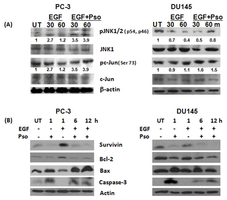
Epidermal growth factor receptor (EGFR) activation is an important event that regulates mitogenic signaling, such as the Raf, mitogen-activated protein kinase (MAPK), and extracellular signal-regulated kinase 1/2 cascades. EGFR activation has been implicated in the transition of prostate cancer from androgen dependence to independence. Therefore, inhibition of EGFR may effectively suppress prostate cancer growth and progression. The goal of this study was to determine whether the natural compound psoralidin alters EGFR-mediated signaling resulting in the inhibition of prostate cancer growth. Results suggest that inhibition of EGFR alone (by serum deprivation) fails to induce stress-mediated protein kinases (SAPK), namely, Jun NH(2)-terminal kinase/c-Jun signaling, in androgen-independent prostate cancer (AIPC) cells. Treatment with psoralidin, however, inhibited both constitutive and EGF-induced EGFR activation and simultaneously triggered SAPK signaling, resulting in the induction of apoptosis in AIPC cells. In addition, psoralidin downregulated EGFR-regulated MAPK signaling and inhibited cell proliferation in AIPC cells. Oral administration of psoralidin effectively suppressed PC-3 xenograft tumors in nude mice. Compared with control tumors, inhibition of pEGFR expression and an increase in the phosphorylation, activation, and nuclear translocation of c-Jun were observed in psoralidin-treated tumor sections. Our studies suggest that psoralidin may be a potent therapeutic agent that modulates EGFR-mediated key epigenetic events in AIPC.
Figures


Similar articles
-
Inhibiting TNF-mediated signaling: a novel therapeutic paradigm for androgen independent prostate cancer.Apoptosis. 2010 Feb;15(2):153-61. doi: 10.1007/s10495-009-0416-9. Apoptosis. 2010. PMID: 19851870 Free PMC article.
-
Grape seed extract inhibits EGF-induced and constitutively active mitogenic signaling but activates JNK in human prostate carcinoma DU145 cells: possible role in antiproliferation and apoptosis.Oncogene. 2003 Mar 6;22(9):1302-16. doi: 10.1038/sj.onc.1206265. Oncogene. 2003. PMID: 12618755
-
Psoralidin, an herbal molecule, inhibits phosphatidylinositol 3-kinase-mediated Akt signaling in androgen-independent prostate cancer cells.Cancer Prev Res (Phila). 2009 Mar;2(3):234-43. doi: 10.1158/1940-6207.CAPR-08-0129. Epub 2009 Feb 17. Cancer Prev Res (Phila). 2009. Retraction in: Cancer Prev Res (Phila). 2011 Nov;4(11):1945. doi: 10.1158/1940-6207.CAPR-11-0467. PMID: 19223576 Free PMC article. Retracted.
-
Combination of Carmustine and Selenite Inhibits EGFR Mediated Growth Signaling in Androgen-Independent Prostate Cancer Cells.J Cell Biochem. 2017 Dec;118(12):4331-4340. doi: 10.1002/jcb.26086. Epub 2017 May 25. J Cell Biochem. 2017. PMID: 28430389
-
Gonadotropin-releasing hormone induces apoptosis of prostate cancer cells: role of c-Jun NH2-terminal kinase, protein kinase B, and extracellular signal-regulated kinase pathways.Cancer Res. 2004 Aug 15;64(16):5736-44. doi: 10.1158/0008-5472.CAN-04-1156. Cancer Res. 2004. PMID: 15313914
Cited by
-
Molecular interplay between cdk4 and p21 dictates G0/G1 cell cycle arrest in prostate cancer cells.Cancer Lett. 2013 Sep 1;337(2):177-83. doi: 10.1016/j.canlet.2013.05.014. Epub 2013 May 16. Cancer Lett. 2013. PMID: 23684928 Free PMC article.
-
KIF15 Promotes Progression of Castration Resistant Prostate Cancer by Activating EGFR Signaling Pathway.Front Oncol. 2021 Nov 4;11:679173. doi: 10.3389/fonc.2021.679173. eCollection 2021. Front Oncol. 2021. PMID: 34804913 Free PMC article.
-
Psoralidin induces autophagy through ROS generation which inhibits the proliferation of human lung cancer A549 cells.PeerJ. 2014 Sep 9;2:e555. doi: 10.7717/peerj.555. eCollection 2014. PeerJ. 2014. PMID: 25250213 Free PMC article.
-
The coumarin psoralidin enhances anticancer effect of tumor necrosis factor-related apoptosis-inducing ligand (TRAIL).Molecules. 2012 May 29;17(6):6449-64. doi: 10.3390/molecules17066449. Molecules. 2012. PMID: 22643355 Free PMC article.
-
Synthesis of Psoralidin derivatives and their anticancer activity: First synthesis of Lespeflorin I1.Tetrahedron. 2016 Jun 9;72(23):3324-3334. doi: 10.1016/j.tet.2016.04.066. Tetrahedron. 2016. PMID: 27698514 Free PMC article.
References
-
- Lee TL, Yeh J, Van Waes C, Chen Z. Epigenetic modification of SOCS-1 differentially regulates STAT3 activation in response to interleukin-6 receptor and epidermal growth factor receptor signaling through JAK and/or MEK in head and neck squamous cell carcinomas. Mol Cancer Ther. 2006;5:8–19. - PubMed
-
- Tillotson JK, Rose DP. Endogenous secretion of epidermal growth factor peptides stimulates growth of DU145 prostate cancer cells. Cancer Lett. 1991;60:109–12. - PubMed
-
- Marks RA, Zhang S, Montironi R, et al. Epidermal growth factor receptor (EGFR) expression in prostatic adenocarcinoma after hormonal therapy: a fluorescence in situ hybridization and immunohistochemical analysis. Prostate. 2008;68:919–23. - PubMed
Publication types
MeSH terms
Substances
Grants and funding
LinkOut - more resources
Full Text Sources
Medical
Research Materials
Miscellaneous
