

Nephromyces, a beneficial apicomplexan symbiont in marine animals
- PMID: 20736348
- PMCID: PMC2941302
- DOI: 10.1073/pnas.1002335107
Nephromyces, a beneficial apicomplexan symbiont in marine animals
Abstract
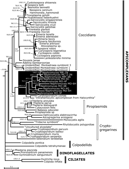
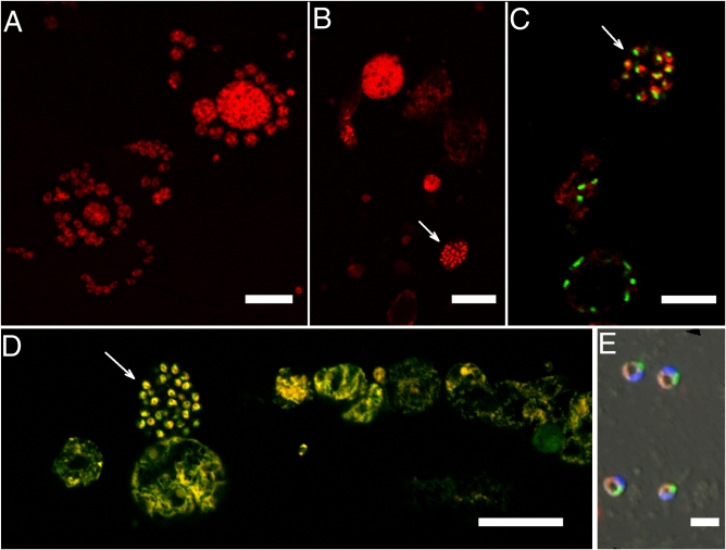
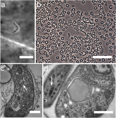
With malaria parasites (Plasmodium spp.), Toxoplasma, and many other species of medical and veterinary importance its iconic representatives, the protistan phylum Apicomplexa has long been defined as a group composed entirely of parasites and pathogens. We present here a report of a beneficial apicomplexan: the mutualistic marine endosymbiont Nephromyces. For more than a century, the peculiar structural and developmental features of Nephromyces, and its unusual habitat, have thwarted characterization of the phylogenetic affinities of this eukaryotic microbe. Using short-subunit ribosomal DNA (SSU rDNA) sequences as key evidence, with sequence identity confirmed by fluorescence in situ hybridization (FISH), we show that Nephromyces, originally classified as a chytrid fungus, is actually an apicomplexan. Inferences from rDNA data are further supported by the several apicomplexan-like structural features in Nephromyces, including especially the strong resemblance of Nephromyces infective stages to apicomplexan sporozoites. The striking emergence of the mutualistic Nephromyces from a quintessentially parasitic clade accentuates the promise of this organism, and the three-partner symbiosis of which it is a part, as a model for probing the factors underlying the evolution of mutualism, pathogenicity, and infectious disease.
Conflict of interest statement
The authors declare no conflict of interest.
Figures
References
-
- Saffo MB. Coming to terms with a field: Words and concepts in symbiosis. Symbiosis. 1992;14:17–31.
-
- Keeling PJ, et al. The tree of eukaryotes. Trends Ecol Evol. 2005;20:670–676. - PubMed
-
- Edman JC, et al. Ribosomal RNA sequence shows Pneumocystis carinii to be a member of the fungi. Nature. 1988;334:519–522. - PubMed
-
- de Lacaze-Duthiers H. Account of solitary ascidians from the coasts of France (Translated from French) Archiv Zool Exp Gen. 1874;3:309–311.
-
- Saffo MB, Lowenstam HA. Calcareous deposits in the renal sac of a molgulid tunicate. Science. 1978;200:1166–1168. - PubMed
Publication types
MeSH terms
Associated data
- Actions
- Actions
- Actions
- Actions
- Actions
- Actions
- Actions
- Actions
- Actions
- Actions
Grants and funding
LinkOut - more resources
Full Text Sources
Molecular Biology Databases
Miscellaneous
