. 2010 Sep 14;107(37):16325-30.
doi: 10.1073/pnas.1003459107. Epub 2010 Aug 24.

Altered distributions of Gemini of coiled bodies and mitochondria in motor neurons of TDP-43 transgenic mice

Affiliations

Altered distributions of Gemini of coiled bodies and mitochondria in motor neurons of TDP-43 transgenic mice

Xiu Shan et al. Proc Natl Acad Sci U S A. .

Abstract

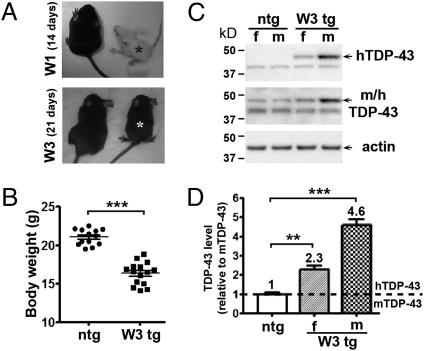
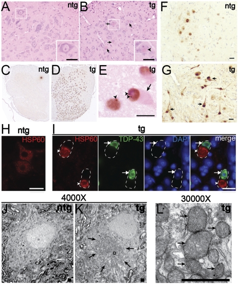
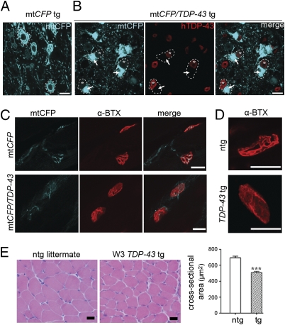
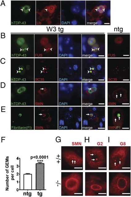
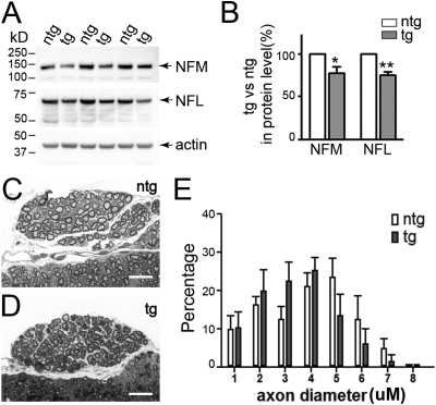
TAR DNA-binding protein-43 (TDP-43), a DNA/RNA-binding protein involved in RNA transcription and splicing, has been associated with the pathophysiology of neurodegenerative diseases, including ALS. However, the function of TDP-43 in motor neurons remains undefined. Here we use both gain- and loss-of-function approaches to determine roles of TDP-43 in motor neurons. Mice expressing human TDP-43 in neurons exhibited growth retardation and premature death that are characterized by abnormal intranuclear inclusions composed of TDP-43 and fused in sarcoma/translocated in liposarcoma (FUS/TLS), and massive accumulation of mitochondria in TDP-43-negative cytoplasmic inclusions in motor neurons, lack of mitochondria in motor axon terminals, and immature neuromuscular junctions. Whereas an elevated level of TDP-43 disrupts the normal nuclear distribution of survival motor neuron (SMN)-associated Gemini of coiled bodies (GEMs) in motor neurons, its absence prevents the formation of GEMs in the nuclei of these cells. Moreover, transcriptome-wide deep sequencing analysis revealed that a decrease in abundance of neurofilament transcripts contributed to the reduction of caliber of motor axons in TDP-43 mice. In concert, our findings indicate that TDP-43 participates in pathways critical for motor neuron physiology, including those that regulate the normal distributions of SMN-associated GEMs in the nucleus and mitochondria in the cytoplasm.


Conflict of interest statement

The authors declare no conflict of interest.

Figures

Fig. 1.
Fig. 1.
Early postnatal growth retardation in mice expressing wild-type TDP-43. (A) Decrease in size of wild-type TDP-43 transgenic (tg) male mice derived from independent founders W1 and W3; asterisks indicate, respectively, transgenic mice at 14 and 21 d of age. (B) Body weights of 4-wk-old male TDP-43 transgenic mice from line W3 were compared with nontransgenic (ntg) littermates. Note significant reduction in body weights of TDP-43 transgenic mice (ntg, n = 13; W3 tg, n = 15; P < 0.0001). Error bars indicate SEM. (C) Accumulation of TDP-43 in spinal cords of W3 mice at 4 wk of age. A mouse monoclonal antibody recognizing human-specific TDP-43 was used to determine the level of transgene expression, whereas a rabbit antibody against both mouse and human TDP-43 was used to compare the level of transgene expression with that of endogenous TDP-43. f, female; m, male. (D) Densitometric analysis of TDP-43 protein levels in 4-wk-old W3 mice using the rabbit antibody against both mouse and human TDP-43 (female tg, n = 3; male tg, n = 6; ntg littermates, n = 4). Compared with that of mouse endogenous TDP-43, the levels of human TDP-43 are, respectively, 1.3- and 3.6-fold in 4-wk-old W3 transgenic females (P = 0.0011) and males (P < 0.0001). Error bars indicate SEM.
Fig. 2.
Fig. 2.
Pathological abnormalities in spinal cords of 3-wk-old W3 TDP-43 mice. (A and B) Hematoxylin and eosin staining reveals eosinophilic aggregates in cell bodies of motor neurons in spinal cords of transgenic (tg) mice (B, arrows); such structures were not identified in nontransgenic (ntg) littermates (A). Boxes at bottom right are enlarged micrographs showing a healthy motor neuron (A) and a neuron bearing a large cytoplasmic aggregate marked by an arrowhead (B). (CE) The antibody recognizing human TDP-43 specifically reveals that the transgene is extensively expressed in spinal cord neurons of transgenic mice (D and E); no immunoreactivity is detected in nontransgenic mice (C). hTDP-43 is localized in the nucleus and forms intranuclear granular structures (E, arrowheads) in some neurons bearing cytoplasmic aggregates (E, arrow), which are identified by eosin counterstain. (F and G) Immunohistochemical analysis with ubiquitin antibody shows that the level of ubiquitination is elevated in the spinal cords of transgenic mice (G) when compared with that in nontransgenic mice (F). Arrows point to neurons with eccentric nuclei, indicating the presence of cytoplasmic aggregates, and those neurons are heavily stained with ubiquitin, particularly within the nuclear compartments. (H and I) Double immunofluorescence analyses using antisera against HSP60, a mitochondrial marker, and hTDP-43 suggest the abnormal accumulation of mitochondria (I, arrowheads) in transgene-expressing neurons delineated by dashed lines. Arrows in I point to staining of hTDP-43 in the nucleus. Note the normal distinct distribution of HSP60 in the cytoplasm of motor neurons of nontransgenic mice (H). (JL) Electron micrographs of spinal motor neurons show massive accumulation of mitochondria (L; arrows denote mitochondria) within large, cytoplasmic aggregates in transgenic mice (K; arrows point to a cytoplasmic aggregate). [Scale bars, 20 μm (A–B and EI), 1 μm (J–L).]
Fig. 3.
Fig. 3.
Altered distribution of mitochondria in motor neurons and abnormal neuromuscular junctions in 3-wk-old W3 TDP-43 mice. (A and B) The distribution of CFP-labeled mitochondria normally observed in motor neurons of mtCFP tg mice (A; two motor neurons are delineated by dashed lines) but reorganized and confined within large cytoplasmic inclusions (B, arrows; asterisks denote nuclei of motor neurons) in compound mtCFP;TDP-43 tg mice (B; three such motor neurons are delineated by dashed lines). (C) A decreased level of mitochondria is observed at nerve terminals of neuromuscular junctions of double transgenic mice (Lower) compared with mtCFP mice (Upper). Note the lack of mitochondria invested into the presynaptic terminals; postsynaptic terminals are marked by staining with α-bungrotoxin (α-BTX). Three mtCFP and four mtCFP;TDP-43 double transgenic mice were examined. (D and E) Alteration of postsynaptic distribution of AChR on muscle fibers (D) and a decrease of muscle size (E) are observed in TDP-43 transgenic mice. Quantitative analysis (E) shows ≈20% reduction of cross-sectional area of muscle fibers in transgenic mice compared with nontransgenic littermate controls (P < 0.001, n = 4). Error bars indicate SEM. (Scale bars, 20 μm.)
Fig. 4.
Fig. 4.
TDP-43 regulates SMN-associated GEMs in motor neurons. (AC) Double immunofluorescence analyses of components of intranuclear TDP-43-immunoreactive granules in spinal motor neurons of 3-wk-old W3 transgenic mice (n = 3) and control littermates (n = 3) using antisera against TDP-43 (green channel) and a series of nuclear markers (red channel). Representative double immunofluorescence staining showed a TDP-43-positive neuron (A) lacked ubiquitin (Ub) immunoreactivity in intranuclear granules (A, arrowheads), but was immunoreactive with antisera against either Fused in Sarcoma (FUS; B, arrowheads) or a marker of non-snRNP splicing speckles (SC35; C, arrowheads). (D and E) Analysis of the number and distribution of the Gemini of coiled bodies (GEMs) using SMN antibody. Whereas motor neurons in nontransgenic mice showed two SMN-containing GEMs (arrows; D, ntg), the number of GEMs increases significantly in TDP-43 transgenic mice (arrow; D, W3 tg). Error bars indicate SEM. In contrast to motor neurons from nontransgenic littermates in which one GEM is associated with the nucleolus [arrows, E (ntg, SMN/Fl)], motor neurons that harbor cytoplasmic aggregates from transgenic mice showed SMN is present diffusely within the nucleolus, identified by fibrillarin antibody (E), and SMN-containing GEMs are confined to the perinucleolar region (E, arrow). All sections are stained with DAPI to mark nuclei. [Scale bars, 10 μm (AE).] (F) Quantitative measurement on the number of GEMs in neurons. Counts were based on 20 serial sections for each spinal cord. P < 0.0001, n = 3. Error bars indicate SEM. (GI) Absence of GEMs in spinal motor neurons lacking TDP-43. Immunofluorescence analysis using antisera against SMN (G), Gemin 2 (H), and Gemin 8 (I) localized two GEMs (indicated by arrows) in the nucleus of motor neurons of control mice (+/+, upper). No GEM can be identified in the nucleus of motor neurons derived from conditional Tardbp knockout mice (−/−, lower) using antisera against SMN (G), Gemin 2 (H), or Gemin 8 (I); 100 motor neurons from three control or three conditional Tardbp knockout mice were examined. [Scale bars, 20 μm (GI).] G2, Gemin 2; G8, Gemin 8.
Fig. 5.
Fig. 5.
Decreased levels of neurofilament proteins and reduction in caliber of motor axons in TDP-43 mice. (A and B) Protein blot analysis of neurofilament from spinal cords of nontransgenic (ntg) and W3 transgenic (tg) mice (A) showed, respectively, a 23% and a 25% reduction in the protein level of NF-M and NF-L in 3-wk-old W3 transgenic mice (B; n = 5; *P < 0.0164 and **P < 0.0001). Error bars indicate SEM. (CE) Analysis of caliber of axons in ventral roots. Note the reduction in number of large-caliber axons and a concomitant increase in the number of small-caliber axons in 3-wk-old W3 transgenic mice (D and E; n = 3) when compared with nontransgenic littermates (C and E; n = 3). Error bars indicate SEM. Scale bars, 20 μm (C and D).

Similar articles

Cited by

References

    1. Forman MS, Trojanowski JQ, Lee VM. TDP-43: A novel neurodegenerative proteinopathy. Curr Opin Neurobiol. 2007;17:548–555. - PMC - PubMed
    1. Lagier-Tourenne C, Cleveland DW. Rethinking ALS: The FUS about TDP-43. Cell. 2009;136:1001–1004. - PMC - PubMed
    1. Buratti E, et al. Nuclear factor TDP-43 and SR proteins promote in vitro and in vivo CFTR exon 9 skipping. EMBO J. 2001;20:1774–1784. - PMC - PubMed
    1. Mercado PA, Ayala YM, Romano M, Buratti E, Baralle FE. Depletion of TDP 43 overrides the need for exonic and intronic splicing enhancers in the human apoA-II gene. Nucleic Acids Res. 2005;33:6000–6010. - PMC - PubMed
    1. Bose JK, Wang IF, Hung L, Tarn WY, Shen CK. TDP-43 overexpression enhances exon 7 inclusion during the survival of motor neuron pre-mRNA splicing. J Biol Chem. 2008;283:28852–28859. - PMC - PubMed

Publication types

Substances

Associated data