. 2010 Sep 1;70(17):6988-98.
doi: 10.1158/0008-5472.CAN-10-0242. Epub 2010 Aug 24.

Matrix metalloproteinase-9 regulates tumor cell invasion through cleavage of protease nexin-1

Affiliations

Matrix metalloproteinase-9 regulates tumor cell invasion through cleavage of protease nexin-1

Danmei Xu et al. Cancer Res. .

Abstract

Matrix metalloproteinase-9 (MMP-9) expression is known to enhance the invasion and metastasis of tumor cells. In previous work based on a proteomic screen, we identified the serpin protease nexin-1 (PN-1) as a potential target of MMP-9. Here, we show that PN-1 is a substrate for MMP-9 and establish a link between PN-1 degradation by MMP-9 and regulation of invasion. PN-1 levels increased in prostate carcinoma cells after downregulation of MMP-9 and in tissues of MMP-9-deficient mice, consistent with PN-1 degradation by MMP-9. We identified three MMP-9 cleavage sites in PN-1 and showed that mutations in those sites made PN-1 more resistant to MMP-9. Urokinase plasminogen activator (uPA) is inhibited by PN-1. MMP-9 augmented uPA activity in the medium of PC3-ML cells by degrading PN-1. Prostate cancer cells, overexpressing PN-1 or treated with MMP-9 shRNA, had reduced cell invasion in Matrigel. PN-1 siRNA restored uPA activity and the invasive capacity. PN-1 mutated in the serpin inhibitory domain, the reactive center loop, failed to inhibit uPA and to reduce Matrigel invasion. This study shows a novel molecular pathway in which MMP-9 regulates uPA activity and tumor cell invasion through cleavage of PN-1.


Figures
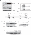
Figure 1
Figure 1. PN-1 is cleaved by MMP-9 in tumor cells
A) Conditioned media (CM) and whole cell lysates (WL) collected from PC-3ML (-) or MMP9 KD PC-3ML (KD) cells with or without transfection of the pcDNA3-PN-1 expression vector, were subjected to SDS-PAGE electrophoresis and blotted with anti-PN-1, anti-MMP-9 or anti-Actin antibodies. B) Within the square an immunoblot after limited cleavage of PN-1 by recombinant MMP-9 in solution. Sizes of cleaved fragments as described (7). Schematic illustration of PN-1: with the black arrow heads indicating the putative cleavage sites and the altered amino acids in the mutants. Known functional domains such as LRP and heparin binding domains, reactive centre loop (RCL) domain are indicated. C) PN-1 in conditioned media collected from PC-3ML cells (i upper panel, ii and iii) or MMP-9 KD PC-3ML cells (i bottom panel), transfected with either WT PN-1 or indicated mutants. was analysed by immunoblot. The most resistant PN-1 mutants against MMP-9 cleavage were marked with asterisks *.
Figure 2
Figure 2. PN-1 is targeted by MMP-9 in vivo
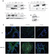
A) Tissue lysates of indicated organs from C57/B6 WT (+/+) or MMP-9 KO (−/−) mice were resolved by SDS-PAGE gel and blotted with anti-mouse PN-1 or anti-β-actin antibody (i and ii). 20ng recombinant mouse PN-1 protein (rPN-1) was a control. Please note β-actin is not present in heart or muscle. Panel iii) shows Panel i) after longer exposure. A PN-1 degradation fragment was now apparent in the lysates from the seminal vesicles and the prostate. B) Primary bone marrow derived cells (BMDC) were grown from C57/B6 WT (+/+) or MMP-9 KO (−/−) mice, serum free conditioned media was subjected to immunoblot with anti-MMP-9 or anti-PN-1 antibody. C) Immunohistochemistry showing the expression of PN-1 (green) in seminal vesicles, pancreas and prostate from WT or MMP-9 KO mice. Cell nuclei were stained by Hoechst (blue).
Figure 3
Figure 3. MMP-9 regulates uPA activity through cleavage of PN-1
A) The uPA activity of conditioned media collected from PC-3ML cells (-) or MMP-9 KD PC-3ML (KD) cells, with or without PN-1 transfection, were measured. Triplicate experiments were plotted and subjected to one-way ANOVA statistical analysis, P<0.0001. B) Recombinant uPA was incubated with active MMP-9 at 37°C for 3h. All lanes have 10U uPA. Lane 1 has no MMP-9, lane 2 has 50ng and lane 3 has 100ng MMP-9. The reaction products were resolved by 15% SDS-PAGE and immunoblotted with anti-MMP-9 or anti-uPA antibody. C) Reactions as in B): uPA activity was assayed and normalized against uPA control. P>0.05 by one-way ANOVA test. D) Conditioned media collected from control (-) or His-PN-1 transfected (+) PC-3ML cells were incubated with Ni beads for 3h and washed. The protein complexes were then resolved by SDS-PAGE gel and immunoblotted with indicated antibodies.
Figure 4
Figure 4. PN-1 and MMP-9 regulate tumor cell invasion by targeting uPA
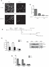
A) 1× 10⁵ PC-3ML or MMP-9 KD PC-3ML cells, mock treated or transfected with 2μg PN-1 vector for 24hrs, were seeded on matrigel invasion inserts. After 48hrs, invaded cells were stained by Hoechst (Shown in Fig 4A-i). Cells from triplicate experiments were counted, normalized against mock control and plotted (ii), followed by ANOVA test and P<0.0001. B) i)-Diagram showing the experimental design. 2×10⁵ PC-3ML cells alone (mock), or treated with Ambion predesigned negative control siRNA (siNEG) or siRNA uPA (siuPA), were allowed to grow for 24hrs on 6-well plates before transfection with 1μg PN-1 vector. 24hrs after transfection, cells were washed and incubated with serum free medium (SFM) for 48hrs, the conditioned media was then collected, subjected to uPA activity assay (ii) and immunoblotting (iii). In parallel, cells were trypsinized, seeded onto matrigel invasion inserts, and allowed to invade for 48hrs. The invaded cells were stained and counted as shown in iv), followed by a two-way ANOVA test (P<0.0001) and Bonferroni posttests for between groups. Comparison of control (Ctrl) and PN-1 overexpression (PN-1), P<0.001 in Mock or siNEG (marked as *), P>0.05 in siuPA. A two-way ANOVA plus Bonferroni posttests were used for analysing uPA activity. P<0.001 in Mock or siNEG (*), P>0.05 in siuPA.
Figure 5
Figure 5. The impact of PN-1 mutations on uPA activity and cell invasiveness
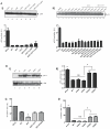
A) PC-3ML cells were transfected with the expression vectors for WT-PN-1 and the indicated mutants. uPA levels in the conditioned media were determined by immunoblot (upper panel). The lower panel shows the uPA activity in the condition media from the transfected cells. The reading of uPA activity was analysed by one-way ANOVA followed by Tukey’s multiple comparison test. Comparison of mock with each mutant PN-1 ectopic expression (P<0.05), however there was no significant difference on uPA activity between WT PN-1 and each mutant. B) Conditioned media were collected from cells transfected with expression vectors for WT PN-1, LRP binding mutants (H48A or D49A), or mutations in the RCL (R365P or S366P), and immunoblotted for PN-1 and uPA levels (i) or assayed for uPA activity (ii). P>0.05 between WT and H48A or D49A (NS), or between mock and R365 or S366P. P<0.05 between WT, H48A or D49A and R365P or S366P (*). C) and D) show invasion arrays after transfection with the indicated plasmids. Invaded cells were counted and plotted. In Fig 5C, no significant difference between WT and I58T+I107T, or I58T+P368H, P<0.05 comparing I58T with the rest of groups. In Fig 5D, P<0.05 between Mock, R365P or S366P and WT, H48A, D49A (*); no significance between WT and H48A or D49A (NS).
Figure 6
Figure 6. PN-1 is essential for MMP-9 regulation of tumor cell invasion
A) Immunoblot (i) showing that 10nM siRNA against PN-1 reduced the expression of PN-1 in conditioned media from both PC-3ML cells (Lane 1-3) and MMP-9KD PC-3ML cells (Lane 4-6). qRT-PCR (ii) showing that the expression of PN-1 RNA in PC-3ML cells was reduced by both of 10nM and 20nM PN-1 siRNA. ANOVA test: P<0.05. B) Conditioned media collected from PC-3ML or MMP-9 KD PC-3ML cells, mock treated or treated with 10nM negative control siRNA (siNeg) or siRNA against PN-1 (siPN-1), were assayed for the uPA activity. The reading from triplicate samples were plotted, analysed by a grouped two-way ANOVA (P<0.05) and Bonferroni posttests. P<0.05 between PC-3ML or MMP-9 KD PC-3ML cells in Mock or siNEG, but P>0.05 in siPN-1. C) In parallel experiments to B), cells were treated with or without 10nM siRNA for 48hrs and then trypsinized, 1×10⁵ cells were seeded on each matrigel chamber. 48hrs later, invaded cells were stained, counted and plotted. Data was analysed a grouped two-way ANOVA (P<0.05) and Bonferroni posttests. P<0.05 between PC-3ML or MMP-9 KD PC-3ML cells in Mock or siNEG, but P>0.05 in siPN-1. D) Schematic model of the pathway that MMP-9 regulates uPA activity and tumor cell invasion through cleaving PN-1. A feedback loop has been suggested between MMP-9 and PN-1 (16). However, we failed to see substantial regulation of MMP-9 by PN-1 (Fig S4).

Similar articles

Cited by

References

    1. Bjorklund M, Koivunen E. Gelatinase-mediated migration and invasion of cancer cells. Biochim Biophys Acta. 2005;1755:37–69. - PubMed
    1. Kaplan RN, Riba RD, Zacharoulis S, et al. VEGFR1-positive haematopoietic bone marrow progenitors initiate the pre-metastatic niche. Nature. 2005;438:820–7. - PMC - PubMed
    1. Coussens LM, Fingleton B, Matrisian LM. Matrix metalloproteinase inhibitors and cancer: trials and tribulations. Science. 2002;295:2387–92. - PubMed
    1. Roy R, Yang J, Moses MA. Matrix metalloproteinases as novel biomarkers and potential therapeutic targets in human cancer. J Clin Oncol. 2009;27:5287–97. - PMC - PubMed
    1. Cauwe B, Van den Steen PE, Opdenakker G. The biochemical, biological, and pathological kaleidoscope of cell surface substrates processed by matrix metalloproteinases. Crit Rev Biochem Mol Biol. 2007;42:113–85. - PubMed

Publication types