. 2010 Dec;9(12):2760-71.
doi: 10.1074/mcp.M110.001875. Epub 2010 Aug 24.

Characterization of macaque pulmonary fluid proteome during monkeypox infection: dynamics of host response

Affiliations

Characterization of macaque pulmonary fluid proteome during monkeypox infection: dynamics of host response

Joseph N Brown et al. Mol Cell Proteomics. 2010 Dec.

Abstract

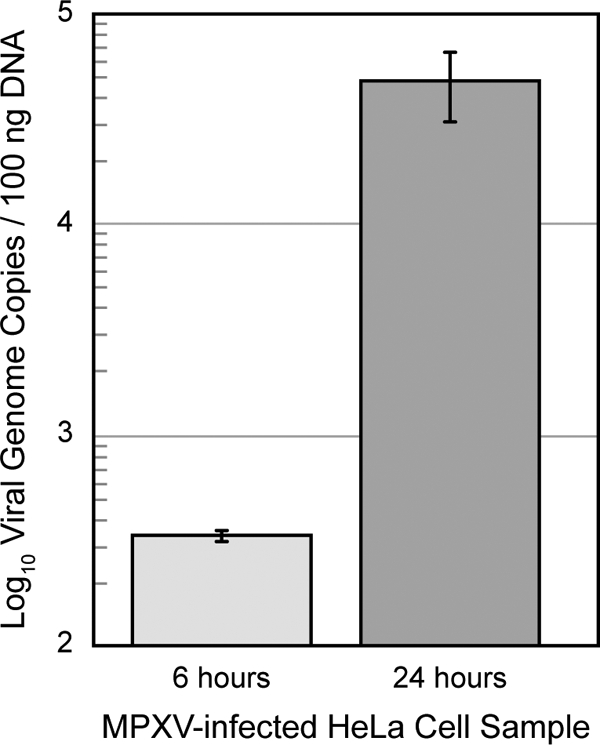
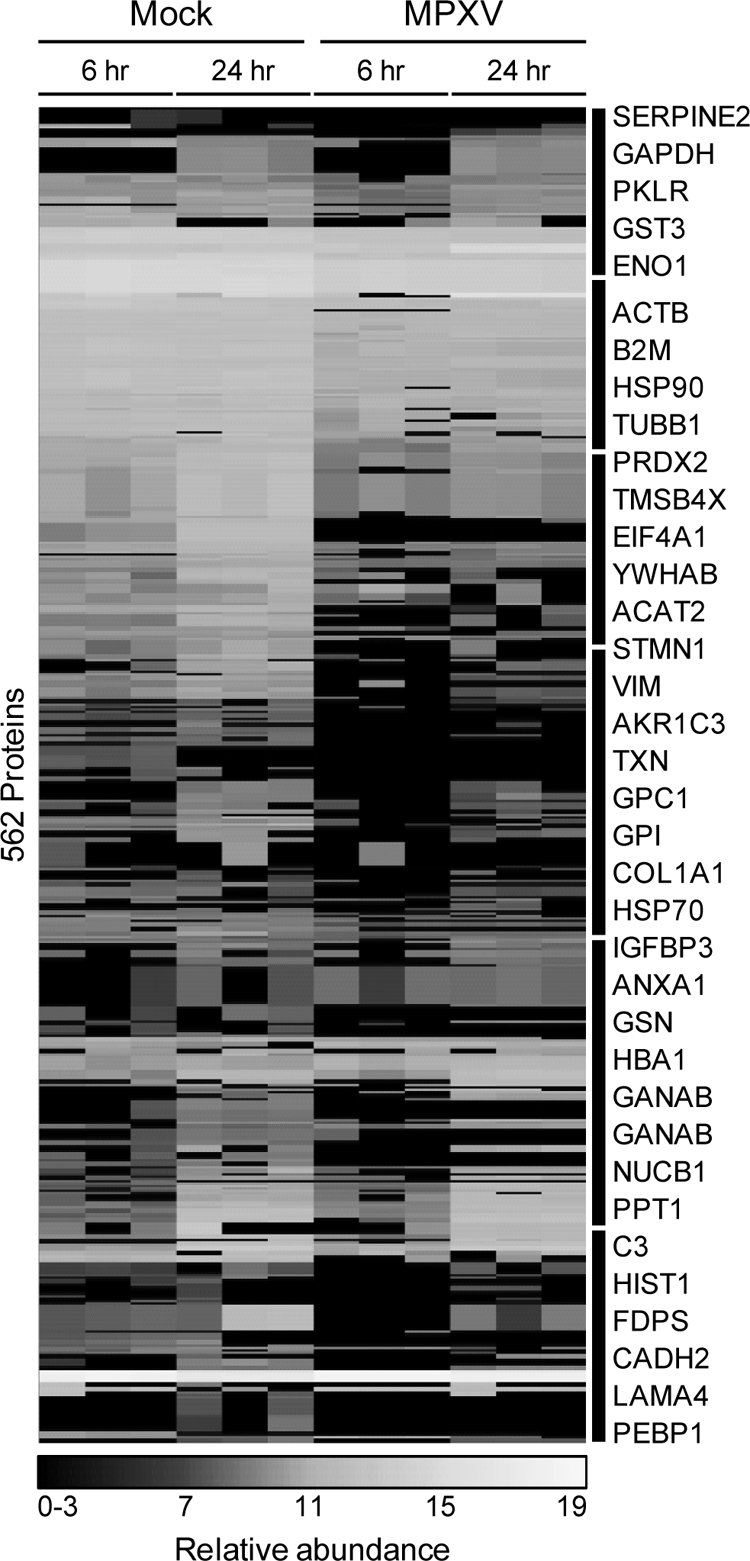
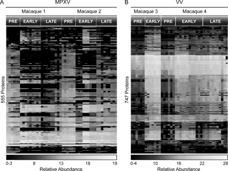
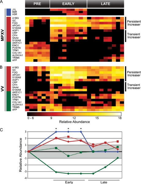
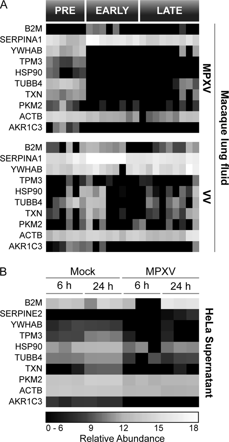
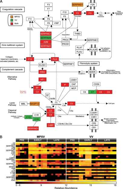
Understanding viral pathogenesis is challenging because of confounding factors, including nonabrasive access to infected tissues and high abundance of inflammatory mediators that may mask mechanistic details. In diseases such as influenza and smallpox where the primary cause of mortality results from complications in the lung, the characterization of lung fluid offers a unique opportunity to study host-pathogen interactions with minimal effect on infected animals. This investigation characterizes the global proteome response in the pulmonary fluid, bronchoalveolar lavage fluid, of macaques during upper respiratory infection by monkeypox virus (MPXV), a close relative of the causative agent of smallpox, variola virus. These results are compared and contrasted against infections by vaccinia virus (VV), a low pathogenic relative of MPXV, and with extracellular fluid from MPXV-infected HeLa cells. To identify changes in the pulmonary protein compartment, macaque lung fluid was sampled twice prior to infection, serving as base line, and up to six times following intrabronchial infection with either MPXV or VV. Increased expression of inflammatory proteins was observed in response to both viruses. Although the increased expression resolved for a subset of proteins, such as C-reactive protein, S100A8, and S100A9, high expression levels persisted for other proteins, including vitamin D-binding protein and fibrinogen γ. Structural and metabolic proteins were substantially decreased in lung fluid exclusively during MPXV and not VV infection. Decreases in structural and metabolic proteins were similarly observed in the extracellular fluid of MPXV-infected HeLa cells. Results from this study suggest that the host inflammatory response may not be the only facilitator of viral pathogenesis, but rather maintaining pulmonary structural integrity could be a key factor influencing disease progression and mortality.


Figures

Fig. 1.
Fig. 1.
MPXV replication kinetics in HeLa cell culture over 24 h. Viral replication kinetics was assessed by using quantitative real-time PCR analysis to determine the number of viral genome copies per 100 ng of total DNA. Error bars represent standard deviations.
Fig. 2.
Fig. 2.
HeLa protein response to monkeypox infection over 24 h. Whole cell lysate proteins (A), supernatant proteins (B), viral proteins observed in the lysate (C), and viral proteins observed in the supernatant (D) are shown. Each row represents an individual protein, and each column represents an individual LC-MS analysis. Relative protein abundance is color-coded relative to the scale at the bottom.
Fig. 3.
Fig. 3.
Representative mass spectra of y and b product ions for viral peptides mapping exclusively to O2L (A), B12R (B), A29L (C), or G4L (D).
Fig. 4.
Fig. 4.
HeLa host supernatant proteins significantly altered by MPXV infection. Clusters of proteins are indicated on the right by black bars and symbols of selected proteins. PKLR, pyruvate kinase 1; GPI, phosphohexose isomerase; VIM, vimentin; FDPS, farnesyl pyrophosphate synthetase; GSN, gelsolin; GANAB, α-glucosidase II α subunit.
Fig. 5.
Fig. 5.
Significantly altered proteins in macaque lung fluid upon inoculation with MPXV (A) or VV (B). Time points above heat maps refer to prior to viral infection (PRE), <16 days (EARLY), or >16 days (LATE).
Fig. 6.
Fig. 6.
Selected proteins from macaque lung fluid observed in both MPXV (A) and VV (B) infections. Viral proteins are designated by a blue bar on the left, proteins that increased during infection are designated by a red bar, and proteins that decreased are designated by a green bar. Persistent and transient increasers are indicated to the right of the heat maps. Averaged and normalized protein abundances are graphed (C) relative to pretreatment. Solid lines with diamonds represent MPXV infection, and dotted lines with squares represent VV infection. Red represents proteins that increased during infection, green represents proteins that decreased, and blue represents the viral proteins. The gray box designates one standard deviation from the mean. Asterisks (*) indicate viral proteins that were only detected in the MPXV infection during the early phase of infection and were given an arbitrary score of +3. GC, vitamin D-binding protein; FGG, fibrinogen γ; CRP, C-reactive protein; CFH, complement factor H; AFP, α-fetoprotein.
Fig. 7.
Fig. 7.
Common interspecies response profile in MPXV- and VV-infected macaque lung fluid (A) and supernatant from MPXV-infected HeLa cells (B).
Fig. 8.
Fig. 8.
MPXV and VV infection impacted the complement cascade in macaque lung fluid. The complement cascade was identified by DAVID as significantly affected (MPXV: p = 1.3 × 10⁻²² and Benjamini = 2.6 × 10⁻²⁰; VV: p = 4.4 × 10⁻³⁰ and Benjamini = 8.9 × 10⁻²⁸) (A). Proteins increased exclusively by MPXV are colored orange, proteins increased exclusive by VV are green, and proteins increased during both infections are red. Expression profiles for proteins involved in the complement pathway are displayed in B. FG, fibrinogen; CF, complement factor H/B, VWF, von Willebrand factor; BDKR, bradykinin receptor; THBD, thrombomodulin; MBL, mannose-binding lectin; PLG, plasminogen; KNG, kininogen; BF, factor B; DF, factor D; IF, factor I; MCP, membrane cofactor protein; DAF, decay-accelerating factor; PLAU, plasminogen activator, urokinase; PLAT, plasminogen activator, tissue; PLAUR, plasminogen activator, urokinase receptor; PROC, protein C; CRRY, CR1-related y.

Similar articles

Cited by

References

    1. Di Giulio D. B., Eckburg P. B. (2004) Human monkeypox: an emerging zoonosis. Lancet Infect. Dis. 4, 15–25 - PMC - PubMed
    1. Weaver J. R., Isaacs S. N. (2008) Monkeypox virus and insights into its immunomodulatory proteins. Immunol. Rev. 225, 96–113 - PMC - PubMed
    1. Henderson D. A., Inglesby T. V., Bartlett J. G., Ascher M. S., Eitzen E., Jahrling P. B., Hauer J., Layton M., McDade J., Osterholm M. T., O'Toole T., Parker G., Perl T., Russell P. K., Tonat K. (1999) Smallpox as a biological weapon: medical and public health management. Working Group on Civilian Biodefense. JAMA 281, 2127–2137 - PubMed
    1. Reed K. D., Melski J. W., Graham M. B., Regnery R. L., Sotir M. J., Wegner M. V., Kazmierczak J. J., Stratman E. J., Li Y., Fairley J. A., Swain G. R., Olson V. A., Sargent E. K., Kehl S. C., Frace M. A., Kline R., Foldy S. L., Davis J. P., Damon I. K. (2004) The detection of monkeypox in humans in the Western Hemisphere. N. Engl. J. Med. 350, 342–350 - PubMed
    1. Centers for Disease Control and Prevention (CDC) (2003) Multistate outbreak of monkeypox—Illinois, Indiana, and Wisconsin, 2003. MMWR Morb. Mortal. Wkly. Rep. 52, 537–540 - PubMed

Publication types