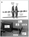
Effects of torsion on intervertebral disc gene expression and biomechanics, using a rat tail model
- PMID: 20736890
- PMCID: PMC3061235
- DOI: 10.1097/BRS.0b013e3181d9b58b
Effects of torsion on intervertebral disc gene expression and biomechanics, using a rat tail model
Abstract
Study design: In vitro and in vivo rat tail model to assess effects of torsion on intervertebral disc biomechanics and gene expression.
Objective: Investigate effects of torsion on promoting biosynthesis and producing injury in rat caudal intervertebral discs.
Summary of background data: Torsion is an important loading mode in the disc and increased torsional range of motion is associated with clinical symptoms from disc disruption. Altered elastin content is implicated in disc degeneration, but its effects on torsional loading are unknown. Although effects of compression have been studied, the effect of torsion on intervertebral disc gene expression is unknown.
Methods: In vitro biomechanical tests were performed in torsion on rat tail motion segments subjected to 4 treatments: elastase, collagenase, genipin, control. In vivo tests were performed on rats with Ilizarov-type fixators implanted to caudal motion segments with five 90 minute loading groups: 1 Hz cyclic torsion to ± 5 ± 15° and ± 30°, static torsion to + 30°, and sham. Anulus and nucleus tissues were separately analyzed using qRT-PCR for gene expression of anabolic, catabolic, and proinflammatory cytokine markers.
Results: In vitro tests showed decreased torsional stiffness following elastase treatment and no changes in stiffness with frequency. In vivo tests showed no significant changes in dynamic stiffness with time. Cyclic torsion upregulated elastin expression in the anulus fibrosus. Up regulation of TNF-α and IL-1β was measured at ±30°.
Conclusion: We conclude that strong differences in the disc response to cyclic torsion and compression are apparent with torsion increasing elastin expression and compression resulting in a more substantial increase in disc metabolism in the nucleus pulposus. Results highlight the importance of elastin in torsional loading and suggest that elastin remodels in response to shearing. Torsional loading can cause injury to the disc at excessive amplitudes that are detectable biologically before they are biomechanically.
Figures


References
-
- Hadjipavlou AG, Tzermiadianos MN, Bogduk N, et al. The pathophysiology of disc degeneration: a critical review. J Bone Joint Surg Br. 2008;90:1261–70. - PubMed
-
- Cloyd JM, Elliott DM. Elastin content correlates with human disc degeneration in the anulus fibrosus and nucleus pulposus. Spine. 2007;32:1826–31. - PubMed
Publication types
MeSH terms
Substances
Grants and funding
LinkOut - more resources
Full Text Sources
Research Materials

