. 2010 Oct 6;29(19):3236-48.
doi: 10.1038/emboj.2010.200. Epub 2010 Aug 24.

Oct-3/4 regulates stem cell identity and cell fate decisions by modulating Wnt/β-catenin signalling

Affiliations

Oct-3/4 regulates stem cell identity and cell fate decisions by modulating Wnt/β-catenin signalling

Monther Abu-Remaileh et al. EMBO J. .

Abstract

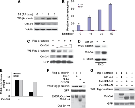
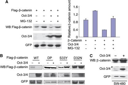
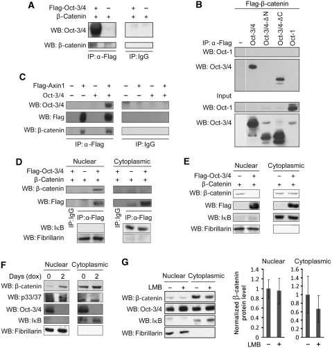
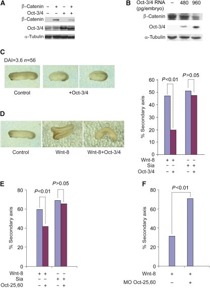
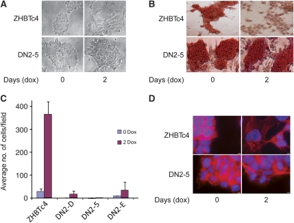
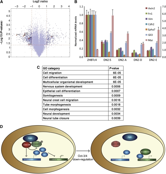
Although the transcriptional regulatory events triggered by Oct-3/4 are well documented, understanding the proteomic networks that mediate the diverse functions of this POU domain homeobox protein remains a major challenge. Here, we present genetic and biochemical studies that suggest an unexpected novel strategy for Oct-3/4-dependent regulation of embryogenesis and cell lineage determination. Our data suggest that Oct-3/4 specifically interacts with nuclear β-catenin and facilitates its proteasomal degradation, resulting in the maintenance of an undifferentiated, early embryonic phenotype both in Xenopus embryos and embryonic stem (ES) cells. Our data also show that Oct-3/4-mediated control of β-catenin stability has an important function in regulating ES cell motility. Down-regulation of Oct-3/4 increases β-catenin protein levels, enhancing Wnt signalling and initiating invasive cellular activity characteristic of epithelial-mesenchymal transition. Our data suggest a novel mode of regulation by which a delicate balance between β-catenin, Tcf3 and Oct-3/4 regulates maintenance of stem cell identity. Altering the balance between these proteins can direct cell fate decisions and differentiation.


Conflict of interest statement

The authors declare that they have no conflict of interest.

Figures

Figure 1
Figure 1
Oct-3/4 promotes β-catenin degradation through its N-terminal domain. (A) Western blot (WB) analysis of Oct-3/4 and β-catenin levels during retinoic acid (RA)-induced ES cell differentiation. Cells were treated with RA for 0–3 days. β-actin served as a loading control. (B) ZHBTc4 ES cells were transfected with TOPFlash or FOPFlash expression vectors. Luciferase activity was assayed in untreated cells and cells treated with dox for the indicated time points. (C) 293T cells were transfected with Flag-β-catenin and GFP expression vectors, together with increasing amounts of Oct-3/4 expression plasmid (1, 5 and 10 μg). β-catenin and Oct-3/4 protein levels were detected using anti-Flag and anti-Oct-3/4, antibodies, respectively. GFP was used as a transfection efficiency control. (D) RKO cells were infected with lentiviral Oct-3/4 expression vector or empty control vector. WB analyses of Oct-3/4 and endogenous β-catenin protein levels were performed using antibodies directed against Oct-3/4 and β-catenin. The α-tubulin served as a loading control. (E) A relative quantification of endogenous β-catenin protein (black bars) and β-catenin mRNA (grey bars) levels in Oct-3/4 transfected (+) or untransfected (−) RKO cells. Ubiquitin C (UBC) and glyceraldehyde 3-phosphate dehydrogenase (Gapdh) were used as loading controls for real-time quantitative PCR. (F) The 293T cells were transfected with Oct-1, Oct-2 and Oct-3/4 expression vectors. β-catenin degradation was shown by WB analysis. GFP served as a loading control. Cell extracts were incubated with P32-end-labelled Octa oligonucleotide, and Oct-1, Oct-2 and Oct-3/4 binding was detected using EMSA. (G) The 293T cells were transfected with the indicated expression vectors. Both Oct-3/4 and β-catenin levels were determined using WB analysis. GFP was used as a transfection efficiency control.
Figure 2
Figure 2
Oct-3/4 facilitates β-catenin proteasomal degradation. (A) The 293T cells were transfected with the indicated expression vectors. Cells were treated with 20 μM of MG-132 for 5 h. Levels of β-catenin and Oct-3/4 were measured by WB analysis using anti-Flag and anti-Oct-3/4, respectively. A relative quantification of β-catenin levels is shown. GFP served as a loading control. (B) The 293T cells were transfected with wt Flag-β-catenin, or the indicated β-catenin mutants, with or without Oct-3/4 expression vector. DP is a dominant-positive mutant in which S33, S37, T40, S45 and S47 were substituted with alanines; D32N and S33Y are single-point mutants (Amit et al, 2002). Protein levels were detected through WB using anti-Flag and anti-Oct-3/4. GFP served as a loading control. (C) SW480 cells were infected with a lentivirus expressing both Oct-3/4 and GFP. Control infections were carried out with a lentivirus expressing only GFP. Oct-3/4 and endogenous β-catenin were analysed by WB. GFP served as a loading control.
Figure 3
Figure 3
Oct-3/4 associates with β-catenin and promotes its phosphorylation and destabilization in the nucleus. (A) The 293T cells were transfected with Flag-Oct-3/4 and β-catenin expression vectors. Cell extracts were subjected to immunoprecipitation using either an antibody directed against the Flag-epitope or a control antibody, and the precipitated material was subjected to WB analysis with antibodies directed against Oct-3/4 and β-catenin. (B) The 293T cells were transfected with Flag-β-catenin expression vector together with wt Oct-3/4 or wt Oct-1 or Oct-3/4 mutants with deletion in either the N- or the C-terminal domains. Cell extracts were subjected to immunoprecipitation using anti-Flag, and the precipitated material was subjected to WB analysis with antibodies directed against Oct-1 and Oct-3/4. (C) The 293T cells were co-transfected with expression vector for β-catenin together with Flag-tagged Axin1 and/or Oct-3/4 expression vectors, as indicated. Cell extracts were subjected to immunoprecipitation using either an antibody directed against the Flag-epitope or a control antibody, and the precipitated material was subjected to WB analysis with antibodies directed against Oct-3/4, Flag-epitope and β-catenin. (D) The 293T cells were transfected with expression vectors for Flag-tagged Oct-3/4 and β-catenin, as indicated. Cytoplasmic and nuclear extracts were prepared and subjected to immunoprecipitation using an antibody directed against the Flag-epitope, or a control antibody, and the precipitated material was subjected to WB analysis with antibodies directed against β-catenin and the Flag-epitope. IκB and Fibrillarin were used as cytoplasmic and nuclear control markers, respectively. (E) 293T cells were transfected with expression vectors for β-catenin and Flag-tagged Oct-3/4, as indicated. Cytoplasmic and nuclear extracts were prepared and levels of β-catenin and Oct-3/4 were measured by WB analysis using anti-β-catenin and anti-Flag antibodies. IκB and Fibrillarin were used as cytoplasmic and nuclear control markers, respectively. (F) ZHBTc4 ES cells were either treated or not with 1 μg/ml of dox for 2 days and cytosolic and nuclear extracts were prepared. The phosphorylation levels of β-catenin, as well as total levels of β-catenin and Oct-3/4, were analysed by WB analysis using the indicated antibodies. (G) ZHBTc4 ES cells were either treated or not with 4 ng/ml of LMB for 12 h and cytosolic and nuclear extracts were prepared, and levels of β-catenin and Oct-3/4 were measured by WB analysis using anti-β-catenin and anti-Oct-3/4 antibodies. IκB and Fibrillarin were used as cytoplasmic and nuclear control markers, respectively. Experiments were repeated four times and a relative quantification of β-catenin levels is shown. Arrow bars depict +/− StDV.
Figure 4
Figure 4
Oct-3/4 interferes with Wnt/β-catenin signalling in vivo in Xenopus embryos. (A) Xenopus embryos were radially injected with β-catenin and/or Oct-3/4 mRNA at the 2–4-cell stage. β-catenin and Oct-3/4 protein levels were detected in embryo extracts using WB. The α-tubulin was used as a loading control. (B) Xenopus embryos were injected with increasing amounts of Oct-3/4 mRNA at the 2–4-cell stage. Embryo extracts were analysed using WB for Oct-3/4 and endogenous β-catenin protein levels. The α-tubulin was used as a loading control. (C) Xenopus embryos at the 2–4-cell stage were radially injected with Oct-3/4 mRNA. Examination of the embryos shows a ventralization phenotype. An un-injected embryo is shown as a control. (D) Summary of secondary-axis assays from Xenopus embryos injected at the 2–4 cell stage. Injection of mRNA encoding Wnt8 or Sia (5 pg) resulted in a high (48–55%) frequency of embryos with secondary axes. Co-injection of Oct-3/4 mRNA (30 ng) with Wnt8 mRNA results in inhibition of secondary-axis formation (P⩽0.01). Oct-3/4 does not inhibit Sia-induced secondary-axis formation. Representative embryos injected with the indicated RNAs are shown. (E) Summary of secondary-axis assays from Xenopus embryos injected at the 2–4 cell stage. Injections of mRNAs and analysis were performed as in (D). Oct-25,60 were used instead of Oct-3/4. Co-injection of Oct-25,60 mRNAs (30 ng) with Wnt8 mRNA results in inhibition of secondary-axis formation (P⩽0.01). Oct-25,60 does not inhibit Sia-induced secondary-axis formation. (F) Xenopus embryos were injected at the 2–4 cell stage with mRNA encoding Wnt8 together with antisense morpholinos oligos (MO) directed against Oct-25,60. A full-colour version of this figure is available at The EMBO Journal Online.
Figure 5
Figure 5
Oct-3/4 repression induces cellular motility in ES cells through activation of the Wnt signalling pathway. (A) Morphological changes in ZHBTc4 and ZHBTc4/DN-5 ES cells upon dox-induced repression of Oct-3/4. Left—photomicrographs of undifferentiated ES cells cultured in the presence of LIF, and the absence of dox. Right—cells were grown in the presence of LIF and dox for 2 days. All photographs are in the same magnification (× 200). (B) Alkaline phosphatase staining for the indicated ES cells before and after dox treatment in the presence of LIF. All photographs are in the same magnification (× 200). (C) Cellular motility of the indicated ES cells grown in the presence of LIF, treated or untreated with dox. Data represent the average number of migrating cells, counting in magnification of × 100. (D) Actin filaments phalloidin immunofluorescent staining of the indicated ES cells, either treated or untreated with dox. Scale bar, 80 μm.
Figure 6
Figure 6
Genome-wide analysis of differentiated ZHBTc4 and ZHBTc4/DN-5 cells. (A) To challenge the statistical significance of the microarray results, we used the data obtained from the four arrays to perform the volcano analysis. Using thresholds of ⩾two-fold decrease or increase in expression and a P-value of ⩽0.05, 878 genes were detected. All dots in the scatter plot above the horizontal line have P-values <0.05 (calculated using the t-test). All points to the left of the left vertical line are down-regulated by at least two-fold in dox-treated ZHBTc4/DN-5 relative to the dox-treated ZHBTc4. The dots to the right of the right vertical line are up-regulated by at least two-fold. (B) Seven indicated genes were measured using quantitative real-time PCR in order to validate the array results. The levels of Myc are shown on the scale to the right. UBC and Gapdh were used as endogenous controls. (C) Gene ontology (GO) analysis of the genes with a differential expression between dox-treated ZHBTc4 and ZHBTc4/DN-5 ES cells. The P-value of this representation is based on a hypergeometric distribution. Lowest P-value GO categories are shown. (D) A model for the functional cross-talk between Oct-3/4 and Wnt/β-catenin signalling, see text.

References

    1. Akiyama H, Lyons JP, Mori-Akiyama Y, Yang X, Zhang R, Zhang Z, Deng JM, Taketo MM, Nakamura T, Behringer RR, McCrea PD, de Crombrugghe B (2004) Interactions between Sox9 and beta-catenin control chondrocyte differentiation. Genes Dev 18: 1072–1087 - PMC - PubMed
    1. Amit S, Hatzubai A, Birman Y, Andersen JS, Ben-Shushan E, Mann M, Ben-Neriah Y, Alkalay I (2002) Axin-mediated CKI phosphorylation of beta-catenin at Ser 45: a molecular switch for the Wnt pathway. Genes Dev 16: 1066–1076 - PMC - PubMed
    1. Aubert J, Dunstan H, Chambers I, Smith A (2002) Functional gene screening in embryonic stem cells implicates Wnt antagonism in neural differentiation. Nat Biotechnol 20: 1240–1245 - PubMed
    1. Ben-Aroya S, Agmon N, Yuen K, Kwok T, McManus K, Kupiec M, Hieter P (2010) Proteasome nuclear activity affects chromosome stability by controlling the turnover of mms22 a protein important for DNA repair. PLoS Genet 6: e1000852. - PMC - PubMed
    1. Ben-Shushan E, Thompson JR, Gudas LJ, Bergman Y (1998) Rex-1, a gene encoding a transcription factor expressed in the early embryo, is regulated via Oct-3/4 and Oct-6 binding to an octamer site and a novel protein, Rox-1, binding to an adjacent site. Mol Cell Biol 18: 1866–1878 - PMC - PubMed

Publication types