. 2010 Sep 28;103(7):1008-18.
doi: 10.1038/sj.bjc.6605839. Epub 2010 Aug 24.

Co-expression of CD147 (EMMPRIN), CD44v3-10, MDR1 and monocarboxylate transporters is associated with prostate cancer drug resistance and progression

Affiliations

Co-expression of CD147 (EMMPRIN), CD44v3-10, MDR1 and monocarboxylate transporters is associated with prostate cancer drug resistance and progression

J Hao et al. Br J Cancer. .

Abstract

Background: The aim of this study is to seek an association between markers of metastatic potential, drug resistance-related protein and monocarboxylate transporters in prostate cancer (CaP).

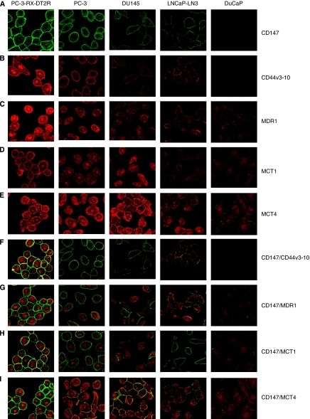
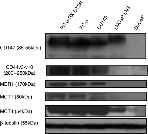
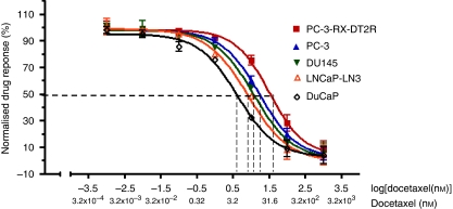
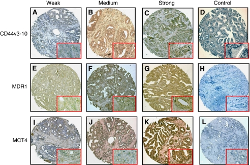
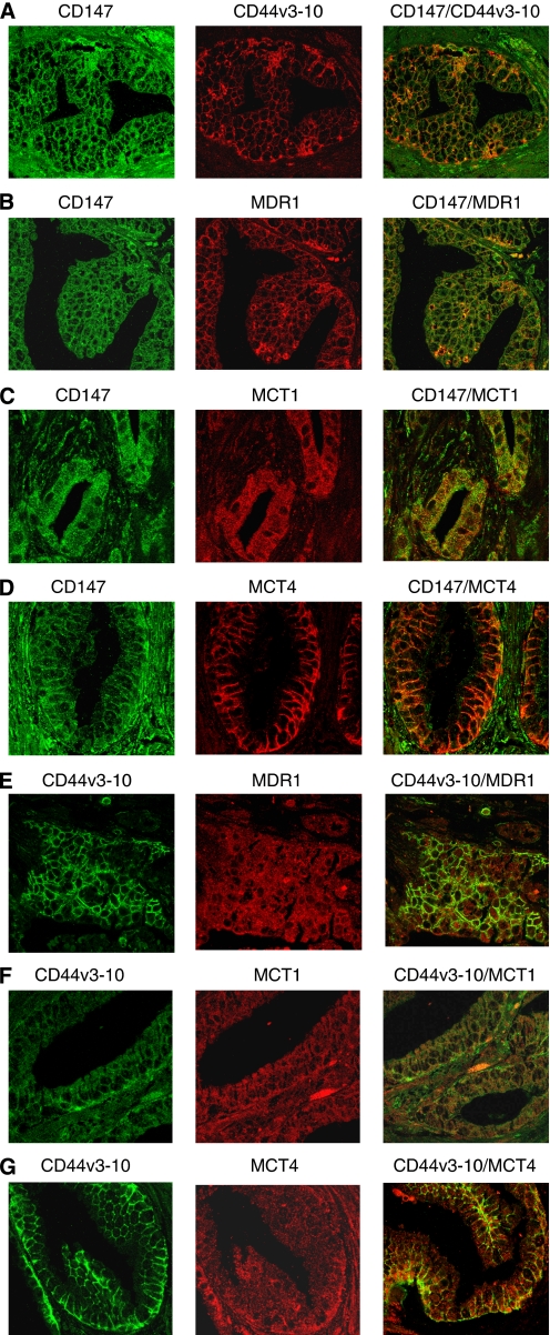
Methods: We evaluated the expression of invasive markers (CD147, CD44v3-10), drug-resistance protein (MDR1) and monocarboxylate transporters (MCT1 and MCT4) in CaP metastatic cell lines and CaP tissue microarrays (n=140) by immunostaining. The co-expression of CD147 and CD44v3-10 with that of MDR1, MCT1 and MCT4 in CaP cell lines was evaluated using confocal microscopy. The relationship between the expression of CD147 and CD44v3-10 and the sensitivity (IC(50)) to docetaxel in CaP cell lines was assessed using MTT assay. The relationship between expression of CD44v3-10, MDR1 and MCT4 and various clinicopathological CaP progression parameters was examined.

Results: CD147 and CD44v3-10 were co-expressed with MDR1, MCT1 and MCT4 in primary and metastatic CaP cells. Both CD147 and CD44v3-10 expression levels were inversely related to docetaxel sensitivity (IC(50)) in metastatic CaP cell lines. Overexpression of CD44v3-10, MDR1 and MCT4 was found in most primary CaP tissues, and was significantly associated with CaP progression.

Conclusions: Our results suggest that the overexpression of CD147, CD44v3-10, MDR1 and MCT4 is associated with CaP progression. Expression of both CD147 and CD44v3-10 is correlated with drug resistance during CaP metastasis and could be a useful potential therapeutic target in advanced disease.


Figures

Figure 1
Figure 1
Co-immunolabelling of CD147, CD44v3-10, MDR1, MCT1 and MCT4 in metastatic prostate cancer (CaP) cell lines. Representative confocal images of CD147 (green), CD44v3-10, MDR1, MCT1 and MCT4 (red) expression are shown. Merged images and red and green channels are shown separately. Membrane expression was found for CD147 and CD44v3-10 antibodies, whereas expressions of both membrane and cytoplasm were seen for MDR1, MCT1 and MCT4 antibodies. All immunostainings are more homogeneous. (A) CD147; (B) CD44v3-10; (C) MDR1; (D) MCT1; (E) MCT4; (F) colocalisation of CD147 with CD44v3-10; (G) colocalisation of CD147 with MDR1; (H) colocalisation of CD147 with MCT1; (I) colocalisation of CD147 with MCT1. Magnification: AI × 400. The colour reproduction of the figure is available on the html full text version of the paper.
Figure 2
Figure 2
Expression of CD147, CD44v3-10, MDR1, MCT1 and MCT4 in prostate cancer (CaP) cell lines by western blotting. A representative western blot showing high levels of CD147, CD44v3-10, MDR1 (F4), MCT1 and MCT4 expression in PC-3-RX-DT2R and PC-3 cell lines, moderate expression in DU 145 cells and low or no expression in LNCaP and DuCaP cells. Equal loading is demonstrated with β-tubulin antibody in the bottom panel (lane 1: RX-DT2R, lane 2: PC-3, lane 3: DU145, lane 4: LNCaP-LN3. lane 5: DuCaP).
Figure 3
Figure 3
Dose response of metastatic prostate cancer (CaP) cell lines to docetaxel measured using 3-(4,5-dimethylthiazol-2-yl)-2,5-diphenyl tetrazolium bromide (MTT) assay. Drug-resistant and metastatic CaP cell lines treated with a range of concentrations of docetaxel (0.001–1000 nM, x axis) showed varying responses. The IC50 value (50% normalised cell response) is related to the expression of CD147 and CD44v3-10. For example, the drug-resistant cell line, PC3-RX-DT2R, displays Grade 3 CD147 and CD44v3-10 expression, and an IC50 of 44.7 nM (x axis); the drug-sensitive CaP cell line, DuCaP, displays Grade 0 CD147 and CD44v3-10 immunostaining and the lowest IC50 of 4 nM (x axis) (n=3, mean±s.d.).
Figure 4
Figure 4
Expression of CD44v3-10, MDR1 and MCT4 in prostate cancer (CaP) tissue microarrays (TMAs). Representative images are shown of Grade 1 (weak) CD44v3-10 (A), MDR1 (E) and MCT4 (I); Grade 2 (medium) CD44v3-10 (B), MDR1(F) and MCT4 (J); and Grade 3 (strong) CD44v3-10 (C), MDR1(G) and MCT4 (K) immunostaining. No immunoreactivity is seen in non-specific negative controls for CD44v3-10 (D), MDR1 (H) and MCT4 (L). Brown colour indicates positive immunostaining. Insets indicate the typical areas of staining at high amplification. Magnification: AL × 10; and insets × 40. The colour reproduction of the figure is available on the html full text version of the paper.
Figure 5
Figure 5
Co-immunolabelling of CD147, CD44v3-10, MDR1, MCT1 and MCT4 in primary prostate cancer (CaP) tissues (different tumours with a range of Gleason scores). Representative confocal images of CD147 and CD44v3-10 (green), CD44v3-10, MDR1, MCT1 and MCT4 (red) immunolabelling are shown. Merged images and red and green channels are shown separately. (A) CD147 and CD44v3-10 in high-grade CaP (Gleason score=8); (B) CD147 and MDR1 in high-grade CaP (Gleason score=8); (C) CD147 and MCT1 in low-grade CaP (Gleason score=6); (D) CD147 and MCT4 in high-grade CaP (Gleason score=8); (E) CD44v3-10 and MDR1 in high-grade CaP (Gleason score=9); (F) CD44v3-10 and MCT1 in high-grade CaP (Gleason score=8); (G) CD44v3-10 and MCT4 in high-grade CaP (Gleason score=8). CD147 immunolabelling is seen on epithelial cell membranes and stromal cells. CD44v3-10, MDR1, MCT1 and MCT4 immunostaining is predominantly epithelial. MCT4 localises mostly to basal epithelial cells and to lateral cell walls (magnification: AG × 400). The colour reproduction of the figure is available on the html full text version of the paper.

Similar articles

Cited by

References

    1. Albers MJ, Bok R, Chen AP, Cunningham CH, Zierhut ML, Zhang VY, Kohler SJ, Tropp J, Hurd RE, Yen YF, Nelson SJ, Vigneron DB, Kurhanewicz J (2008) Hyperpolarized 13C lactate, pyruvate, and alanine: noninvasive biomarkers for prostate cancer detection and grading. Cancer Res 68: 8607–8615 - PMC - PubMed
    1. Auzenne E, Ghosh SC, Khodadadian M, Rivera B, Farquhar D, Price RE, Ravoori M, Kundra V, Freedman RS, Klostergaard J (2007) Hyaluronic acid-paclitaxel: antitumour efficacy against CD44(+) human ovarian carcinoma xenografts. Neoplasia 9: 479–486 - PMC - PubMed
    1. Chen CT, Gan Y, Au JL, Wientjes MG (1998) Androgen-dependent and -independent human prostate xenograft tumours as models for drug activity evaluation. Cancer Res 58: 2777–2783 - PubMed
    1. Colone M, Calcabrini A, Toccacieli L, Bozzuto G, Stringaro A, Gentile M, Cianfriglia M, Ciervo A, Caraglia M, Budillon A, Meo G, Arancia G, Molinari A (2008) The multidrug transporter P-glycoprotein: a mediator of melanoma invasion? J Invest Dermatol 128: 957–971 - PubMed
    1. Cozzi PJ, Wang J, Delprado W, Madigan MC, Fairy S, Russell PJ, Li Y (2006) Evaluation of urokinase plasminogen activator and its receptor in different grades of human prostate cancer. Human Pathol 37: 1442–1451 - PubMed

Publication types

MeSH terms